
Recommended
More Related Content
Similar to Chronic Periodontitis- the malaise of population
Similar to Chronic Periodontitis- the malaise of population (20)
More from AshokKp4
More from AshokKp4 (20)
Recently uploaded
Recently uploaded (20)
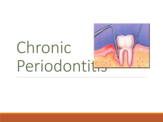
Chronic Periodontitis- the malaise of population
- 2. Definition Chronic Periodontitis can be defined as “an infectious disease resulting in inflammation within the supporting tissues of the teeth, progressive attachment loss, and bone loss.” - Previously known as adult periodontitis or slowly progressive periodontitis. - Occur as a result of extension of inflammation from the gingiva into deeper periodontal tissue. 2
- 3. Common Characteristics Onset - any age; most common in adults Plaque initiates condition Subgingival calculus common finding Slow-mod progression; periods of rapid progression possible Modified by local factors/systemic factors/stress/smoking 3
- 4. Extent & Severity Extent: ◦ Localized: <30% of sites affected ◦ Generalized: > 30% of sites affected Severity: entire dentition or individual teeth/site ◦ Slight = 1-2 mm CAL ◦ Moderate = 3-4 mm CAL ◦ Severe = 5 mm CAL 4
- 5. Clinical Characteristics Gingiva moderately swollen Deep red to bluish-red tissues Blunted and rolled gingival margin Cratered papilla Bleeding and/or suppuration 5
- 6. Clinical Characteristics Plaque/calculus deposits Variable pocket depths Loss of periodontal attachment Horizontal/vertical bone loss Tooth mobility 6
- 7. CLASSIFICATION 7 A) Based on Disease Distribution: Localized: Periodontitis is considered localized when <30% of the sites assessed in mouth demonstrate attachment loss and bone loss. Generalized: Periodontitis is considered generalized when >30% of the sites assessed demonstrate attachment loss and bone loss. The pattern of bone loss in chronic periodontitis can be vertical or horizontal.
- 8. Sub classification of Chronic Periodontitis Severity Pocket Depths CAL Bone Loss Furcation Early 4-5 mm 1-2 mm Slight horizontal Moderate 5-7 mm 3-4 mm Sl – mod horizontal Advanced > 7 mm 5 mm Mod- severe horizontal vertical 8
- 9. DISEASE DISTRIBUTION : It is a site-specific disease CLINICAL SIGNS - - Inflammation ,pocket formation ,attachment loss ,bone loss - All caused by site specific effects of a sub-gingival plaque accumulation - That is why the effect are on one side only –other surface may maintain normal attachment level. - Eg.-proximal surface with plaque may have C.A.L. - And plaque free surface –FACIAL surface of same tooth may be without disease.
- 10. SYMPTOMS Patient notices-- 1. gum bleed 2. space appear between teeth due to tooth movement 3. May be painless (sleeping disease )goes unnoticed 4. Some time pain due to caries , root hypersensitivity 5. To cold /hot or both 6. PAIN-may be-- dull—deep radiating in the jaw 7. Area of food impaction can cause more discomfort 8. May be gingival tenderness or itchiness found
- 11. Periodontal Pathogens •Gram negative organism dominate •P.g., P.i., A.a. may infiltrate: • - Intercellular spaces of the epithelium • - Between deeper epithelial cells • - Basement lamina 11
- 12. Periodontal Pathogens Contn. Pathogens include: Nonmotile rods: Facultative: Actinobacillus a. E.c. Anaerobic: P. g., P. i., B.f., F.n. Motile rods: Facultative: C.r. Spirochetes: Anaerobic, motile: Treponema denticola 12
- 13. Pathogenesis – Pocket Formation Bacterial challenge initiates initial lesion of gingivitis With disease progression & change in microorganisms development of periodontitis 13
- 14. Pocket Formation Cellular & fluid inflammatory exudate degenerates CT Gingival fibers destroyed Collagen fibers apical to JE destroyed infiltration of inflammatory cells & edema Apical migration of junctional epithelium along root Coronal portion of JE detaches 14
- 15. Pocket Formation Continued extension of JE requires healthy epithelial cells! Necrotic JE slows down pocket formation Pocket base degeneration less severe than lateral 15
- 16. Pocket Formation Continue inflammation: ◦ Coronal extension of gingival margin ◦ JE migrates apically & separates from root ◦ Lateral pocket wall proliferates & extends into CT ◦ Leukocytes & edema ◦ Infiltrate lining epithelium ◦ Varying degrees of degeneration & necrosis 16
- 18. Continuous Cycle! Plaque gingival inflammation pocket formation more plaque 18
- 19. Classification of Pockets Gingival: ◦ Coronal migration of gingival margin Periodontal: ◦ Apical migration of epithelial attachment ◦ Suprabony: ◦ Base of pocket coronal to height of alveolar crest ◦ Infrabony: ◦ Base of pocket apical to height of alveolar crest ◦ Characterized by angular bony defects 19
- 20. Histopathology Connective Tissue: ◦ Edematous ◦ Dense infiltrate: ◦ Plasma cells (80%) ◦ Lymphocytes, PMNs ◦ Blood vessels proliferate, dilate & are engorged. ◦ Varying degrees of degeneration in addition to newly formed capillaries, fibroblasts, collagen fibers in some areas. 20
- 21. Histopathology Periodontal pocket: ◦ Lateral wall shows most severe degeneration ◦ Epithelial proliferation & degeneration ◦ Rete pegs protrude deep within CT ◦ Dense infiltrate of leukocytes & fluid found in rete pegs & epithelium ◦ Degeneration & necrosis of epithelium leads to ulceration of lateral wall, exposure of CT, suppuration 21
- 22. Clinical & Histopathologic Features Clinical : 1. Pocket wall bluish-red 2. Smooth, shiny surface 3. Pitting on pressure Histopathology: 1. Vasodilation & vasostagnation 2. Epithelial proliferation, edema 3. Edema & degeneration of epithelium 22
- 23. Clinical & Histopathologic Features Contn… Clinical: 1. Pocket wall may be pink & firm 2. Bleeding with probing 3. Pain with instrumentation Histopathology: 1. Fibrotic changes dominate 2. blood flow, degenerated, thin epithelium 3. Ulceration of pocket epithelium 23
- 24. Clinical & Histopathologic Features Contn… Clinical : 1. Exudate 2. Flaccid tissues Histopathology: 1. Accumulation of inflammatory products 2. Destruction of gingival fibers 24
- 25. Stages of Periodontal Disease 25
- 26. Root Surface Wall Periodontal disease affects root surface: ◦ Perpetuates disease ◦ Decay, sensitivity ◦ Complicates treatment Embedded collagen fibers degenerate cementum exposed to environment Bacteria penetrate unprotected root 26
- 27. Root Surface Wall Contn… Necrotic areas of cementum form; clinically soft Act as reservoir for bacteria Root planing may remove necrotic areas firmer surface 27
- 28. Inflammatory Pathway Stages I-III – inflammation degrades gingival fibers ◦ Spreads via blood vessels: Interproximal: Loose CT transseptal fibers marrow spaces of cancellous bone periodontal ligament suprabony pockets & horizontal bone loss transseptal fibers transverse horizontally 28
- 29. Inflammatory Pathway Interproximal: ◦ Loose CT periodontal ligament bone infrabony pockets & vertical bone loss transseptal fibers transverse in oblique direction 29
- 30. Inflammatory Pathway Facial & Lingual: ◦ Loose CT along periosteum marrow spaces of cancellous bone supporting bone destroyed first alvoelar bone proper periodontal ligament suprabony pocket & horizontal bone loss 30
- 31. Inflammatory Pathway Facial & Lingual: ◦ Loose CT periodontal ligament destruction of periodontal ligament fibers infrabony pockets & vertical or angular bone loss 31
- 32. Periodontal Disease Activity Bursts of activity followed by periods of quiescence characterized by: ◦ Reduced inflammatory response ◦ Little to no bone loss & CT loss Accumulation of Gram negative organisms leads to: ◦ Bone & attachment loss ◦ Bleeding, exudates ◦ May last days, weeks, months 32
- 33. Periodontal Disease Activity Period of activity followed by period of remission: ◦ Accumulation of Gram positive bacteria ◦ Condition somewhat stabilized Periodontal destruction is site specific PD affects few teeth at one time, or some surfaces of given teeth 33
- 34. Prevalence: Chronic Periodontitis increases in prevalence & severity with age. Affect both the sexes equally. It is an age-associated, not age related disease.
- 35. RISK FACTORS FOR DISEASE: 1) PRIOR HISTORY OF PERIODONTITIS—predictor-more risk for developing damage to periodontium. 2) LOCAL FACTORS: Plaque Accumulation Oral Hygiene Tooth Malposition Restoration Preserve & Quantity of certain bacteria Host defences Subgingival Restoration Environment Calculus, smoking Connective Tissue destruction Genetic influence Inflammation Periodontopathic bacteria Smoking, Calculus Loss of Attachment M O D I F Y I N G F A C T O R S
- 36. 3) SYSTEMIC FACTORS: Type II or Non – Insulin dependent Diabetes Mellitus (NIIDDM) 4) ENVIRONMENTAL & BEHAVIORAL FACTORS: ◦ Smoking ◦ Emotional Stress 5) GENETIC FACTORS: ◦ Frequent among family members and across different generations. GENERAL CONCEPT FOR ETIOLOGY OF CHRONIC PERIODONTITIS Plaque accumulation Maturation of Plaque Quality & Quantity of periodontopathic Plaque accumulation Maturation of Plaque Quality & Quantity of periodontopathic bacteria InflammationPlaque accumulation Maturation of Plaque Quality & Quantity of periodontopathic bacteria Inflammation Connective tissue destruction. Connective tissue destruction. bacteria Inflammation Connective tissue destruction. Host status and defences Plaque accumulation Maturation of Plaque Quality & Quantity of periodontopathic bacteria Inflammation
- 37. MANAGEMENT The treatment consists of – 1. Non-surgical procedures ◦ Scaling ◦ Root planing ◦ Curettage 2. Surgical procedure ◦ Pocket reduction surgery ◦ Resective ◦ Regenerative ◦ Correction of morphological / anatomic defects
- 38. Overall Prognosis Dependent on: ◦ Client compliance ◦ Systemic involvement ◦ Severity of condition ◦ # of remaining teeth 38
- 39. Prognosis of Individual Teeth Dependent on: ◦ Attachment levels, bone height ◦ Status of adjacent teeth ◦ Type of pockets: suprabony, infrabony ◦ Furcation involvement ◦ Root resorption 39
- 40. Thank You 40
Editor's Notes
- Algonquin College