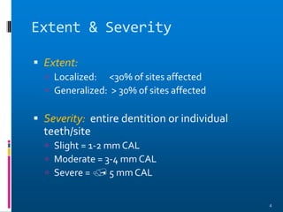
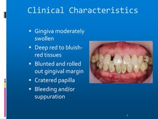
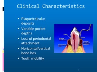
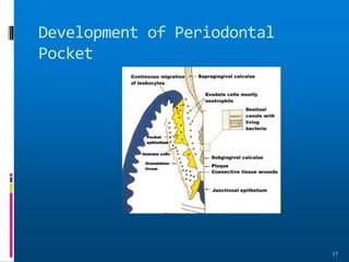
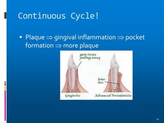
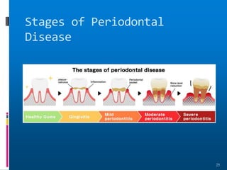
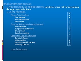
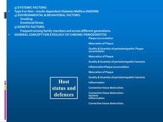
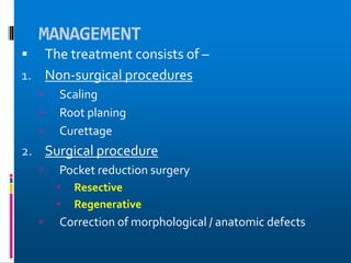
SlideShare a Scribd company logo
CHRONIC
PERIODONTITIS
Dr.D.Navya, MDS
1
Definition
 Chronic Periodontitis can be defined as “an
infectious disease resulting in inflammation
within the supporting tissues of the teeth,
progressive attachment loss, and bone loss.”
- Previously known as adult periodontitis or
slowly progressive periodontitis.
- Occur as a result of extension of inflammation
from the gingiva into deeper periodontal tissue.
2
Common Characteristics
 Onset - any age; most common in adults
 Plaque initiates condition
 Subgingival calculus common finding
 Slow-mod progression; periods of rapid
progression possible
 Modified by local factors/systemic
factors/stress/smoking
3
Extent & Severity
 Extent:
 Localized: <30% of sites affected
 Generalized: > 30% of sites affected
 Severity: entire dentition or individual
teeth/site
 Slight = 1-2 mm CAL
 Moderate = 3-4 mm CAL
 Severe =  5 mm CAL
4
Clinical Characteristics
 Gingiva moderately
swollen
 Deep red to bluish-
red tissues
 Blunted and rolled
out gingival margin
 Cratered papilla
 Bleeding and/or
suppuration
5
Clinical Characteristics
 Plaque/calculus
deposits
 Variable pocket
depths
 Loss of periodontal
attachment
 Horizontal/vertical
bone loss
 Tooth mobility
6
CLASSIFICATION
7
A) Based on Disease Distribution:
Localized:
Periodontitis is considered localized when <30% of
the sites assessed in mouth demonstrate attachment
loss and bone loss.
Generalized:
Periodontitis is considered generalized when >30% of
the sites assessed demonstrate attachment loss and
bone loss.
The pattern of bone loss in chronic periodontitis can
be vertical or horizontal.
Sub classification of Chronic
Periodontitis
8
 DISEASE DISTRIBUTION : It is a site-specific disease
 CLINICAL SIGNS -
- Inflammation ,pocket formation ,attacment loss ,bone loss
- All caused by site specific effects of a sub-gingival plaque
accumulation
-That is why the effect are on one side only –other surface
may maintain normal attachment level.
- Eg.-proximal surface with plaque may have C.A.L.
- And plaque free surface –FACIALsurface of same tooth
may be without disease.
SYMPTOMS
Patient notices--
1. Bleeding gums
2. spaces appear between teeth due to tooth movement
3. May be painless (sleeping disease )goes unnoticed
4. Some time pain due to caries , root hypersensitivity
5. To cold /hot or both
6. PAIN-may be-- dull—deep radiating in the jaw
7. Area of food impaction can cause more discomfort
8. May be gingival tenderness or itchiness found
Periodontal Pathogens
• Gram negative organism dominate
• P.g., P.i., A.a. may infiltrate:
• - Intercellular spaces of the epithelium
• - Between deeper epithelial cells
• - Basement lamina
11
Periodontal Pathogens Contn.
 Pathogens include:
 Nonmotile rods:
 Facultative:
 Actinobacillus a. E.c.
 Anaerobic:
 P. g., P. i., B.f., F.n.
 Motile rods:
 Facultative:
 C.r.
 Spirochetes:
 Anaerobic, motile:
 Treponema denticola
12
Pathogenesis – Pocket
Formation
 Bacterial challenge
initiates initial lesion
of gingivitis
 With disease
progression &
change in
microorganisms 
development of
periodontitis
13
Pocket Formation
 Cellular & fluid inflammatory exudate 
degenerates CT
 Gingival fibers destroyed
 Collagen fibers apical to JE destroyed 
infiltration of inflammatory cells & edema
 Apical migration of junctional epithelium
along root
 Coronal portion of JE detaches
14
Pocket Formation
 Continued extension
of JE requires
healthy epithelial
cells!
 Necrotic JE slows
down pocket
formation
 Pocket base
degeneration less
severe than lateral
15
Pocket Formation
 Continue inflammation:
 Coronal extension of gingival margin
 JE migrates apically & separates from root
 Lateral pocket wall proliferates & extends into CT
 Leukocytes & edema
 Infiltrate lining epithelium
 Varying degrees of degeneration & necrosis
16
Development of Periodontal
Pocket
17
Continuous Cycle!
 Plaque  gingival inflammation  pocket
formation  more plaque
18
Classification of Pockets
 Gingival:
 Coronal migration of gingival margin
 Periodontal:
 Apical migration of epithelial attachment
 Suprabony:
 Base of pocket coronal to height of alveolar crest
 Infrabony:
 Base of pocket apical to height of alveolar crest
 Characterized by angular bony defects
19
Histopathology
 ConnectiveTissue:
 Edematous
 Dense infiltrate:
 Plasma cells (80%)
 Lymphocytes, PMNs
 Blood vessels proliferate, dilate & are engorged.
 Varying degrees of degeneration in addition to newly
formed capillaries, fibroblasts, collagen fibers in some
areas.
20
Histopathology
 Periodontal pocket:
 Lateral wall shows most severe degeneration
 Epithelial proliferation & degeneration
 Rete pegs protrude deep within CT
 Dense infiltrate of leukocytes & fluid found in rete
pegs & epithelium
 Degeneration & necrosis of epithelium leads to
ulceration of lateral wall, exposure of CT,
suppuration
21
Clinical &
Histopathologic
Features
 Clinical :
1. Pocket wall
bluish-red
2. Smooth, shiny
surface
3. Pitting on
pressure
 Histopathology:
1. Vasodilation &
vasostagnation
2. Epithelial
proliferation,
edema
3. Edema &
degeneration of
epithelium
22
Clinical & Histopathologic
Features
Contn…
 Clinical:
1. Pocket wall may be
pink & firm
2. Bleeding with probing
3. Pain with
instrumentation
 Histopathology:
1. Fibrotic changes
dominate
2.  blood flow,
degenerated, thin
epithelium
3. Ulceration of pocket
epithelium
23
Clinical & Histopathologic
Features
Contn…
 Clinical :
1. Exudate
2. Flaccid tissues
 Histopathology:
1. Accumulation of
inflammatory
products
2. Destruction of gingival
fibers
24
Stages of Periodontal
Disease
25
Root Surface Wall
 Periodontal disease affects root surface:
 Perpetuates disease
 Decay, sensitivity
 Complicates treatment
 Embedded collagen fibers degenerate 
cementum exposed to environment
 Bacteria penetrate unprotected root
26
Root Surface Wall Contn…
 Necrotic areas of cementum form; clinically
soft
 Act as reservoir for bacteria
 Root planing may remove necrotic areas 
firmer surface
27
Inflammatory Pathway
 Stages I-III – inflammation degrades gingival
fibers
 Spreads via blood vessels:
 Interproximal:
 Loose CT  transseptal fibers  marrow spaces
of cancellous bone  periodontal ligament 
suprabony pockets & horizontal bone loss
transseptal fibers transverse horizontally
28
Inflammatory Pathway
 Interproximal:
 Loose CT  periodontal ligament  bone 
infrabony pockets & vertical bone loss 
transseptal fibers transverse in oblique direction
29
Inflammatory Pathway
 Facial & Lingual:
 Loose CT  along periosteum  marrow spaces
of cancellous bone  supporting bone destroyed
first  alvoelar bone proper  periodontal
ligament  suprabony pocket & horizontal bone
loss
30
Inflammatory Pathway
 Facial & Lingual:
 Loose CT  periodontal ligament  destruction
of periodontal ligament fibers  infrabony pockets
& vertical or angular bone loss
31
Periodontal Disease Activity
 Bursts of activity followed by periods of
quiescence characterized by:
 Reduced inflammatory response
 Little to no bone loss & CT loss
 Accumulation of Gram negative organisms leads
to:
 Bone & attachment loss
 Bleeding, exudates
 May last days, weeks, months
32
Periodontal Disease Activity
 Period of activity followed by period of
remission:
 Accumulation of Gram positive bacteria
 Condition somewhat stabilized
 Periodontal destruction is site specific
 PD affects few teeth at one time, or some
surfaces of given teeth
33
Prevalence:
 Chronic Periodontitis increases in prevalence & severity with
age.
 Affect both the sexes equally.
 It is an age-associated, not age related disease.
RISK FACTORS FOR DISEASE:
1) PRIOR HISTORY OF PERIODONTITIS—predictor-more risk for developing
damage to periodontium.
2) LOCAL FACTORS:
Plaque Accumulation
Oral Hygiene
Tooth Malposition
Restoration
Preserve & Quantity of certain bacteria
Host defences
Subgingival Restoration
Environment
Calculus, smoking
ConnectiveTissue destruction
Genetic influence
Inflammation
Periodontopathic bacteria
Smoking, Calculus
Loss of Attachment
M
O
D
I
F
Y
I
N
G
F
A
C
T
O
R
S
3) SYSTEMIC FACTORS:
 Type II or Non – Insulin dependent Diabetes Mellitus (NIIDDM)
4) ENVIRONMENTAL & BEHAVIORAL FACTORS:
 Smoking
 Emotional Stress
5) GENETIC FACTORS:
 Frequent among family members and across different generations.
 GENERAL CONCEPT FOR ETIOLOGY OF CHRONIC PERIODONTITIS
Plaque accumulation
Maturation of Plaque
Quality & Quantity of periodontopathic Plaque
accumulation
Maturation of Plaque
Quality & Quantity of periodontopathic bacteria
InflammationPlaque accumulation
Maturation of Plaque
Quality & Quantity of periodontopathic bacteria
Inflammation
Connective tissue destruction.
bacteria
Inflammation
Connective tissue destruction.
Host
status and
defences
MANAGEMENT
 The treatment consists of –
1. Non-surgical procedures
 Scaling
 Root planing
 Curettage
2. Surgical procedure
 Pocket reduction surgery
 Resective
 Regenerative
 Correction of morphological / anatomic defects
Overall Prognosis
 Dependent on:
 Patient compliance
 Systemic involvement
 Severity of condition
 status of remaining teeth
38
Prognosis of Individual
Teeth
 Dependent on:
 Attachment levels, bone height
 Status of adjacent teeth
 Type of pockets: suprabony, infrabony
 Furcation involvement
 Root resorption
39
conclusion

More Related Content

Similar to 12.Chronic Periodontitis.pptx

Periodontal disease
Periodontal diseasePeriodontal disease
Periodontal disease
Hermie Culeen Flores
 
Gingival enlargement and recession
Gingival enlargement and recessionGingival enlargement and recession
Gingival enlargement and recession
Dr. Ashif Iqbal
 
Necrotizing ulcerative gingivitis & periodontits
Necrotizing ulcerative gingivitis & periodontitsNecrotizing ulcerative gingivitis & periodontits
Necrotizing ulcerative gingivitis & periodontits
DrGhadooRa
 
clinical features of gingivitis.pdf
clinical features of gingivitis.pdfclinical features of gingivitis.pdf
clinical features of gingivitis.pdf
Rama Dental College Hospital and Research Center
 
Acute Necrotizing Ulcerative Gingivitis.pptx
Acute Necrotizing Ulcerative Gingivitis.pptxAcute Necrotizing Ulcerative Gingivitis.pptx
Acute Necrotizing Ulcerative Gingivitis.pptx
soonhanmemon2k20
 
chronic periodontitis.pptx
chronic periodontitis.pptxchronic periodontitis.pptx
chronic periodontitis.pptx
Maria Antony Dhivyan
 
presentation 2.docx
presentation 2.docxpresentation 2.docx
presentation 2.docx
MohammadAlkhasawneh4
 
Gingival enlargement
Gingival enlargementGingival enlargement
Gingival enlargement
yaskodental
 
PERIODONTAL POCKET.pptx
PERIODONTAL POCKET.pptxPERIODONTAL POCKET.pptx
PERIODONTAL POCKET.pptx
veena621629
 
Chronic periodontitis
Chronic periodontitisChronic periodontitis
Chronic periodontitis
DR. OINAM MONICA DEVI
 
The periodontic endodontic continuum.
The  periodontic endodontic continuum.The  periodontic endodontic continuum.
The periodontic endodontic continuum.
Anushri Gupta
 
chronic periodontitis
chronic periodontitischronic periodontitis
chronic periodontitis
Dr. Vartika Srivastava
 
Periodontal pockets.pptx
Periodontal  pockets.pptxPeriodontal  pockets.pptx
Periodontal pockets.pptx
abhijeetshete6
 
PERIODONTAL POCKET.pptx
PERIODONTAL POCKET.pptxPERIODONTAL POCKET.pptx
PERIODONTAL POCKET.pptx
veena621629
 
Periodontal pocket
Periodontal pocketPeriodontal pocket
Periodontal pocket
lobna elsaadawy
 
Gingival pathology
Gingival pathologyGingival pathology
Gingival pathology
Dr.Shraddha Kode
 
Tissue space infection ‫‬.ppt
Tissue space infection ‫‬.pptTissue space infection ‫‬.ppt
Tissue space infection ‫‬.ppt
adel mohammed
 
Gingival inflammation- Group B presentation.pptx
Gingival inflammation- Group B presentation.pptxGingival inflammation- Group B presentation.pptx
Gingival inflammation- Group B presentation.pptx
ManuelKituzi
 
pulpal and periapical lesions.pptx
pulpal and periapical lesions.pptxpulpal and periapical lesions.pptx
pulpal and periapical lesions.pptx
SamBradleyDavidson
 
The Periodontal Pocket
The Periodontal Pocket The Periodontal Pocket
The Periodontal Pocket
sambhav7
 

Similar to 12.Chronic Periodontitis.pptx (20)

Periodontal disease
Periodontal diseasePeriodontal disease
Periodontal disease
 
Gingival enlargement and recession
Gingival enlargement and recessionGingival enlargement and recession
Gingival enlargement and recession
 
Necrotizing ulcerative gingivitis & periodontits
Necrotizing ulcerative gingivitis & periodontitsNecrotizing ulcerative gingivitis & periodontits
Necrotizing ulcerative gingivitis & periodontits
 
clinical features of gingivitis.pdf
clinical features of gingivitis.pdfclinical features of gingivitis.pdf
clinical features of gingivitis.pdf
 
Acute Necrotizing Ulcerative Gingivitis.pptx
Acute Necrotizing Ulcerative Gingivitis.pptxAcute Necrotizing Ulcerative Gingivitis.pptx
Acute Necrotizing Ulcerative Gingivitis.pptx
 
chronic periodontitis.pptx
chronic periodontitis.pptxchronic periodontitis.pptx
chronic periodontitis.pptx
 
presentation 2.docx
presentation 2.docxpresentation 2.docx
presentation 2.docx
 
Gingival enlargement
Gingival enlargementGingival enlargement
Gingival enlargement
 
PERIODONTAL POCKET.pptx
PERIODONTAL POCKET.pptxPERIODONTAL POCKET.pptx
PERIODONTAL POCKET.pptx
 
Chronic periodontitis
Chronic periodontitisChronic periodontitis
Chronic periodontitis
 
The periodontic endodontic continuum.
The  periodontic endodontic continuum.The  periodontic endodontic continuum.
The periodontic endodontic continuum.
 
chronic periodontitis
chronic periodontitischronic periodontitis
chronic periodontitis
 
Periodontal pockets.pptx
Periodontal  pockets.pptxPeriodontal  pockets.pptx
Periodontal pockets.pptx
 
PERIODONTAL POCKET.pptx
PERIODONTAL POCKET.pptxPERIODONTAL POCKET.pptx
PERIODONTAL POCKET.pptx
 
Periodontal pocket
Periodontal pocketPeriodontal pocket
Periodontal pocket
 
Gingival pathology
Gingival pathologyGingival pathology
Gingival pathology
 
Tissue space infection ‫‬.ppt
Tissue space infection ‫‬.pptTissue space infection ‫‬.ppt
Tissue space infection ‫‬.ppt
 
Gingival inflammation- Group B presentation.pptx
Gingival inflammation- Group B presentation.pptxGingival inflammation- Group B presentation.pptx
Gingival inflammation- Group B presentation.pptx
 
pulpal and periapical lesions.pptx
pulpal and periapical lesions.pptxpulpal and periapical lesions.pptx
pulpal and periapical lesions.pptx
 
The Periodontal Pocket
The Periodontal Pocket The Periodontal Pocket
The Periodontal Pocket
 

More from DrNavyadidla

16.Role of dental calculus.ppt
16.Role of dental calculus.ppt16.Role of dental calculus.ppt
16.Role of dental calculus.ppt
DrNavyadidla
 
13.Aggressive Peridontitis.pptx
13.Aggressive Peridontitis.pptx13.Aggressive Peridontitis.pptx
13.Aggressive Peridontitis.pptx
DrNavyadidla
 
14.HIV and periodontium.pptx
14.HIV and periodontium.pptx14.HIV and periodontium.pptx
14.HIV and periodontium.pptx
DrNavyadidla
 
20.Halitosis.pptx
20.Halitosis.pptx20.Halitosis.pptx
20.Halitosis.pptx
DrNavyadidla
 
17.smoking.pptx
17.smoking.pptx17.smoking.pptx
17.smoking.pptx
DrNavyadidla
 
10.Pathological migration.ppt
10.Pathological migration.ppt10.Pathological migration.ppt
10.Pathological migration.ppt
DrNavyadidla
 
19.periodontal microbiology.ppt
19.periodontal microbiology.ppt19.periodontal microbiology.ppt
19.periodontal microbiology.ppt
DrNavyadidla
 
15.Gingival Inflammation.pptx
15.Gingival Inflammation.pptx15.Gingival Inflammation.pptx
15.Gingival Inflammation.pptx
DrNavyadidla
 
9.Tooth mobility.ppt
9.Tooth mobility.ppt9.Tooth mobility.ppt
9.Tooth mobility.ppt
DrNavyadidla
 
8.TFO.pptx
8.TFO.pptx8.TFO.pptx
8.TFO.pptx
DrNavyadidla
 
6.desquamative gingivitis.ppt
6.desquamative gingivitis.ppt6.desquamative gingivitis.ppt
6.desquamative gingivitis.ppt
DrNavyadidla
 
3.gingival enlargement.ppt
3.gingival enlargement.ppt3.gingival enlargement.ppt
3.gingival enlargement.ppt
DrNavyadidla
 
7.Bone loss n patterns of bone destruction.pptx
7.Bone loss n patterns of bone destruction.pptx7.Bone loss n patterns of bone destruction.pptx
7.Bone loss n patterns of bone destruction.pptx
DrNavyadidla
 
11.Aging and periodontium.ppt
11.Aging and periodontium.ppt11.Aging and periodontium.ppt
11.Aging and periodontium.ppt
DrNavyadidla
 
4.acute gingival infections.ppt
4.acute gingival infections.ppt4.acute gingival infections.ppt
4.acute gingival infections.ppt
DrNavyadidla
 
5.pocket.pptx
5.pocket.pptx5.pocket.pptx
5.pocket.pptx
DrNavyadidla
 
1.introduction to periodontics.pptx
1.introduction to periodontics.pptx1.introduction to periodontics.pptx
1.introduction to periodontics.pptx
DrNavyadidla
 
2.clinical features of gingivitis.ppt
2.clinical features of gingivitis.ppt2.clinical features of gingivitis.ppt
2.clinical features of gingivitis.ppt
DrNavyadidla
 

More from DrNavyadidla (18)

16.Role of dental calculus.ppt
16.Role of dental calculus.ppt16.Role of dental calculus.ppt
16.Role of dental calculus.ppt
 
13.Aggressive Peridontitis.pptx
13.Aggressive Peridontitis.pptx13.Aggressive Peridontitis.pptx
13.Aggressive Peridontitis.pptx
 
14.HIV and periodontium.pptx
14.HIV and periodontium.pptx14.HIV and periodontium.pptx
14.HIV and periodontium.pptx
 
20.Halitosis.pptx
20.Halitosis.pptx20.Halitosis.pptx
20.Halitosis.pptx
 
17.smoking.pptx
17.smoking.pptx17.smoking.pptx
17.smoking.pptx
 
10.Pathological migration.ppt
10.Pathological migration.ppt10.Pathological migration.ppt
10.Pathological migration.ppt
 
19.periodontal microbiology.ppt
19.periodontal microbiology.ppt19.periodontal microbiology.ppt
19.periodontal microbiology.ppt
 
15.Gingival Inflammation.pptx
15.Gingival Inflammation.pptx15.Gingival Inflammation.pptx
15.Gingival Inflammation.pptx
 
9.Tooth mobility.ppt
9.Tooth mobility.ppt9.Tooth mobility.ppt
9.Tooth mobility.ppt
 
8.TFO.pptx
8.TFO.pptx8.TFO.pptx
8.TFO.pptx
 
6.desquamative gingivitis.ppt
6.desquamative gingivitis.ppt6.desquamative gingivitis.ppt
6.desquamative gingivitis.ppt
 
3.gingival enlargement.ppt
3.gingival enlargement.ppt3.gingival enlargement.ppt
3.gingival enlargement.ppt
 
7.Bone loss n patterns of bone destruction.pptx
7.Bone loss n patterns of bone destruction.pptx7.Bone loss n patterns of bone destruction.pptx
7.Bone loss n patterns of bone destruction.pptx
 
11.Aging and periodontium.ppt
11.Aging and periodontium.ppt11.Aging and periodontium.ppt
11.Aging and periodontium.ppt
 
4.acute gingival infections.ppt
4.acute gingival infections.ppt4.acute gingival infections.ppt
4.acute gingival infections.ppt
 
5.pocket.pptx
5.pocket.pptx5.pocket.pptx
5.pocket.pptx
 
1.introduction to periodontics.pptx
1.introduction to periodontics.pptx1.introduction to periodontics.pptx
1.introduction to periodontics.pptx
 
2.clinical features of gingivitis.ppt
2.clinical features of gingivitis.ppt2.clinical features of gingivitis.ppt
2.clinical features of gingivitis.ppt
 

Recently uploaded

The Electrocardiogram - Physiologic Principles
The Electrocardiogram - Physiologic PrinciplesThe Electrocardiogram - Physiologic Principles
The Electrocardiogram - Physiologic Principles
MedicoseAcademics
 
Aortic Association CBL Pilot April 19 – 20 Bern
Aortic Association CBL Pilot April 19 – 20 BernAortic Association CBL Pilot April 19 – 20 Bern
Aortic Association CBL Pilot April 19 – 20 Bern
suvadeepdas911
 
Clinic ^%[+27633867063*Abortion Pills For Sale In Tembisa Central
Clinic ^%[+27633867063*Abortion Pills For Sale In Tembisa CentralClinic ^%[+27633867063*Abortion Pills For Sale In Tembisa Central
Clinic ^%[+27633867063*Abortion Pills For Sale In Tembisa Central
19various
 
Artificial Intelligence Symposium (THAIS)
Artificial Intelligence Symposium (THAIS)Artificial Intelligence Symposium (THAIS)
Artificial Intelligence Symposium (THAIS)
Josep Vidal-Alaball
 
Integrating Ayurveda into Parkinson’s Management: A Holistic Approach
Integrating Ayurveda into Parkinson’s Management: A Holistic ApproachIntegrating Ayurveda into Parkinson’s Management: A Holistic Approach
Integrating Ayurveda into Parkinson’s Management: A Holistic Approach
Ayurveda ForAll
 
Histopathology of Rheumatoid Arthritis: Visual treat
Histopathology of Rheumatoid Arthritis: Visual treatHistopathology of Rheumatoid Arthritis: Visual treat
Histopathology of Rheumatoid Arthritis: Visual treat
DIVYANSHU740006
 
Complementary feeding in infant IAP PROTOCOLS
Complementary feeding in infant IAP PROTOCOLSComplementary feeding in infant IAP PROTOCOLS
Complementary feeding in infant IAP PROTOCOLS
chiranthgowda16
 
Histololgy of Female Reproductive System.pptx
Histololgy of Female Reproductive System.pptxHistololgy of Female Reproductive System.pptx
Histololgy of Female Reproductive System.pptx
AyeshaZaid1
 
Cardiac Assessment for B.sc Nursing Student.pdf
Cardiac Assessment for B.sc Nursing Student.pdfCardiac Assessment for B.sc Nursing Student.pdf
Cardiac Assessment for B.sc Nursing Student.pdf
shivalingatalekar1
 
Vestibulocochlear Nerve by Dr. Rabia Inam Gandapore.pptx
Vestibulocochlear Nerve by Dr. Rabia Inam Gandapore.pptxVestibulocochlear Nerve by Dr. Rabia Inam Gandapore.pptx
Vestibulocochlear Nerve by Dr. Rabia Inam Gandapore.pptx
Dr. Rabia Inam Gandapore
 
Ear and its clinical correlations By Dr. Rabia Inam Gandapore.pptx
Ear and its clinical correlations By Dr. Rabia Inam Gandapore.pptxEar and its clinical correlations By Dr. Rabia Inam Gandapore.pptx
Ear and its clinical correlations By Dr. Rabia Inam Gandapore.pptx
Dr. Rabia Inam Gandapore
 
REGULATION FOR COMBINATION PRODUCTS AND MEDICAL DEVICES.pptx
REGULATION FOR COMBINATION PRODUCTS AND MEDICAL DEVICES.pptxREGULATION FOR COMBINATION PRODUCTS AND MEDICAL DEVICES.pptx
REGULATION FOR COMBINATION PRODUCTS AND MEDICAL DEVICES.pptx
LaniyaNasrink
 
Local Advanced Lung Cancer: Artificial Intelligence, Synergetics, Complex Sys...
Local Advanced Lung Cancer: Artificial Intelligence, Synergetics, Complex Sys...Local Advanced Lung Cancer: Artificial Intelligence, Synergetics, Complex Sys...
Local Advanced Lung Cancer: Artificial Intelligence, Synergetics, Complex Sys...
Oleg Kshivets
 
Role of Mukta Pishti in the Management of Hyperthyroidism
Role of Mukta Pishti in the Management of HyperthyroidismRole of Mukta Pishti in the Management of Hyperthyroidism
Role of Mukta Pishti in the Management of Hyperthyroidism
Dr. Jyothirmai Paindla
 
Diabetic nephropathy diagnosis treatment
Diabetic nephropathy diagnosis treatmentDiabetic nephropathy diagnosis treatment
Diabetic nephropathy diagnosis treatment
arahmanzai5
 
Cell Therapy Expansion and Challenges in Autoimmune Disease
Cell Therapy Expansion and Challenges in Autoimmune DiseaseCell Therapy Expansion and Challenges in Autoimmune Disease
Cell Therapy Expansion and Challenges in Autoimmune Disease
Health Advances
 
CHEMOTHERAPY_RDP_CHAPTER 6_Anti Malarial Drugs.pdf
CHEMOTHERAPY_RDP_CHAPTER 6_Anti Malarial Drugs.pdfCHEMOTHERAPY_RDP_CHAPTER 6_Anti Malarial Drugs.pdf
CHEMOTHERAPY_RDP_CHAPTER 6_Anti Malarial Drugs.pdf
rishi2789
 
Muscles of Mastication by Dr. Rabia Inam Gandapore.pptx
Muscles of Mastication by Dr. Rabia Inam Gandapore.pptxMuscles of Mastication by Dr. Rabia Inam Gandapore.pptx
Muscles of Mastication by Dr. Rabia Inam Gandapore.pptx
Dr. Rabia Inam Gandapore
 
Top-Vitamin-Supplement-Brands-in-India List
Top-Vitamin-Supplement-Brands-in-India ListTop-Vitamin-Supplement-Brands-in-India List
Top-Vitamin-Supplement-Brands-in-India List
SwisschemDerma
 
CHEMOTHERAPY_RDP_CHAPTER 4_ANTI VIRAL DRUGS.pdf
CHEMOTHERAPY_RDP_CHAPTER 4_ANTI VIRAL DRUGS.pdfCHEMOTHERAPY_RDP_CHAPTER 4_ANTI VIRAL DRUGS.pdf
CHEMOTHERAPY_RDP_CHAPTER 4_ANTI VIRAL DRUGS.pdf
rishi2789
 

Recently uploaded (20)

The Electrocardiogram - Physiologic Principles
The Electrocardiogram - Physiologic PrinciplesThe Electrocardiogram - Physiologic Principles
The Electrocardiogram - Physiologic Principles
 
Aortic Association CBL Pilot April 19 – 20 Bern
Aortic Association CBL Pilot April 19 – 20 BernAortic Association CBL Pilot April 19 – 20 Bern
Aortic Association CBL Pilot April 19 – 20 Bern
 
Clinic ^%[+27633867063*Abortion Pills For Sale In Tembisa Central
Clinic ^%[+27633867063*Abortion Pills For Sale In Tembisa CentralClinic ^%[+27633867063*Abortion Pills For Sale In Tembisa Central
Clinic ^%[+27633867063*Abortion Pills For Sale In Tembisa Central
 
Artificial Intelligence Symposium (THAIS)
Artificial Intelligence Symposium (THAIS)Artificial Intelligence Symposium (THAIS)
Artificial Intelligence Symposium (THAIS)
 
Integrating Ayurveda into Parkinson’s Management: A Holistic Approach
Integrating Ayurveda into Parkinson’s Management: A Holistic ApproachIntegrating Ayurveda into Parkinson’s Management: A Holistic Approach
Integrating Ayurveda into Parkinson’s Management: A Holistic Approach
 
Histopathology of Rheumatoid Arthritis: Visual treat
Histopathology of Rheumatoid Arthritis: Visual treatHistopathology of Rheumatoid Arthritis: Visual treat
Histopathology of Rheumatoid Arthritis: Visual treat
 
Complementary feeding in infant IAP PROTOCOLS
Complementary feeding in infant IAP PROTOCOLSComplementary feeding in infant IAP PROTOCOLS
Complementary feeding in infant IAP PROTOCOLS
 
Histololgy of Female Reproductive System.pptx
Histololgy of Female Reproductive System.pptxHistololgy of Female Reproductive System.pptx
Histololgy of Female Reproductive System.pptx
 
Cardiac Assessment for B.sc Nursing Student.pdf
Cardiac Assessment for B.sc Nursing Student.pdfCardiac Assessment for B.sc Nursing Student.pdf
Cardiac Assessment for B.sc Nursing Student.pdf
 
Vestibulocochlear Nerve by Dr. Rabia Inam Gandapore.pptx
Vestibulocochlear Nerve by Dr. Rabia Inam Gandapore.pptxVestibulocochlear Nerve by Dr. Rabia Inam Gandapore.pptx
Vestibulocochlear Nerve by Dr. Rabia Inam Gandapore.pptx
 
Ear and its clinical correlations By Dr. Rabia Inam Gandapore.pptx
Ear and its clinical correlations By Dr. Rabia Inam Gandapore.pptxEar and its clinical correlations By Dr. Rabia Inam Gandapore.pptx
Ear and its clinical correlations By Dr. Rabia Inam Gandapore.pptx
 
REGULATION FOR COMBINATION PRODUCTS AND MEDICAL DEVICES.pptx
REGULATION FOR COMBINATION PRODUCTS AND MEDICAL DEVICES.pptxREGULATION FOR COMBINATION PRODUCTS AND MEDICAL DEVICES.pptx
REGULATION FOR COMBINATION PRODUCTS AND MEDICAL DEVICES.pptx
 
Local Advanced Lung Cancer: Artificial Intelligence, Synergetics, Complex Sys...
Local Advanced Lung Cancer: Artificial Intelligence, Synergetics, Complex Sys...Local Advanced Lung Cancer: Artificial Intelligence, Synergetics, Complex Sys...
Local Advanced Lung Cancer: Artificial Intelligence, Synergetics, Complex Sys...
 
Role of Mukta Pishti in the Management of Hyperthyroidism
Role of Mukta Pishti in the Management of HyperthyroidismRole of Mukta Pishti in the Management of Hyperthyroidism
Role of Mukta Pishti in the Management of Hyperthyroidism
 
Diabetic nephropathy diagnosis treatment
Diabetic nephropathy diagnosis treatmentDiabetic nephropathy diagnosis treatment
Diabetic nephropathy diagnosis treatment
 
Cell Therapy Expansion and Challenges in Autoimmune Disease
Cell Therapy Expansion and Challenges in Autoimmune DiseaseCell Therapy Expansion and Challenges in Autoimmune Disease
Cell Therapy Expansion and Challenges in Autoimmune Disease
 
CHEMOTHERAPY_RDP_CHAPTER 6_Anti Malarial Drugs.pdf
CHEMOTHERAPY_RDP_CHAPTER 6_Anti Malarial Drugs.pdfCHEMOTHERAPY_RDP_CHAPTER 6_Anti Malarial Drugs.pdf
CHEMOTHERAPY_RDP_CHAPTER 6_Anti Malarial Drugs.pdf
 
Muscles of Mastication by Dr. Rabia Inam Gandapore.pptx
Muscles of Mastication by Dr. Rabia Inam Gandapore.pptxMuscles of Mastication by Dr. Rabia Inam Gandapore.pptx
Muscles of Mastication by Dr. Rabia Inam Gandapore.pptx
 
Top-Vitamin-Supplement-Brands-in-India List
Top-Vitamin-Supplement-Brands-in-India ListTop-Vitamin-Supplement-Brands-in-India List
Top-Vitamin-Supplement-Brands-in-India List
 
CHEMOTHERAPY_RDP_CHAPTER 4_ANTI VIRAL DRUGS.pdf
CHEMOTHERAPY_RDP_CHAPTER 4_ANTI VIRAL DRUGS.pdfCHEMOTHERAPY_RDP_CHAPTER 4_ANTI VIRAL DRUGS.pdf
CHEMOTHERAPY_RDP_CHAPTER 4_ANTI VIRAL DRUGS.pdf
 

12.Chronic Periodontitis.pptx

  • 2. Definition  Chronic Periodontitis can be defined as “an infectious disease resulting in inflammation within the supporting tissues of the teeth, progressive attachment loss, and bone loss.” - Previously known as adult periodontitis or slowly progressive periodontitis. - Occur as a result of extension of inflammation from the gingiva into deeper periodontal tissue. 2
  • 3. Common Characteristics  Onset - any age; most common in adults  Plaque initiates condition  Subgingival calculus common finding  Slow-mod progression; periods of rapid progression possible  Modified by local factors/systemic factors/stress/smoking 3
  • 4. Extent & Severity  Extent:  Localized: <30% of sites affected  Generalized: > 30% of sites affected  Severity: entire dentition or individual teeth/site  Slight = 1-2 mm CAL  Moderate = 3-4 mm CAL  Severe =  5 mm CAL 4
  • 5. Clinical Characteristics  Gingiva moderately swollen  Deep red to bluish- red tissues  Blunted and rolled out gingival margin  Cratered papilla  Bleeding and/or suppuration 5
  • 6. Clinical Characteristics  Plaque/calculus deposits  Variable pocket depths  Loss of periodontal attachment  Horizontal/vertical bone loss  Tooth mobility 6
  • 7. CLASSIFICATION 7 A) Based on Disease Distribution: Localized: Periodontitis is considered localized when <30% of the sites assessed in mouth demonstrate attachment loss and bone loss. Generalized: Periodontitis is considered generalized when >30% of the sites assessed demonstrate attachment loss and bone loss. The pattern of bone loss in chronic periodontitis can be vertical or horizontal.
  • 8. Sub classification of Chronic Periodontitis 8
  • 9.  DISEASE DISTRIBUTION : It is a site-specific disease  CLINICAL SIGNS - - Inflammation ,pocket formation ,attacment loss ,bone loss - All caused by site specific effects of a sub-gingival plaque accumulation -That is why the effect are on one side only –other surface may maintain normal attachment level. - Eg.-proximal surface with plaque may have C.A.L. - And plaque free surface –FACIALsurface of same tooth may be without disease.
  • 10. SYMPTOMS Patient notices-- 1. Bleeding gums 2. spaces appear between teeth due to tooth movement 3. May be painless (sleeping disease )goes unnoticed 4. Some time pain due to caries , root hypersensitivity 5. To cold /hot or both 6. PAIN-may be-- dull—deep radiating in the jaw 7. Area of food impaction can cause more discomfort 8. May be gingival tenderness or itchiness found
  • 11. Periodontal Pathogens • Gram negative organism dominate • P.g., P.i., A.a. may infiltrate: • - Intercellular spaces of the epithelium • - Between deeper epithelial cells • - Basement lamina 11
  • 12. Periodontal Pathogens Contn.  Pathogens include:  Nonmotile rods:  Facultative:  Actinobacillus a. E.c.  Anaerobic:  P. g., P. i., B.f., F.n.  Motile rods:  Facultative:  C.r.  Spirochetes:  Anaerobic, motile:  Treponema denticola 12
  • 13. Pathogenesis – Pocket Formation  Bacterial challenge initiates initial lesion of gingivitis  With disease progression & change in microorganisms  development of periodontitis 13
  • 14. Pocket Formation  Cellular & fluid inflammatory exudate  degenerates CT  Gingival fibers destroyed  Collagen fibers apical to JE destroyed  infiltration of inflammatory cells & edema  Apical migration of junctional epithelium along root  Coronal portion of JE detaches 14
  • 15. Pocket Formation  Continued extension of JE requires healthy epithelial cells!  Necrotic JE slows down pocket formation  Pocket base degeneration less severe than lateral 15
  • 16. Pocket Formation  Continue inflammation:  Coronal extension of gingival margin  JE migrates apically & separates from root  Lateral pocket wall proliferates & extends into CT  Leukocytes & edema  Infiltrate lining epithelium  Varying degrees of degeneration & necrosis 16
  • 18. Continuous Cycle!  Plaque  gingival inflammation  pocket formation  more plaque 18
  • 19. Classification of Pockets  Gingival:  Coronal migration of gingival margin  Periodontal:  Apical migration of epithelial attachment  Suprabony:  Base of pocket coronal to height of alveolar crest  Infrabony:  Base of pocket apical to height of alveolar crest  Characterized by angular bony defects 19
  • 20. Histopathology  ConnectiveTissue:  Edematous  Dense infiltrate:  Plasma cells (80%)  Lymphocytes, PMNs  Blood vessels proliferate, dilate & are engorged.  Varying degrees of degeneration in addition to newly formed capillaries, fibroblasts, collagen fibers in some areas. 20
  • 21. Histopathology  Periodontal pocket:  Lateral wall shows most severe degeneration  Epithelial proliferation & degeneration  Rete pegs protrude deep within CT  Dense infiltrate of leukocytes & fluid found in rete pegs & epithelium  Degeneration & necrosis of epithelium leads to ulceration of lateral wall, exposure of CT, suppuration 21
  • 22. Clinical & Histopathologic Features  Clinical : 1. Pocket wall bluish-red 2. Smooth, shiny surface 3. Pitting on pressure  Histopathology: 1. Vasodilation & vasostagnation 2. Epithelial proliferation, edema 3. Edema & degeneration of epithelium 22
  • 23. Clinical & Histopathologic Features Contn…  Clinical: 1. Pocket wall may be pink & firm 2. Bleeding with probing 3. Pain with instrumentation  Histopathology: 1. Fibrotic changes dominate 2.  blood flow, degenerated, thin epithelium 3. Ulceration of pocket epithelium 23
  • 24. Clinical & Histopathologic Features Contn…  Clinical : 1. Exudate 2. Flaccid tissues  Histopathology: 1. Accumulation of inflammatory products 2. Destruction of gingival fibers 24
  • 26. Root Surface Wall  Periodontal disease affects root surface:  Perpetuates disease  Decay, sensitivity  Complicates treatment  Embedded collagen fibers degenerate  cementum exposed to environment  Bacteria penetrate unprotected root 26
  • 27. Root Surface Wall Contn…  Necrotic areas of cementum form; clinically soft  Act as reservoir for bacteria  Root planing may remove necrotic areas  firmer surface 27
  • 28. Inflammatory Pathway  Stages I-III – inflammation degrades gingival fibers  Spreads via blood vessels:  Interproximal:  Loose CT  transseptal fibers  marrow spaces of cancellous bone  periodontal ligament  suprabony pockets & horizontal bone loss transseptal fibers transverse horizontally 28
  • 29. Inflammatory Pathway  Interproximal:  Loose CT  periodontal ligament  bone  infrabony pockets & vertical bone loss  transseptal fibers transverse in oblique direction 29
  • 30. Inflammatory Pathway  Facial & Lingual:  Loose CT  along periosteum  marrow spaces of cancellous bone  supporting bone destroyed first  alvoelar bone proper  periodontal ligament  suprabony pocket & horizontal bone loss 30
  • 31. Inflammatory Pathway  Facial & Lingual:  Loose CT  periodontal ligament  destruction of periodontal ligament fibers  infrabony pockets & vertical or angular bone loss 31
  • 32. Periodontal Disease Activity  Bursts of activity followed by periods of quiescence characterized by:  Reduced inflammatory response  Little to no bone loss & CT loss  Accumulation of Gram negative organisms leads to:  Bone & attachment loss  Bleeding, exudates  May last days, weeks, months 32
  • 33. Periodontal Disease Activity  Period of activity followed by period of remission:  Accumulation of Gram positive bacteria  Condition somewhat stabilized  Periodontal destruction is site specific  PD affects few teeth at one time, or some surfaces of given teeth 33
  • 34. Prevalence:  Chronic Periodontitis increases in prevalence & severity with age.  Affect both the sexes equally.  It is an age-associated, not age related disease.
  • 35. RISK FACTORS FOR DISEASE: 1) PRIOR HISTORY OF PERIODONTITIS—predictor-more risk for developing damage to periodontium. 2) LOCAL FACTORS: Plaque Accumulation Oral Hygiene Tooth Malposition Restoration Preserve & Quantity of certain bacteria Host defences Subgingival Restoration Environment Calculus, smoking ConnectiveTissue destruction Genetic influence Inflammation Periodontopathic bacteria Smoking, Calculus Loss of Attachment M O D I F Y I N G F A C T O R S
  • 36. 3) SYSTEMIC FACTORS:  Type II or Non – Insulin dependent Diabetes Mellitus (NIIDDM) 4) ENVIRONMENTAL & BEHAVIORAL FACTORS:  Smoking  Emotional Stress 5) GENETIC FACTORS:  Frequent among family members and across different generations.  GENERAL CONCEPT FOR ETIOLOGY OF CHRONIC PERIODONTITIS Plaque accumulation Maturation of Plaque Quality & Quantity of periodontopathic Plaque accumulation Maturation of Plaque Quality & Quantity of periodontopathic bacteria InflammationPlaque accumulation Maturation of Plaque Quality & Quantity of periodontopathic bacteria Inflammation Connective tissue destruction. Connective tissue destruction. bacteria Inflammation Connective tissue destruction. Host status and defences
  • 37. MANAGEMENT  The treatment consists of – 1. Non-surgical procedures  Scaling  Root planing  Curettage 2. Surgical procedure  Pocket reduction surgery  Resective  Regenerative  Correction of morphological / anatomic defects
  • 38. Overall Prognosis  Dependent on:  Patient compliance  Systemic involvement  Severity of condition  status of remaining teeth 38
  • 39. Prognosis of Individual Teeth  Dependent on:  Attachment levels, bone height  Status of adjacent teeth  Type of pockets: suprabony, infrabony  Furcation involvement  Root resorption 39

Editor's Notes

  1. Algonquin College