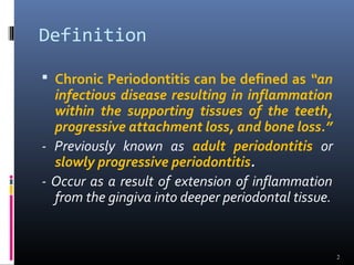
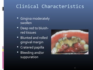
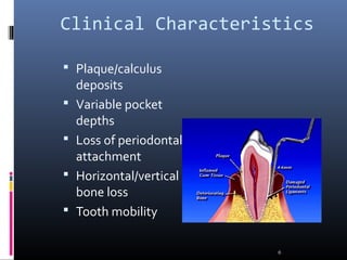
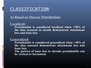
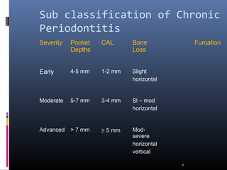
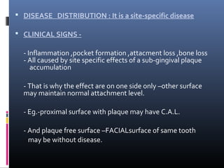
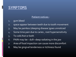
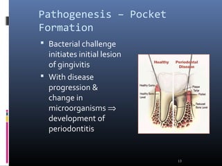
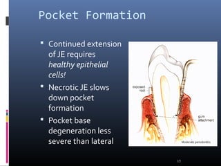
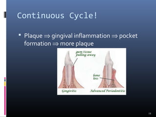
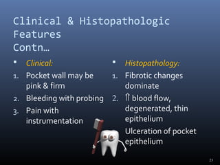
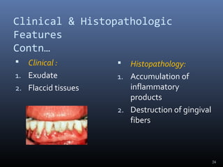
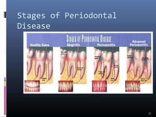
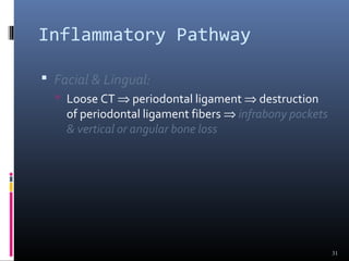
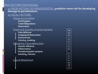
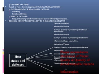
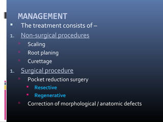
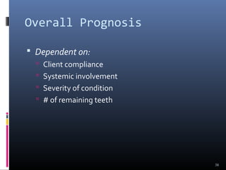
1
Definition
 Chronic Periodontitis can be defined as “an
infectious disease resulting in inflammation
within the supporting tissues of the teeth,
progressive attachment loss, and bone loss.”
- Previously known as adult periodontitis or
slowly progressive periodontitis.
- Occur as a result of extension of inflammation
from the gingiva into deeper periodontal tissue.
2
Common Characteristics
 Onset - any age; most common in adults
 Plaque initiates condition
 Subgingival calculus common finding
 Slow-mod progression; periods of rapid
progression possible
 Modified by local factors/systemic
factors/stress/smoking
3
Extent & Severity
 Extent:
 Localized: <30% of sites affected
 Generalized: > 30% of sites affected
 Severity: entire dentition or individual
teeth/site
 Slight = 1-2 mm CAL
 Moderate = 3-4 mm CAL
 Severe =  5 mm CAL
4
Clinical Characteristics
 Gingiva moderately
swollen
 Deep red to bluish-
red tissues
 Blunted and rolled
gingival margin
 Cratered papilla
 Bleeding and/or
suppuration
5
Clinical Characteristics
 Plaque/calculus
deposits
 Variable pocket
depths
 Loss of periodontal
attachment
 Horizontal/vertical
bone loss
 Tooth mobility
6
CLASSIFICATION
7
A) Based on Disease Distribution:
Localized:
Periodontitis is considered localized when <30% of
the sites assessed in mouth demonstrate attachment
loss and bone loss.
Generalized:
Periodontitis is considered generalized when >30% of
the sites assessed demonstrate attachment loss and
bone loss.
The pattern of bone loss in chronic periodontitis can
be vertical or horizontal.
Sub classification of Chronic
Periodontitis
Severity Pocket
Depths
CAL Bone
Loss
Furcation
Early 4-5 mm 1-2 mm Slight
horizontal
Moderate 5-7 mm 3-4 mm Sl – mod
horizontal
Advanced > 7 mm ≥ 5 mm Mod-
severe
horizontal
vertical
8
 DISEASE DISTRIBUTION : It is a site-specific disease
 CLINICAL SIGNS -
- Inflammation ,pocket formation ,attacment loss ,bone loss
- All caused by site specific effects of a sub-gingival plaque
accumulation
- That is why the effect are on one side only –other surface
may maintain normal attachment level.
- Eg.-proximal surface with plaque may have C.A.L.
- And plaque free surface –FACIALsurface of same tooth
may be without disease.
SYMPTOMS
Patient notices--
1. gum bleed
2. space appear between teeth due to tooth movement
3. May be painless (sleeping disease )goes unnoticed
4. Some time pain due to caries , root hypersensitivity
5. To cold /hot or both
6. PAIN-may be-- dull—deep radiating in the jaw
7. Area of food impaction can cause more discomfort
8. May be gingival tenderness or itchiness found
Periodontal Pathogens
• Gram negative organism dominate
• P.g., P.i., A.a. may infiltrate:
• - Intercellular spaces of the epithelium
• - Between deeper epithelial cells
• - Basement lamina
11
Periodontal Pathogens
Contn.
 Pathogens include:
 Nonmotile rods:
 Facultative:
 Actinobacillus a. E.c.
 Anaerobic:
 P. g., P. i., B.f., F.n.
 Motile rods:
 Facultative:
 C.r.
 Spirochetes:
 Anaerobic, motile:
 Treponema denticola
12
Pathogenesis – Pocket
Formation
 Bacterial challenge
initiates initial lesion
of gingivitis
 With disease
progression &
change in
microorganisms ⇒
development of
periodontitis
13
Pocket Formation
 Cellular & fluid inflammatory exudate ⇒
degenerates CT
 Gingival fibers destroyed
 Collagen fibers apical to JE destroyed ⇒
infiltration of inflammatory cells & edema
 Apical migration of junctional epithelium
along root
 Coronal portion of JE detaches
14
Pocket Formation
 Continued extension
of JE requires
healthy epithelial
cells!
 Necrotic JE slows
down pocket
formation
 Pocket base
degeneration less
severe than lateral
15
Pocket Formation
 Continue inflammation:
 Coronal extension of gingival margin
 JE migrates apically & separates from root
 Lateral pocket wall proliferates & extends into CT
 Leukocytes & edema
 Infiltrate lining epithelium
 Varying degrees of degeneration & necrosis
16
Development of Periodontal
Pocket
17
Continuous Cycle!
 Plaque ⇒ gingival inflammation ⇒ pocket
formation ⇒ more plaque
18
Classification of Pockets
 Gingival:
 Coronal migration of gingival margin
 Periodontal:
 Apical migration of epithelial attachment
 Suprabony:
 Base of pocket coronal to height of alveolar crest
 Infrabony:
 Base of pocket apical to height of alveolar crest
 Characterized by angular bony defects
19
Histopathology
 Connective Tissue:
 Edematous
 Dense infiltrate:
 Plasma cells (80%)
 Lymphocytes, PMNs
 Blood vessels proliferate, dilate & are engorged.
 Varying degrees of degeneration in addition to newly
formed capillaries, fibroblasts, collagen fibers in some
areas.
20
Histopathology
 Periodontal pocket:
 Lateral wall shows most severe degeneration
 Epithelial proliferation & degeneration
 Rete pegs protrude deep within CT
 Dense infiltrate of leukocytes & fluid found in rete
pegs & epithelium
 Degeneration & necrosis of epithelium leads to
ulceration of lateral wall, exposure of CT,
suppuration
21
Clinical & Histopathologic
Features
 Clinical :
1. Pocket wall bluish-red
2. Smooth, shiny surface
3. Pitting on pressure
 Histopathology:
1. Vasodilation &
vasostagnation
2. Epithelial
proliferation, edema
3. Edema &
degeneration of
epithelium
22
Clinical & Histopathologic
Features
Contn…
 Clinical:
1. Pocket wall may be
pink & firm
2. Bleeding with probing
3. Pain with
instrumentation
 Histopathology:
1. Fibrotic changes
dominate
2. ⇑ blood flow,
degenerated, thin
epithelium
3. Ulceration of pocket
epithelium
23
Clinical & Histopathologic
Features
Contn…
 Clinical :
1. Exudate
2. Flaccid tissues
 Histopathology:
1. Accumulation of
inflammatory
products
2. Destruction of gingival
fibers
24
Stages of Periodontal
Disease
25
Root Surface Wall
 Periodontal disease affects root surface:
 Perpetuates disease
 Decay, sensitivity
 Complicates treatment
 Embedded collagen fibers degenerate ⇒
cementum exposed to environment
 Bacteria penetrate unprotected root
26
Root Surface Wall Contn…
 Necrotic areas of cementum form; clinically
soft
 Act as reservoir for bacteria
 Root planing may remove necrotic areas ⇒
firmer surface
27
Inflammatory Pathway
 Stages I-III – inflammation degrades gingival
fibers
 Spreads via blood vessels:
 Interproximal:
 Loose CT ⇒ transseptal fibers ⇒ marrow spaces
of cancellous bone ⇒ periodontal ligament ⇒
suprabony pockets & horizontal bone loss
⇒transseptal fibers transverse horizontally
28
Inflammatory Pathway
 Interproximal:
 Loose CT ⇒ periodontal ligament ⇒ bone ⇒
infrabony pockets & vertical bone loss ⇒
transseptal fibers transverse in oblique direction
29
Inflammatory Pathway
 Facial & Lingual:
 Loose CT ⇒ along periosteum ⇒ marrow spaces
of cancellous bone ⇒ supporting bone destroyed
first ⇒ alvoelar bone proper ⇒ periodontal
ligament ⇒ suprabony pocket & horizontal bone
loss
30
Inflammatory Pathway
 Facial & Lingual:
 Loose CT ⇒ periodontal ligament ⇒ destruction
of periodontal ligament fibers ⇒ infrabony pockets
& vertical or angular bone loss
31
Periodontal Disease
Activity
 Bursts of activity followed by periods of
quiescence characterized by:
 Reduced inflammatory response
 Little to no bone loss & CT loss
 Accumulation of Gram negative organisms leads
to:
 Bone & attachment loss
 Bleeding, exudates
 May last days, weeks, months
32
Periodontal Disease
Activity
 Period of activity followed by period of
remission:
 Accumulation of Gram positive bacteria
 Condition somewhat stabilized
 Periodontal destruction is site specific
 PD affects few teeth at one time, or some
surfaces of given teeth
33
Prevalence:
 Chronic Periodontitis increases in prevalence
& severity with age.
 Affect both the sexes equally.
 It is an age-associated, not age related
disease.
RISK FACTORS FOR DISEASE:
1) PRIOR HISTORY OF PERIODONTITIS—predictor-more risk for developing
damage to periodontium.
2) LOCAL FACTORS:
Plaque Accumulation
Oral Hygiene
Tooth Malposition
Restoration
Preserve & Quantity of certain bacteria
Host defences
Subgingival Restoration
Environment
Calculus, smoking
Connective Tissue destruction
Genetic influence
Inflammation
Periodontopathic bacteria
Smoking, Calculus
Loss of Attachment
M
O
D
I
F
Y
I
N
G
F
A
C
T
O
R
S
3) SYSTEMIC FACTORS:
 Type II or Non – Insulin dependent Diabetes Mellitus (NIIDDM)
4) ENVIRONMENTAL & BEHAVIORAL FACTORS:
 Smoking
 Emotional Stress
5) GENETIC FACTORS:
 Frequent among family members and across different generations.
 GENERAL CONCEPT FOR ETIOLOGY OF CHRONIC PERIODONTITIS
Plaque accumulation
Maturation of Plaque
Quality & Quantity of periodontopathic Plaque
accumulation
Maturation of Plaque
Quality & Quantity of periodontopathic bacteria
InflammationPlaque accumulation
Maturation of Plaque
Quality & Quantity of periodontopathic bacteria
Inflammation
Connective tissue destruction.
bacteria
Inflammation
Connective tissue destruction.
Host
status and
defences
Plaque accumulationPlaque accumulation
Maturation of PlaqueMaturation of Plaque
Quality & Quantity ofQuality & Quantity of
periodontopathic bacteriaperiodontopathic bacteria
InflammationInflammation
MANAGEMENT
 The treatment consists of –
1. Non-surgical procedures
 Scaling
 Root planing
 Curettage
1. Surgical procedure
 Pocket reduction surgery
 Resective
 Regenerative
 Correction of morphological / anatomic defects
Overall Prognosis
 Dependent on:
 Client compliance
 Systemic involvement
 Severity of condition
 # of remaining teeth
38
Prognosis of Individual
Teeth
 Dependent on:
 Attachment levels, bone height
 Status of adjacent teeth
 Type of pockets: suprabony, infrabony
 Furcation involvement
 Root resorption
39

periodontitis

  • 1.
  • 2.
    Definition  Chronic Periodontitiscan be defined as “an infectious disease resulting in inflammation within the supporting tissues of the teeth, progressive attachment loss, and bone loss.” - Previously known as adult periodontitis or slowly progressive periodontitis. - Occur as a result of extension of inflammation from the gingiva into deeper periodontal tissue. 2
  • 3.
    Common Characteristics  Onset- any age; most common in adults  Plaque initiates condition  Subgingival calculus common finding  Slow-mod progression; periods of rapid progression possible  Modified by local factors/systemic factors/stress/smoking 3
  • 4.
    Extent & Severity Extent:  Localized: <30% of sites affected  Generalized: > 30% of sites affected  Severity: entire dentition or individual teeth/site  Slight = 1-2 mm CAL  Moderate = 3-4 mm CAL  Severe =  5 mm CAL 4
  • 5.
    Clinical Characteristics  Gingivamoderately swollen  Deep red to bluish- red tissues  Blunted and rolled gingival margin  Cratered papilla  Bleeding and/or suppuration 5
  • 6.
    Clinical Characteristics  Plaque/calculus deposits Variable pocket depths  Loss of periodontal attachment  Horizontal/vertical bone loss  Tooth mobility 6
  • 7.
    CLASSIFICATION 7 A) Based onDisease Distribution: Localized: Periodontitis is considered localized when <30% of the sites assessed in mouth demonstrate attachment loss and bone loss. Generalized: Periodontitis is considered generalized when >30% of the sites assessed demonstrate attachment loss and bone loss. The pattern of bone loss in chronic periodontitis can be vertical or horizontal.
  • 8.
    Sub classification ofChronic Periodontitis Severity Pocket Depths CAL Bone Loss Furcation Early 4-5 mm 1-2 mm Slight horizontal Moderate 5-7 mm 3-4 mm Sl – mod horizontal Advanced > 7 mm ≥ 5 mm Mod- severe horizontal vertical 8
  • 9.
     DISEASE DISTRIBUTION: It is a site-specific disease  CLINICAL SIGNS - - Inflammation ,pocket formation ,attacment loss ,bone loss - All caused by site specific effects of a sub-gingival plaque accumulation - That is why the effect are on one side only –other surface may maintain normal attachment level. - Eg.-proximal surface with plaque may have C.A.L. - And plaque free surface –FACIALsurface of same tooth may be without disease.
  • 10.
    SYMPTOMS Patient notices-- 1. gumbleed 2. space appear between teeth due to tooth movement 3. May be painless (sleeping disease )goes unnoticed 4. Some time pain due to caries , root hypersensitivity 5. To cold /hot or both 6. PAIN-may be-- dull—deep radiating in the jaw 7. Area of food impaction can cause more discomfort 8. May be gingival tenderness or itchiness found
  • 11.
    Periodontal Pathogens • Gramnegative organism dominate • P.g., P.i., A.a. may infiltrate: • - Intercellular spaces of the epithelium • - Between deeper epithelial cells • - Basement lamina 11
  • 12.
    Periodontal Pathogens Contn.  Pathogensinclude:  Nonmotile rods:  Facultative:  Actinobacillus a. E.c.  Anaerobic:  P. g., P. i., B.f., F.n.  Motile rods:  Facultative:  C.r.  Spirochetes:  Anaerobic, motile:  Treponema denticola 12
  • 13.
    Pathogenesis – Pocket Formation Bacterial challenge initiates initial lesion of gingivitis  With disease progression & change in microorganisms ⇒ development of periodontitis 13
  • 14.
    Pocket Formation  Cellular& fluid inflammatory exudate ⇒ degenerates CT  Gingival fibers destroyed  Collagen fibers apical to JE destroyed ⇒ infiltration of inflammatory cells & edema  Apical migration of junctional epithelium along root  Coronal portion of JE detaches 14
  • 15.
    Pocket Formation  Continuedextension of JE requires healthy epithelial cells!  Necrotic JE slows down pocket formation  Pocket base degeneration less severe than lateral 15
  • 16.
    Pocket Formation  Continueinflammation:  Coronal extension of gingival margin  JE migrates apically & separates from root  Lateral pocket wall proliferates & extends into CT  Leukocytes & edema  Infiltrate lining epithelium  Varying degrees of degeneration & necrosis 16
  • 17.
  • 18.
    Continuous Cycle!  Plaque⇒ gingival inflammation ⇒ pocket formation ⇒ more plaque 18
  • 19.
    Classification of Pockets Gingival:  Coronal migration of gingival margin  Periodontal:  Apical migration of epithelial attachment  Suprabony:  Base of pocket coronal to height of alveolar crest  Infrabony:  Base of pocket apical to height of alveolar crest  Characterized by angular bony defects 19
  • 20.
    Histopathology  Connective Tissue: Edematous  Dense infiltrate:  Plasma cells (80%)  Lymphocytes, PMNs  Blood vessels proliferate, dilate & are engorged.  Varying degrees of degeneration in addition to newly formed capillaries, fibroblasts, collagen fibers in some areas. 20
  • 21.
    Histopathology  Periodontal pocket: Lateral wall shows most severe degeneration  Epithelial proliferation & degeneration  Rete pegs protrude deep within CT  Dense infiltrate of leukocytes & fluid found in rete pegs & epithelium  Degeneration & necrosis of epithelium leads to ulceration of lateral wall, exposure of CT, suppuration 21
  • 22.
    Clinical & Histopathologic Features Clinical : 1. Pocket wall bluish-red 2. Smooth, shiny surface 3. Pitting on pressure  Histopathology: 1. Vasodilation & vasostagnation 2. Epithelial proliferation, edema 3. Edema & degeneration of epithelium 22
  • 23.
    Clinical & Histopathologic Features Contn… Clinical: 1. Pocket wall may be pink & firm 2. Bleeding with probing 3. Pain with instrumentation  Histopathology: 1. Fibrotic changes dominate 2. ⇑ blood flow, degenerated, thin epithelium 3. Ulceration of pocket epithelium 23
  • 24.
    Clinical & Histopathologic Features Contn… Clinical : 1. Exudate 2. Flaccid tissues  Histopathology: 1. Accumulation of inflammatory products 2. Destruction of gingival fibers 24
  • 25.
  • 26.
    Root Surface Wall Periodontal disease affects root surface:  Perpetuates disease  Decay, sensitivity  Complicates treatment  Embedded collagen fibers degenerate ⇒ cementum exposed to environment  Bacteria penetrate unprotected root 26
  • 27.
    Root Surface WallContn…  Necrotic areas of cementum form; clinically soft  Act as reservoir for bacteria  Root planing may remove necrotic areas ⇒ firmer surface 27
  • 28.
    Inflammatory Pathway  StagesI-III – inflammation degrades gingival fibers  Spreads via blood vessels:  Interproximal:  Loose CT ⇒ transseptal fibers ⇒ marrow spaces of cancellous bone ⇒ periodontal ligament ⇒ suprabony pockets & horizontal bone loss ⇒transseptal fibers transverse horizontally 28
  • 29.
    Inflammatory Pathway  Interproximal: Loose CT ⇒ periodontal ligament ⇒ bone ⇒ infrabony pockets & vertical bone loss ⇒ transseptal fibers transverse in oblique direction 29
  • 30.
    Inflammatory Pathway  Facial& Lingual:  Loose CT ⇒ along periosteum ⇒ marrow spaces of cancellous bone ⇒ supporting bone destroyed first ⇒ alvoelar bone proper ⇒ periodontal ligament ⇒ suprabony pocket & horizontal bone loss 30
  • 31.
    Inflammatory Pathway  Facial& Lingual:  Loose CT ⇒ periodontal ligament ⇒ destruction of periodontal ligament fibers ⇒ infrabony pockets & vertical or angular bone loss 31
  • 32.
    Periodontal Disease Activity  Burstsof activity followed by periods of quiescence characterized by:  Reduced inflammatory response  Little to no bone loss & CT loss  Accumulation of Gram negative organisms leads to:  Bone & attachment loss  Bleeding, exudates  May last days, weeks, months 32
  • 33.
    Periodontal Disease Activity  Periodof activity followed by period of remission:  Accumulation of Gram positive bacteria  Condition somewhat stabilized  Periodontal destruction is site specific  PD affects few teeth at one time, or some surfaces of given teeth 33
  • 34.
    Prevalence:  Chronic Periodontitisincreases in prevalence & severity with age.  Affect both the sexes equally.  It is an age-associated, not age related disease.
  • 35.
    RISK FACTORS FORDISEASE: 1) PRIOR HISTORY OF PERIODONTITIS—predictor-more risk for developing damage to periodontium. 2) LOCAL FACTORS: Plaque Accumulation Oral Hygiene Tooth Malposition Restoration Preserve & Quantity of certain bacteria Host defences Subgingival Restoration Environment Calculus, smoking Connective Tissue destruction Genetic influence Inflammation Periodontopathic bacteria Smoking, Calculus Loss of Attachment M O D I F Y I N G F A C T O R S
  • 36.
    3) SYSTEMIC FACTORS: Type II or Non – Insulin dependent Diabetes Mellitus (NIIDDM) 4) ENVIRONMENTAL & BEHAVIORAL FACTORS:  Smoking  Emotional Stress 5) GENETIC FACTORS:  Frequent among family members and across different generations.  GENERAL CONCEPT FOR ETIOLOGY OF CHRONIC PERIODONTITIS Plaque accumulation Maturation of Plaque Quality & Quantity of periodontopathic Plaque accumulation Maturation of Plaque Quality & Quantity of periodontopathic bacteria InflammationPlaque accumulation Maturation of Plaque Quality & Quantity of periodontopathic bacteria Inflammation Connective tissue destruction. Connective tissue destruction. bacteria Inflammation Connective tissue destruction. Host status and defences Plaque accumulationPlaque accumulation Maturation of PlaqueMaturation of Plaque Quality & Quantity ofQuality & Quantity of periodontopathic bacteriaperiodontopathic bacteria InflammationInflammation
  • 37.
    MANAGEMENT  The treatmentconsists of – 1. Non-surgical procedures  Scaling  Root planing  Curettage 1. Surgical procedure  Pocket reduction surgery  Resective  Regenerative  Correction of morphological / anatomic defects
  • 38.
    Overall Prognosis  Dependenton:  Client compliance  Systemic involvement  Severity of condition  # of remaining teeth 38
  • 39.
    Prognosis of Individual Teeth Dependent on:  Attachment levels, bone height  Status of adjacent teeth  Type of pockets: suprabony, infrabony  Furcation involvement  Root resorption 39