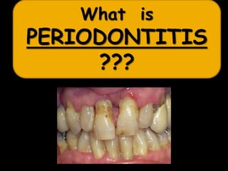
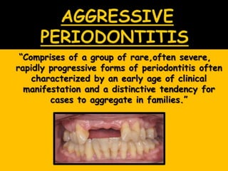
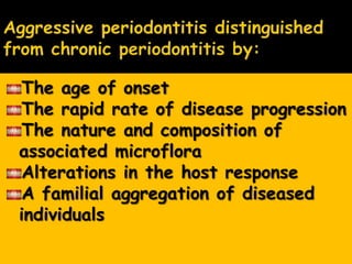
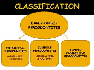
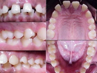
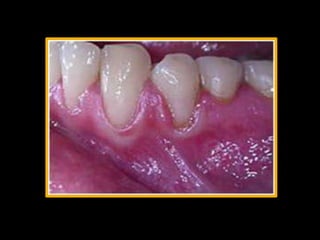
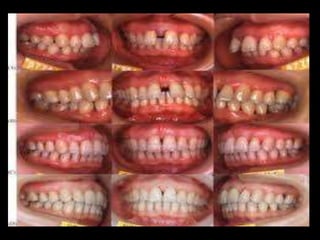
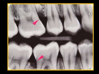
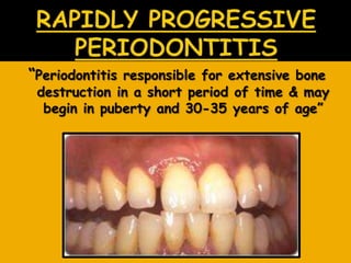
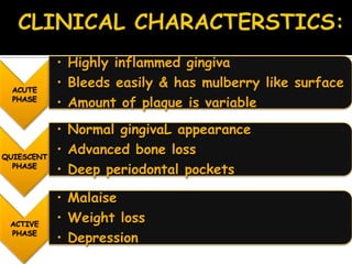
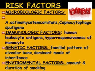
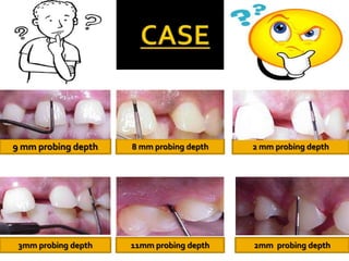
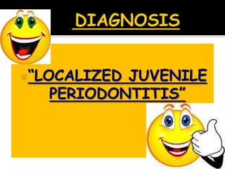
SHAISTA ZAFAR
What is
PERIODONTITIS
???
―Comprises of a group of rare,often severe,
rapidly progressive forms of periodontitis often
characterized by an early age of clinical
manifestation and a distinctive tendency for
cases to aggregate in families.‖
The age of onset
The rapid rate of disease progression
The nature and composition of
associated microflora
Alterations in the host response
A familial aggregation of diseased
individuals
EARLY ONSET
PERIODONTITIS
PREPUBERTAL
PERIODONTITIS
GENERALIZED
LOCALIZED
JUVENILE
PERIODONTITIS
GENERALIZED
LOCALIZED
RAPIDLY
PROGRESSIVE
PERIODONTITIS
“periodontitis that results in resorption of
pdl in young children during or shortly
following the eruption of the primary
teeth‖
It may also affect permanent dentition
Affected patients are usually diagnosed by age 4
Child may has affected leukocytes
-Minimal clincal signs of gingival
inflammation
-Only some of teeth are
involved
-Rate of tissue destruction is
slower
-Leukocytes defects involve
polymorphonucear or
mononuclear leukocytes, but
not both.
LOCALIZED PREPUBERTAL
PERIODONTITIS
GENERALIZED PREPUBERTAL
PERIODONTITIS
CLINICAL FEATURES
ETIOLOGY
 Associated with abnormal
cementum formation &
defective pdl attachment
results in decreased
resistance of periodontal
tissues to microbial
infections & allow rapid
tissue destruction.
-Local mechanical
debridement
-Antibiotic therapy(penicillin
or erythromycin –QD 250mg
for 3 weeks)
-improved oral hygiene
ASSOCIATED FEATURES
TREATMENT
 “uncommon form of periodontitis seen
in children & adolescents‖
Characterised by rapid
alveolar bone destruction
with minimal signs of
gingival inflammation
LOCALIZED
JUVENILE
PERIODONTITIS
GENERALIZED
JUVENILE
PERIODONTITIS
CHARACTERISED BY:
loss of collagen fibers in pdl
Replacement by loose connective tissue
Extensive bone resorption
Widened periodontal ligament space
Gingiva is not involved
In 1928, Gottlieb termed the disease
―Deep cementopathia‖
disease of eruption & cementum
initiated a foreign body response
Host attempted to exfoliate the
tooth
Bone resorption
Pocket formation
In 1938 Wannermacher described incisor-
first molar involvement and called the
disease ―parodontitis marginalis
progressiva‖
Many author considered this to be a
degenerative, non-inflammatory disease
process & therefore gave it the name
―periodontosis‖
Age of onset is around puberty
Localized involvement of 1st molar/incisor
Interproximal attachment loss on atleast two
permanent teeth, one of which is a 1st molar
Involves no more than two teeth other than 1st
molar & incisors
Distolabial migration of maxillary incisors with
concomitant diastema formation
Increase mobility of first molars
Sensitivity of denuded root surfaces to thermal &
tactile stimuli.
Deep,dull,radiating pain during mastication,probably
caused by irritation of the supporting structures by
mobile teeth & impacted food
Periodontal abscess may form at this stage &
regional lymph node enlargement may occur.
Classic diagnostic sign:
Vertical loss of alveolar bone around 1st
molars & incisors
Beginning around puberty in an otherwise
healthy teenagers
―Arc shaped loss of alveolar bone
extending from distal surface of 2nd
premolar to mesial surface of second
molar‖
Colonization of Actinobacillus
actinomycetemcomitans
Impaired neutrophils chemotaxis lowers
patient’s resistance to bacterial infection
Intense immune responses
Patient education
Oral hygiene instructions in plaque control
& reinforcement
Selective extraction & replacement
scaling & root planing of teeth
Surgical curretement of periodontal
pocket
Systemic administration of antibiotic
(tetracycline—250mg—1 tab—6 hours for
3 weeks)
LJP affects both males and
females
Most frequently between puberty
& 20 yrs of age
Affects white females more &
black males more
Affects individuals under the age of
30,but older patients may also be
affected
Involves entire dentition, frequently
associated with down's syndrome and
papillon-lefevre syndrome.
Can also occur in individuals with no
systemic disease
CHARACTERISED BY:
Generalized interproximal
attachment loss affecting atleast 3
permanent teeth other than 1st
molars & incisors
Small amount of bacterial plaque with
affected teeth
Quantitatively—amount of plaque seems
inconsistent with the amount of
periodontal desruction
Qualitatively-
A.Actinomycetemcomitants,Bacteroides
are detected in plaque
DESTRUCTIVE STAGE:
severe acutely inflammed tissue
Ulcerative
fiery red
bleeding may occur spontaneously or on
stimulation pressure
Suppuration maybe an important feature
Attachment & bone are actively lost
OTHER CASES:
Gingival tissues may appear pink
Free of inflammation
Absence of some degree of stippling
Deep pockets demonstrated by probing
Systemic manifestations:
Weight loss
Mental depression
Malaise
Severe bone loss associated with minimal
number of teeth, to advanced bone loss
affecting the majority of teeth in
dentition
Subgingival plaque from affected
site
Impaired neutrophils chemotaxis.
Familial involvement.
Medical histories updated & reviewed
Patient education
Oral hygiene instructions in plaque control
& reinforcement
Periodic scaling & curettage
Antibiotic therapy
Surgical pocket elimination(periodontal flap
procedure,osseous recontouring,root
amputation)
Extraction of all teeth & replacement with
complete dentures
Follow up
Blacks are at high risk than
whites
Males were more likely to
have GAP then females
―Periodontitis responsible for extensive bone
destruction in a short period of time & may
begin in puberty and 30-35 years of age‖
ACUTE
PHASE
• Highly inflammed gingiva
• Bleeds easily & has mulberry like surface
• Amount of plaque is variable
QUIESCENT
PHASE
• Normal gingivaL appearance
• Advanced bone loss
• Deep periodontal pockets
ACTIVE
PHASE
• Malaise
• Weight loss
• Depression
Immunocompromised host
Defects in neutrophils & monocytes
Bacterial flora
e.g:Actinobacillus,Capnocytophaga
Diabetes mellitus
Down’s syndrome
Neutropenia
Crohn’s disease
Agranulocytosis
SYSTEMIC INVOLVEMENT
Scaling
Open or closed curettage
Antibiotic therapy
Osseous grafts
MICROBIOLOGIC FACTORS:
A.actinomycetemcomitans,Capnocytophaga
sputigena
IMMUNOLOGIC FACTORS: human
leukocyte antigens,hyperresponsiveness of
monocyte
GENETIC FACTORS: familial pattern of
alveolar bone,dominant mode of
inheritance
ENVIROMENTAL FACTORS: amount &
duration of smoking
9 mm probing depth 8 mm probing depth 2 mm probing depth
3mm probing depth 11mm probing depth 2mm probing depth
―LOCALIZED JUVENILE
PERIODONTITIS‖
Clinically, localized juvenile periodontitis (LJP)
patients rarely show calculus or plaque formation
and often exhibit little or no gingivitis.
However, deep probing, attachment loss,
radiographic bone loss are found. Deep
interproximal vertical bone loss on first molars
and incisors are characteristic of LJP. Juvenile
periodontits should be identified and treated
early with antimicrobial therapy, scaling and
root planing, and also surgery according to
extent of destruction.
1. Generalized: affecting most of the dentition.
2. Localized: affecting only first molars and
incisors.
 CARRANZA’S CLINICAL PERIODONTOLOGY
 ESSENTIALS OF PERIODONTOLOGY
 IMAGES FROM GOOGLE
 CASE-http://www.drbui.com/perio.html
 F
 L
 O
 B R U S H
I S
N T
S M I L E
E E
T H A N KY O U
H

Aggressive periodontitis

  • 2.
  • 3.
  • 4.
    ―Comprises of agroup of rare,often severe, rapidly progressive forms of periodontitis often characterized by an early age of clinical manifestation and a distinctive tendency for cases to aggregate in families.‖
  • 5.
    The age ofonset The rapid rate of disease progression The nature and composition of associated microflora Alterations in the host response A familial aggregation of diseased individuals
  • 6.
  • 7.
    “periodontitis that resultsin resorption of pdl in young children during or shortly following the eruption of the primary teeth‖ It may also affect permanent dentition Affected patients are usually diagnosed by age 4 Child may has affected leukocytes
  • 8.
    -Minimal clincal signsof gingival inflammation -Only some of teeth are involved -Rate of tissue destruction is slower -Leukocytes defects involve polymorphonucear or mononuclear leukocytes, but not both. LOCALIZED PREPUBERTAL PERIODONTITIS GENERALIZED PREPUBERTAL PERIODONTITIS CLINICAL FEATURES ETIOLOGY
  • 9.
     Associated withabnormal cementum formation & defective pdl attachment results in decreased resistance of periodontal tissues to microbial infections & allow rapid tissue destruction. -Local mechanical debridement -Antibiotic therapy(penicillin or erythromycin –QD 250mg for 3 weeks) -improved oral hygiene ASSOCIATED FEATURES TREATMENT
  • 10.
     “uncommon formof periodontitis seen in children & adolescents‖ Characterised by rapid alveolar bone destruction with minimal signs of gingival inflammation
  • 11.
  • 12.
    CHARACTERISED BY: loss ofcollagen fibers in pdl Replacement by loose connective tissue Extensive bone resorption Widened periodontal ligament space Gingiva is not involved
  • 13.
    In 1928, Gottliebtermed the disease ―Deep cementopathia‖ disease of eruption & cementum initiated a foreign body response Host attempted to exfoliate the tooth Bone resorption Pocket formation
  • 14.
    In 1938 Wannermacherdescribed incisor- first molar involvement and called the disease ―parodontitis marginalis progressiva‖ Many author considered this to be a degenerative, non-inflammatory disease process & therefore gave it the name ―periodontosis‖
  • 15.
    Age of onsetis around puberty Localized involvement of 1st molar/incisor Interproximal attachment loss on atleast two permanent teeth, one of which is a 1st molar Involves no more than two teeth other than 1st molar & incisors Distolabial migration of maxillary incisors with concomitant diastema formation Increase mobility of first molars Sensitivity of denuded root surfaces to thermal & tactile stimuli. Deep,dull,radiating pain during mastication,probably caused by irritation of the supporting structures by mobile teeth & impacted food Periodontal abscess may form at this stage & regional lymph node enlargement may occur.
  • 19.
    Classic diagnostic sign: Verticalloss of alveolar bone around 1st molars & incisors Beginning around puberty in an otherwise healthy teenagers ―Arc shaped loss of alveolar bone extending from distal surface of 2nd premolar to mesial surface of second molar‖
  • 21.
    Colonization of Actinobacillus actinomycetemcomitans Impairedneutrophils chemotaxis lowers patient’s resistance to bacterial infection Intense immune responses
  • 22.
    Patient education Oral hygieneinstructions in plaque control & reinforcement Selective extraction & replacement scaling & root planing of teeth Surgical curretement of periodontal pocket Systemic administration of antibiotic (tetracycline—250mg—1 tab—6 hours for 3 weeks)
  • 23.
    LJP affects bothmales and females Most frequently between puberty & 20 yrs of age Affects white females more & black males more
  • 24.
    Affects individuals underthe age of 30,but older patients may also be affected Involves entire dentition, frequently associated with down's syndrome and papillon-lefevre syndrome. Can also occur in individuals with no systemic disease
  • 25.
    CHARACTERISED BY: Generalized interproximal attachmentloss affecting atleast 3 permanent teeth other than 1st molars & incisors
  • 26.
    Small amount ofbacterial plaque with affected teeth Quantitatively—amount of plaque seems inconsistent with the amount of periodontal desruction Qualitatively- A.Actinomycetemcomitants,Bacteroides are detected in plaque
  • 27.
    DESTRUCTIVE STAGE: severe acutelyinflammed tissue Ulcerative fiery red bleeding may occur spontaneously or on stimulation pressure Suppuration maybe an important feature Attachment & bone are actively lost
  • 28.
    OTHER CASES: Gingival tissuesmay appear pink Free of inflammation Absence of some degree of stippling Deep pockets demonstrated by probing Systemic manifestations: Weight loss Mental depression Malaise
  • 30.
    Severe bone lossassociated with minimal number of teeth, to advanced bone loss affecting the majority of teeth in dentition
  • 32.
    Subgingival plaque fromaffected site Impaired neutrophils chemotaxis. Familial involvement.
  • 33.
    Medical histories updated& reviewed Patient education Oral hygiene instructions in plaque control & reinforcement Periodic scaling & curettage Antibiotic therapy Surgical pocket elimination(periodontal flap procedure,osseous recontouring,root amputation) Extraction of all teeth & replacement with complete dentures Follow up
  • 34.
    Blacks are athigh risk than whites Males were more likely to have GAP then females
  • 35.
    ―Periodontitis responsible forextensive bone destruction in a short period of time & may begin in puberty and 30-35 years of age‖
  • 36.
    ACUTE PHASE • Highly inflammedgingiva • Bleeds easily & has mulberry like surface • Amount of plaque is variable QUIESCENT PHASE • Normal gingivaL appearance • Advanced bone loss • Deep periodontal pockets ACTIVE PHASE • Malaise • Weight loss • Depression
  • 37.
    Immunocompromised host Defects inneutrophils & monocytes Bacterial flora e.g:Actinobacillus,Capnocytophaga Diabetes mellitus Down’s syndrome Neutropenia Crohn’s disease Agranulocytosis SYSTEMIC INVOLVEMENT
  • 38.
    Scaling Open or closedcurettage Antibiotic therapy Osseous grafts
  • 39.
    MICROBIOLOGIC FACTORS: A.actinomycetemcomitans,Capnocytophaga sputigena IMMUNOLOGIC FACTORS:human leukocyte antigens,hyperresponsiveness of monocyte GENETIC FACTORS: familial pattern of alveolar bone,dominant mode of inheritance ENVIROMENTAL FACTORS: amount & duration of smoking
  • 40.
    9 mm probingdepth 8 mm probing depth 2 mm probing depth 3mm probing depth 11mm probing depth 2mm probing depth
  • 41.
  • 42.
    Clinically, localized juvenileperiodontitis (LJP) patients rarely show calculus or plaque formation and often exhibit little or no gingivitis. However, deep probing, attachment loss, radiographic bone loss are found. Deep interproximal vertical bone loss on first molars and incisors are characteristic of LJP. Juvenile periodontits should be identified and treated early with antimicrobial therapy, scaling and root planing, and also surgery according to extent of destruction. 1. Generalized: affecting most of the dentition. 2. Localized: affecting only first molars and incisors.
  • 43.
     CARRANZA’S CLINICALPERIODONTOLOGY  ESSENTIALS OF PERIODONTOLOGY  IMAGES FROM GOOGLE  CASE-http://www.drbui.com/perio.html
  • 44.
     F  L O  B R U S H I S N T S M I L E E E T H A N KY O U H