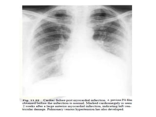
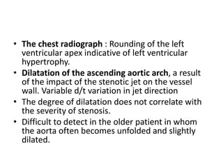
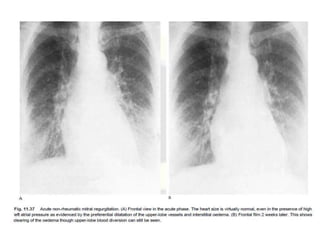
SlideShare a Scribd company logo
Ischemic and
Valvular heart disease
Prashant Gupta
NAMS, Radiodiagnosis and Imaging
Coronary Heart disease
• Commonest cause of premature death in the
developed world
• Commonest cause of acute medical admission
to hospital in developed countries.
• Coronary atheroma development process
complex and multifactorial
• Risk factors:
1. Smoking history,
2. lipid profile,
3. family history,
4. obesity, diet and exercise,
5. hypertension, diabetes and a number of others.
• Chronic increase in the size and occlusive
nature of the plaques stable angina or
ischaemic cardiomyopathy.
• Acute changes, especially plaque rupture will
lead to a variety of acute coronary syndromes,
unstable angina and myocardial infarction.
Stable Angina
• The chest radiograph usually normal, unless
previous events such as myocardial infarction
or other coexisting heart disease have altered
the heart size and pulmonary vascularity.
• Careful examination of the penetrated film
may reveal some abnormalities.
• Coronary artery calcification is best seen in
the proximal left coronary artery and may be
identified near the aortic root on both PA and
lateral views
• The significance of this finding is dependent
on the patient's age.
• >70 years: common, may not relate directly to
plaque calcification but may simply be due to
degenerative calcification in the vessel walls.
• <50 years : highly significant and will usually
indicate calcified atheromatous plaques.
• between 50 and 70 years of age
intermediate significance
• Prominent calcified plaques in the aortic root
just above the sinuses of Valsalva: In some
cases of hyperlipidaemia. Normally an
uncommon site for aortic calcification and if
present, should raise the probability of
coronary heart disease.
• Stable angina for many years can also present
as left ventricular damage, presumably due to
chronic ischemia and fibrosis, which may he
manifest as a dilated and impaired left
ventricle.
Acute myocardial infarction
• Normal in the acute phase (less than 24 hours
after onset of symptoms) in the majority of
patients if they have had a normal preceding
film.
• The chest radiograph: important piece,
provides some insight into the severity of the
myocardial infarction.
• In addition, over the course of the acute
illness serial films can provide important
diagnostic and prognostic information.
• The larger the infarct and the more acute its
onset, the more likely early changes are seen.
• The most common feature: development of
pulmonary edema - vary, with haziness of the
pulmonary arteries at the lung bases and
prominence of upper lobe vessels indicating
pulmonary venous hypertension.
• May progress to perihilar and peripheral
parenchymal clouding, leading to the formation
of septal lines and alveolar pulmonary oedema.
Pleural effusions can develop if the left heart
failure is prolonged.
Mortality has been estimated from radiographic
features.
• The presence of pulmonary edema indicates a
one-year mortality of 44%.
• If there is no evidence of heart failure, this
indicates a good prognosis with an 8% one-
year mortality.
• Also includes progressive enlargement of the
heart, more often identified in anterior MI
• If the serial films taken over the first few days
or weeks following MI: progressive cardiac
enlargement, an important adverse
prognostic sign that should be further
evaluated.
• Several of the important complications of an
acute myocardial infarction can be suggested
from the plain chest radiograph, an useful
adjunct.
Complications of MI
• Acute MR
• Rupture of interventricular septum
• Left ventricular rupture
• Left ventricular aneurysm
• Pericardial effusion
Acute mitral regurgitation
Mechanisms: 2 mechanisms
• Acute dilatation of the left ventricle will cause
annular dilatation and consequent 'functional'
mitral regurgitation.
• An infarct affecting the papillary muscle(s)
can lead to malfunction of the mitral closure
mechanism and consequent mitral
regurgitation.
• Depending on the severity and the speed of
onset, the MR may lead to cardiomegaly or
pulmonary venous hypertension.
• Papillary muscle rupture is rare , in about 1 %
of acute MI.
• leads to acute mitral valve insufficiency; on
the chest radiograph, pulmonary edema
develops with little increase in the cardiac
size or more particularly the left atrium.
• Final diagnosis is clinical and
echocardiographic.
Rupture of the interventricular
septum
• Most likely to rupture between 4 and 21 days
postinfarction rapid-onset left to right shunting
and heart failure.
• rare complication, occurs in up to 2% of all acute
cases
• Engorgement of the pulmonary vasculature
(pulmonary plethora) and pulmonary edema
frequently seen.
• The definitive diagnosis is clinical and
echocardiographic.
Left ventricular rupture
• If the profoundly damaged segment of the left
ventricle affected by an acute infarct lies in
the free wall, then acute rupture into the
pericardium can occur
• an acute fatal complication but in rare
instances the rupture may be contained in the
pericardium and this can lead to the late
development of a false aneurysm with
cardiomegaly and possible heart failure. The
diagnosis of this condition is rarely made
acutely.
Left ventricular aneurysm
• large infarcted segment and sustains full-
thickness ischemia- may develop into a left
ventricular aneurysm (over a few weeks).
• most commonly at the cardiac apex in
association with an anterior infarct , but it can
also occur in the posterior position.
• Several complications : heart failure,
intracardiac thrombus and possible systemic
embolism.
• The chest radiograph: localised bulge on the
left heart border but if the aneurysm is not
well demarcated or if it lies in a less prominent
position it may not be identified on the plain
film.
• The wall of a longstanding aneurysm (or a
non-aneurysmal infarct) may show
calcification.
Pericardial effusion
• Acute pericardial effusion has a prognostic
significance, being most commonly associated
with partial ventricular rupture
• Later in the course of condition, pericardial or
pleural effusion can occur with Dressler’s
syndrome (an inflammatory reaction to the
infarct). Mild cardiomegaly can also occur with
it.
Acquired Valve disease
• On their function, valvular disease can either
be pure stenosis or pure regurgitation, or
more likely a combination of both.
Aortic valve disease
Aortic stenosis:
LVH+aortic dilatation+aortic valve calcification
• Rheumatic heart fever :inflammatory fusion
of the commissures, less likely cause of aortic
stenosis in the current era,
• The most common, in adult life being changes
secondary to a congenital bicuspid valve.
• Significant aortic stenosis may be present with
a virtually normal heart shadow
• The chest radiograph : Rounding of the left
ventricular apex indicative of left ventricular
hypertrophy.
• Dilatation of the ascending aortic arch, a result
of the impact of the stenotic jet on the vessel
wall. Variable d/t variation in jet direction
• The degree of dilatation does not correlate with
the severity of stenosis.
• Difficult to detect in the older patient in whom
the aorta often becomes unfolded and slightly
dilated.
On the lateral film:
• the presence of calcification in the position of
the aortic valve is an important sign, usually
indicating important valve stenosis.
• Some authors suggest this calcification
represents severe aortic stenosis with a
gradient of at least 50 mmHg.
• In most cases, the pulmonary vascularity is
normal
• In advanced cases, there will be left
ventricular impairment and associated
changes of heart failure.
• In patients with aortic stenosis, normal
pulmonary vascularity should not be taken as
an indication that the stenosis is mild.
Aortic regurgitation
• Damage to the valvular cusps as a result of either
1. endocarditis or rheumatic fever,
2. dilatation of the aortic root due to Marfan's syndrome or
degenerative ectasia of the root,
3. and also as a solitary consequence of bicuspid aortic valve.
4. Occasionally aortic regurgitation can develop as a complication of a
primary disease of the aortic wall, such as aortitis.
5. A dissection of the aorta can produce valvular regurgitation if the
false lumen dissects towards the aortic valve ring.
• In chronic aortic regurgitation: The plain
radiograph demonstrates a large heart with a
predominantly left ventricular configuration.
The heart size reflects the severity of the
disease.
• Calcification of the aortic valve is not a feature
of pure aortic regurgitation but can be
visualised if there is a combination of
regurgitation and stenosis.
• The ascending aorta and often the aortic arch are
large and can sometimes be visualized as a bulge
on the right of the mediastinum.
• In many patients with pure aortic regurgitation:
excellent compensation for the increased flow in
the left ventricle and there is normal pulmonary
vasculature.
• The combination of a large left ventricle, no other
chamber enlargement and normal pulmonary
vessels is very suggestive of severe chronic aortic
regurgitation
Mitral valve disease
Mitral stenosis
• Most common cause of mitral stenosis is
rheumatic fever.
• The symptoms of flow restriction (dyspnoea
and heart failure) may be few until the valve
becomes critically narrowed.
• Predispose to thrombus formation in the left
atrium and consequent systemic embolus.
May present with a systemic embolism.
• The chest radiograph : selective left atrial
enlargement, which can vary from trivial to
gross.
• If the mitral stenosis is a result of rheumatic
fever there is often marked enlargement of
the left atrial appendage, which forms a bulge
on the left heart border just below the main
pulmonary artery {THE THIRD MOGUL}: trivial
to the marked with a large protrusion.
• In early years : often a normal heart size with
only subtle signs of left atrial enlargement
being evident
• Unexplained heart failure with a normal heart
size : not to overlook mitral stenosis.
• Late stages of the condition, the atria may be
large but the left ventricular contour is still of
small radius, indicating the small size of the
ventricular chamber.
• Severe and longstanding mitral stenosis:
calcification of the valve can develop.
• Best visualised in the lateral position but can
sometimes be visualised in the PA projection if
the film is penetrated.
• This calcification needs to be differentiated
from the far more common C- or J-shaped
calcification that occurs in the valve annulus.
• In severe mitral valve disease with associated
atrial fibrillation, calcification of the left
atrium, or thrombus within the left atrium,
can occur.
• Marked changes in the pulmonary circulation
can be identified on the plain film.
• Often there is upper lobe blood diversion,
with enlargement of the main and central
pulmonary arteries indicating pulmonary
arterial hypertension.
• Cephalization of pulmonary veins = stag antler
sign
• The right-sided cardiac chambers will often be
considerably enlarged ,the presence of a
'double right heart border.
• A very large left atrium, aneurysmal if it
reaches to within 2.5 cm of the thoracic wall,
may be associated with segmental or even
lobar collapse. This is more common on the
right side.
• longstanding pulmonary venous hypertension
and thus chronic features: Haemosiderosis
and pulmonary ossific nodules may
occasionally be seen.
Mitral regurgitation
• The commonest cause in west is degeneration
of the valve. several variants of this
degeneration, including myxomatous
degeneration of the valve tissue with prolapse
of part or all of the valve (Barlow syndrome,
billowing mitral leaftlet syndorme).
• Degeneration of the chordae can lead to
rupture and a consequent flail portion of
leaflet: occur with abrupt onset of symptoms
• Infective endocarditis will also cause damage
of the valve with consequent regurgitation but
this will usually be on an already abnormal
valve.
• In the case of an impaired left ventricle,
mitral regurgitation can occur due to annular
dilatation or papillary muscle dysfunction.
• Plain film appearances :In chronic phase, the heart
tends to enlarge with a left ventricular configuration,
left atrial enlargement being proportionately less
prominent with enlargement of the left atrial
appendage occurring rarely.
• In longstanding cases, however, there can still be very
marked left atrial enlargement.
• Calcification does not occur.
• If the mitral valve prolapse is associated with Marfan's
disease there may also be enlargement of the aortic
root.
• In the acute phase, the heart size is likely to
remain normal even in the presence of a high
left atrial pressure.
• This high pressure in the acute phase often
leads to the formation of acute pulmonary
edema
• The regurgitation associated with rheumatic
fever results from the destruction of the
actual cusps, usually at the free edges leading
to leakage.
• The commonest result of rheumatic valve
disease is a combination of both stenosis and
regurgitation. The valve fails to open fully in
diastole, but the thickened and rolled edges of
the cusps do not fully coapt (seal).
• The pulmonary vascular appearances are very
similar to those of mitral stenosis but the heart is
often larger.
• There is often greater enlargement of the left
atrium, which can be massive or even
aneurysmal.
• The presence of left ventricular dilatation in
mitral regurgitation (seen as enlargement of the
left ventricular contour with a larger radius curve)
will indicate left ventricular volume overload or
end-stage left ventricular dilatation
Tricuspid valve disease
• TV commonly regugitant as a consequence of
left-sided heart disease but it is unusual for
the secondary effects to cause diagnostic
changes on the chest radiograph.
• It is relatively rare to see primary tricuspid
valve disease, which can occur as a
complication of bacterial endocarditis or as a
late feature of rheumatic heart disease.
• A number of congenital conditions will affect
tricuspid valve function.
• Metastatic carcinoid disease will also affect
the right-sided heart valves, producing
deformity and some regurgitation of both
tricuspid and pulmonary valves.
• Often the endocarditis in tricuspid valve
disease is a complication of intravenous drug
abuse and the most common pathogen is
staphylococcal. As well as cardiac
manifestations there is often consolidation
within the lungs, this often progressing to
cavitation.
• Important tricuspid valve disease will cause
enlargement of the right atrium, producing a
prominent, bulging or elongated right heart border
• This appearance has a single margin and is distinct
from the 'double heart border' produced by left atrial
enlargement.
• The difference can be distinguished by the position of
entry of the IVC, this structure limiting the expansion
of the right atrium. Significant cardiomegaly can be
caused by right atrial enlargement
Pulmonary valve disease
• It is very rare to see acquired disease of the
pulmonary valve.
• Carcinoid disease and endocarditis can
occasionally affect the valve.
References
• Textbook of Radiology and imaging, David
Sutton
• Fundamentals of diagnostic radiology, Bryant
and Helms
Thank you

More Related Content

What's hot

Diseases of the aorta
Diseases of the aortaDiseases of the aorta
Diseases of the aorta
Mohamed Ashraf
 
Coarc2
Coarc2Coarc2
Coarctation of Aorta by Dr Kuntal Surana
Coarctation of Aorta by Dr Kuntal SuranaCoarctation of Aorta by Dr Kuntal Surana
Coarctation of Aorta by Dr Kuntal Surana
Kuntal Surana
 
Aortic anurysm
Aortic anurysmAortic anurysm
Aortic anurysm
Monika Devi NR
 
Aortic aneurys mppt
Aortic aneurys mpptAortic aneurys mppt
Aortic aneurys mppt
Cerin Mathew Mathew
 
Imaging of Aortic Dissection
Imaging of Aortic DissectionImaging of Aortic Dissection
Imaging of Aortic Dissection
Sakher Alkhaderi
 
Aortic dissection dr.tapu
Aortic dissection dr.tapuAortic dissection dr.tapu
Aortic dissection dr.tapu
Nizam Uddin
 
CAROTID DOPPLER STUDY
CAROTID DOPPLER STUDYCAROTID DOPPLER STUDY
CAROTID DOPPLER STUDY
Dr I Gurubharath .
 
Coarctation of aorta
Coarctation of aortaCoarctation of aorta
Coarctation of aorta
S. Ismat
 
Aortic aneurysm and stenosis
Aortic aneurysm and stenosisAortic aneurysm and stenosis
Aortic aneurysm and stenosis
monika204
 
Mitral regurgitation
Mitral regurgitationMitral regurgitation
Mitral regurgitation
Uphar Gupta
 
Aortic disection
Aortic disectionAortic disection
Aortic disection
Monika Devi NR
 
Coarctation of the Aorta
Coarctation of the AortaCoarctation of the Aorta
Coarctation of the Aorta
Dr.Sayeedur Rumi
 
Brain aneurysms in general
Brain aneurysms in generalBrain aneurysms in general
Brain aneurysms in general
Avinash Km
 
Diseases of the aorta
Diseases of the aortaDiseases of the aorta
Diseases of the aortaFuad Farooq
 
Unruptured brain aneurysm
Unruptured brain aneurysmUnruptured brain aneurysm
Unruptured brain aneurysm
Avinash Km
 

What's hot (20)

Diseases of the aorta
Diseases of the aortaDiseases of the aorta
Diseases of the aorta
 
Coarc2
Coarc2Coarc2
Coarc2
 
Coarctation of Aorta by Dr Kuntal Surana
Coarctation of Aorta by Dr Kuntal SuranaCoarctation of Aorta by Dr Kuntal Surana
Coarctation of Aorta by Dr Kuntal Surana
 
Aortic anurysm
Aortic anurysmAortic anurysm
Aortic anurysm
 
Aneurysm
AneurysmAneurysm
Aneurysm
 
Aortic aneurys mppt
Aortic aneurys mpptAortic aneurys mppt
Aortic aneurys mppt
 
Imaging of Aortic Dissection
Imaging of Aortic DissectionImaging of Aortic Dissection
Imaging of Aortic Dissection
 
Aortic dissection dr.tapu
Aortic dissection dr.tapuAortic dissection dr.tapu
Aortic dissection dr.tapu
 
CAROTID DOPPLER STUDY
CAROTID DOPPLER STUDYCAROTID DOPPLER STUDY
CAROTID DOPPLER STUDY
 
Coarctation of aorta
Coarctation of aortaCoarctation of aorta
Coarctation of aorta
 
Carotid Artery Stroke
Carotid Artery StrokeCarotid Artery Stroke
Carotid Artery Stroke
 
Coarctation Of Aorta
Coarctation Of AortaCoarctation Of Aorta
Coarctation Of Aorta
 
Aortic aneurysm and stenosis
Aortic aneurysm and stenosisAortic aneurysm and stenosis
Aortic aneurysm and stenosis
 
Mitral regurgitation
Mitral regurgitationMitral regurgitation
Mitral regurgitation
 
Aortic disection
Aortic disectionAortic disection
Aortic disection
 
Coarctation of the Aorta
Coarctation of the AortaCoarctation of the Aorta
Coarctation of the Aorta
 
Brain aneurysms in general
Brain aneurysms in generalBrain aneurysms in general
Brain aneurysms in general
 
Diseases of the aorta
Diseases of the aortaDiseases of the aorta
Diseases of the aorta
 
Unruptured brain aneurysm
Unruptured brain aneurysmUnruptured brain aneurysm
Unruptured brain aneurysm
 
Cerebral aneurysm
Cerebral aneurysmCerebral aneurysm
Cerebral aneurysm
 

Similar to Ischemic and valvular heart disease

RHEUMATIC HEART disease presentation.pptx
RHEUMATIC HEART disease presentation.pptxRHEUMATIC HEART disease presentation.pptx
RHEUMATIC HEART disease presentation.pptx
sarathrajum17
 
Valvular heart disease
Valvular heart disease Valvular heart disease
Valvular heart disease
BPT4thyearJamiaMilli
 
Aortic Regurgitation - Rivin
Aortic Regurgitation - RivinAortic Regurgitation - Rivin
Aortic Regurgitation - Rivin
Rivindu Wickramanayake
 
Valvular Heart Disease.pptx
Valvular Heart Disease.pptxValvular Heart Disease.pptx
Valvular Heart Disease.pptx
Shilpasree Saha
 
Acquired valvular heart diseases with x ray findings
Acquired valvular heart diseases with x ray findingsAcquired valvular heart diseases with x ray findings
Acquired valvular heart diseases with x ray findingsairwave12
 
Aortic stenosis 11 4
Aortic stenosis 11 4Aortic stenosis 11 4
Aortic stenosis 11 4
azza mokhtar
 
Valvular heart disease
Valvular heart diseaseValvular heart disease
Valvular heart disease
MR. JAGDISH SAMBAD
 
valvularheartdisease-180524125726 (1).pdf
valvularheartdisease-180524125726 (1).pdfvalvularheartdisease-180524125726 (1).pdf
valvularheartdisease-180524125726 (1).pdf
jiregnaetichadako
 
Valvular heart disease
Valvular heart diseaseValvular heart disease
Valvular heart disease
Amir Mahmoud
 
valvularheartdisease-180524125726 (1).pptx
valvularheartdisease-180524125726 (1).pptxvalvularheartdisease-180524125726 (1).pptx
valvularheartdisease-180524125726 (1).pptx
ArpitaHalder8
 
3. ARRHYTHMIAS.pptx
3. ARRHYTHMIAS.pptx3. ARRHYTHMIAS.pptx
3. ARRHYTHMIAS.pptx
mariaidrees3
 
Aortic regurgitation
Aortic regurgitationAortic regurgitation
Aortic regurgitation
Vitrag Shah
 
Pericardial diseases
Pericardial diseasesPericardial diseases
Pericardial diseases
Dilmo Yeldo
 
Valvular heart diseases
Valvular heart diseasesValvular heart diseases
Valvular heart diseases
abelfelege
 
Valvular heart disease
Valvular heart diseaseValvular heart disease
Valvular heart disease
MR. JAGDISH SAMBAD
 
Congenital heart diseases
Congenital heart diseasesCongenital heart diseases
Congenital heart diseases
Tigreentertainment
 
Hypertrophic Cardiomyopathy
Hypertrophic CardiomyopathyHypertrophic Cardiomyopathy
Hypertrophic Cardiomyopathy
Joisy Aloor
 
Echo and CAD-2.pptx
Echo and CAD-2.pptxEcho and CAD-2.pptx
Echo and CAD-2.pptx
AnayaAnaya14
 
Radiography of acquired Heart Disease.pptx
Radiography of acquired Heart Disease.pptxRadiography of acquired Heart Disease.pptx
Radiography of acquired Heart Disease.pptx
ssuser227d6b
 
aorticregurgitation-131030042922-phpapp02 (1).pptx
aorticregurgitation-131030042922-phpapp02 (1).pptxaorticregurgitation-131030042922-phpapp02 (1).pptx
aorticregurgitation-131030042922-phpapp02 (1).pptx
gfcbfd
 

Similar to Ischemic and valvular heart disease (20)

RHEUMATIC HEART disease presentation.pptx
RHEUMATIC HEART disease presentation.pptxRHEUMATIC HEART disease presentation.pptx
RHEUMATIC HEART disease presentation.pptx
 
Valvular heart disease
Valvular heart disease Valvular heart disease
Valvular heart disease
 
Aortic Regurgitation - Rivin
Aortic Regurgitation - RivinAortic Regurgitation - Rivin
Aortic Regurgitation - Rivin
 
Valvular Heart Disease.pptx
Valvular Heart Disease.pptxValvular Heart Disease.pptx
Valvular Heart Disease.pptx
 
Acquired valvular heart diseases with x ray findings
Acquired valvular heart diseases with x ray findingsAcquired valvular heart diseases with x ray findings
Acquired valvular heart diseases with x ray findings
 
Aortic stenosis 11 4
Aortic stenosis 11 4Aortic stenosis 11 4
Aortic stenosis 11 4
 
Valvular heart disease
Valvular heart diseaseValvular heart disease
Valvular heart disease
 
valvularheartdisease-180524125726 (1).pdf
valvularheartdisease-180524125726 (1).pdfvalvularheartdisease-180524125726 (1).pdf
valvularheartdisease-180524125726 (1).pdf
 
Valvular heart disease
Valvular heart diseaseValvular heart disease
Valvular heart disease
 
valvularheartdisease-180524125726 (1).pptx
valvularheartdisease-180524125726 (1).pptxvalvularheartdisease-180524125726 (1).pptx
valvularheartdisease-180524125726 (1).pptx
 
3. ARRHYTHMIAS.pptx
3. ARRHYTHMIAS.pptx3. ARRHYTHMIAS.pptx
3. ARRHYTHMIAS.pptx
 
Aortic regurgitation
Aortic regurgitationAortic regurgitation
Aortic regurgitation
 
Pericardial diseases
Pericardial diseasesPericardial diseases
Pericardial diseases
 
Valvular heart diseases
Valvular heart diseasesValvular heart diseases
Valvular heart diseases
 
Valvular heart disease
Valvular heart diseaseValvular heart disease
Valvular heart disease
 
Congenital heart diseases
Congenital heart diseasesCongenital heart diseases
Congenital heart diseases
 
Hypertrophic Cardiomyopathy
Hypertrophic CardiomyopathyHypertrophic Cardiomyopathy
Hypertrophic Cardiomyopathy
 
Echo and CAD-2.pptx
Echo and CAD-2.pptxEcho and CAD-2.pptx
Echo and CAD-2.pptx
 
Radiography of acquired Heart Disease.pptx
Radiography of acquired Heart Disease.pptxRadiography of acquired Heart Disease.pptx
Radiography of acquired Heart Disease.pptx
 
aorticregurgitation-131030042922-phpapp02 (1).pptx
aorticregurgitation-131030042922-phpapp02 (1).pptxaorticregurgitation-131030042922-phpapp02 (1).pptx
aorticregurgitation-131030042922-phpapp02 (1).pptx
 

More from Milan Silwal

Urinary tract infections
Urinary tract infectionsUrinary tract infections
Urinary tract infections
Milan Silwal
 
Retroperitoneal masses
Retroperitoneal masses Retroperitoneal masses
Retroperitoneal masses
Milan Silwal
 
Renal artery Doppler and renal transplant
Renal artery Doppler and renal transplantRenal artery Doppler and renal transplant
Renal artery Doppler and renal transplant
Milan Silwal
 
RGU (Retrograde urethrogram), MCU (Micturating cystourethrogram) and its inte...
RGU (Retrograde urethrogram), MCU (Micturating cystourethrogram) and its inte...RGU (Retrograde urethrogram), MCU (Micturating cystourethrogram) and its inte...
RGU (Retrograde urethrogram), MCU (Micturating cystourethrogram) and its inte...
Milan Silwal
 
Mammography
MammographyMammography
Mammography
Milan Silwal
 
Intravenous urography
Intravenous urographyIntravenous urography
Intravenous urography
Milan Silwal
 
Imaging of urinary bladder carcinoma
Imaging of urinary bladder carcinomaImaging of urinary bladder carcinoma
Imaging of urinary bladder carcinoma
Milan Silwal
 
Imaging in prostatic patholgy
Imaging in prostatic patholgyImaging in prostatic patholgy
Imaging in prostatic patholgy
Milan Silwal
 
Genito-urinary trauma
Genito-urinary traumaGenito-urinary trauma
Genito-urinary trauma
Milan Silwal
 
Endometrial abnormalities
Endometrial abnormalitiesEndometrial abnormalities
Endometrial abnormalities
Milan Silwal
 
Embryology of genitourinary system
Embryology of genitourinary systemEmbryology of genitourinary system
Embryology of genitourinary system
Milan Silwal
 
Diseases of ureters
Diseases of uretersDiseases of ureters
Diseases of ureters
Milan Silwal
 
An apporach to ovarian pathology
An apporach to ovarian pathologyAn apporach to ovarian pathology
An apporach to ovarian pathology
Milan Silwal
 
X ray c-spine
X ray c-spineX ray c-spine
X ray c-spine
Milan Silwal
 
Non traumatic Subarachnoid hemorrhage (SAH)
Non traumatic Subarachnoid hemorrhage (SAH)Non traumatic Subarachnoid hemorrhage (SAH)
Non traumatic Subarachnoid hemorrhage (SAH)
Milan Silwal
 
Imaging in orbital pathology
Imaging in orbital pathologyImaging in orbital pathology
Imaging in orbital pathology
Milan Silwal
 
Neoplastic disorders of spinal cord
Neoplastic disorders of spinal cordNeoplastic disorders of spinal cord
Neoplastic disorders of spinal cord
Milan Silwal
 
Neoplastic disorders of spinal cord
Neoplastic disorders of spinal cordNeoplastic disorders of spinal cord
Neoplastic disorders of spinal cord
Milan Silwal
 
Neonatal transcranial USG
Neonatal transcranial USGNeonatal transcranial USG
Neonatal transcranial USG
Milan Silwal
 
Mastoid diseases imaging
Mastoid diseases imagingMastoid diseases imaging
Mastoid diseases imaging
Milan Silwal
 

More from Milan Silwal (20)

Urinary tract infections
Urinary tract infectionsUrinary tract infections
Urinary tract infections
 
Retroperitoneal masses
Retroperitoneal masses Retroperitoneal masses
Retroperitoneal masses
 
Renal artery Doppler and renal transplant
Renal artery Doppler and renal transplantRenal artery Doppler and renal transplant
Renal artery Doppler and renal transplant
 
RGU (Retrograde urethrogram), MCU (Micturating cystourethrogram) and its inte...
RGU (Retrograde urethrogram), MCU (Micturating cystourethrogram) and its inte...RGU (Retrograde urethrogram), MCU (Micturating cystourethrogram) and its inte...
RGU (Retrograde urethrogram), MCU (Micturating cystourethrogram) and its inte...
 
Mammography
MammographyMammography
Mammography
 
Intravenous urography
Intravenous urographyIntravenous urography
Intravenous urography
 
Imaging of urinary bladder carcinoma
Imaging of urinary bladder carcinomaImaging of urinary bladder carcinoma
Imaging of urinary bladder carcinoma
 
Imaging in prostatic patholgy
Imaging in prostatic patholgyImaging in prostatic patholgy
Imaging in prostatic patholgy
 
Genito-urinary trauma
Genito-urinary traumaGenito-urinary trauma
Genito-urinary trauma
 
Endometrial abnormalities
Endometrial abnormalitiesEndometrial abnormalities
Endometrial abnormalities
 
Embryology of genitourinary system
Embryology of genitourinary systemEmbryology of genitourinary system
Embryology of genitourinary system
 
Diseases of ureters
Diseases of uretersDiseases of ureters
Diseases of ureters
 
An apporach to ovarian pathology
An apporach to ovarian pathologyAn apporach to ovarian pathology
An apporach to ovarian pathology
 
X ray c-spine
X ray c-spineX ray c-spine
X ray c-spine
 
Non traumatic Subarachnoid hemorrhage (SAH)
Non traumatic Subarachnoid hemorrhage (SAH)Non traumatic Subarachnoid hemorrhage (SAH)
Non traumatic Subarachnoid hemorrhage (SAH)
 
Imaging in orbital pathology
Imaging in orbital pathologyImaging in orbital pathology
Imaging in orbital pathology
 
Neoplastic disorders of spinal cord
Neoplastic disorders of spinal cordNeoplastic disorders of spinal cord
Neoplastic disorders of spinal cord
 
Neoplastic disorders of spinal cord
Neoplastic disorders of spinal cordNeoplastic disorders of spinal cord
Neoplastic disorders of spinal cord
 
Neonatal transcranial USG
Neonatal transcranial USGNeonatal transcranial USG
Neonatal transcranial USG
 
Mastoid diseases imaging
Mastoid diseases imagingMastoid diseases imaging
Mastoid diseases imaging
 

Recently uploaded

Evaluation of antidepressant activity of clitoris ternatea in animals
Evaluation of antidepressant activity of clitoris ternatea in animalsEvaluation of antidepressant activity of clitoris ternatea in animals
Evaluation of antidepressant activity of clitoris ternatea in animals
Shweta
 
basicmodesofventilation2022-220313203758.pdf
basicmodesofventilation2022-220313203758.pdfbasicmodesofventilation2022-220313203758.pdf
basicmodesofventilation2022-220313203758.pdf
aljamhori teaching hospital
 
Pulmonary Thromboembolism - etilogy, types, medical- Surgical and nursing man...
Pulmonary Thromboembolism - etilogy, types, medical- Surgical and nursing man...Pulmonary Thromboembolism - etilogy, types, medical- Surgical and nursing man...
Pulmonary Thromboembolism - etilogy, types, medical- Surgical and nursing man...
VarunMahajani
 
THOA 2.ppt Human Organ Transplantation Act
THOA 2.ppt Human Organ Transplantation ActTHOA 2.ppt Human Organ Transplantation Act
THOA 2.ppt Human Organ Transplantation Act
DrSathishMS1
 
The hemodynamic and autonomic determinants of elevated blood pressure in obes...
The hemodynamic and autonomic determinants of elevated blood pressure in obes...The hemodynamic and autonomic determinants of elevated blood pressure in obes...
The hemodynamic and autonomic determinants of elevated blood pressure in obes...
Catherine Liao
 
New Directions in Targeted Therapeutic Approaches for Older Adults With Mantl...
New Directions in Targeted Therapeutic Approaches for Older Adults With Mantl...New Directions in Targeted Therapeutic Approaches for Older Adults With Mantl...
New Directions in Targeted Therapeutic Approaches for Older Adults With Mantl...
i3 Health
 
Ozempic: Preoperative Management of Patients on GLP-1 Receptor Agonists
Ozempic: Preoperative Management of Patients on GLP-1 Receptor Agonists  Ozempic: Preoperative Management of Patients on GLP-1 Receptor Agonists
Ozempic: Preoperative Management of Patients on GLP-1 Receptor Agonists
Saeid Safari
 
Cervical & Brachial Plexus By Dr. RIG.pptx
Cervical & Brachial Plexus By Dr. RIG.pptxCervical & Brachial Plexus By Dr. RIG.pptx
Cervical & Brachial Plexus By Dr. RIG.pptx
Dr. Rabia Inam Gandapore
 
KDIGO 2024 guidelines for diabetologists
KDIGO 2024 guidelines for diabetologistsKDIGO 2024 guidelines for diabetologists
KDIGO 2024 guidelines for diabetologists
د.محمود نجيب
 
Antiulcer drugs Advance Pharmacology .pptx
Antiulcer drugs Advance Pharmacology .pptxAntiulcer drugs Advance Pharmacology .pptx
Antiulcer drugs Advance Pharmacology .pptx
Rohit chaurpagar
 
Charaka Samhita Sutra Sthana 9 Chapter khuddakachatuspadadhyaya
Charaka Samhita Sutra Sthana 9 Chapter khuddakachatuspadadhyayaCharaka Samhita Sutra Sthana 9 Chapter khuddakachatuspadadhyaya
Charaka Samhita Sutra Sthana 9 Chapter khuddakachatuspadadhyaya
Dr KHALID B.M
 
Factory Supply Best Quality Pmk Oil CAS 28578–16–7 PMK Powder in Stock
Factory Supply Best Quality Pmk Oil CAS 28578–16–7 PMK Powder in StockFactory Supply Best Quality Pmk Oil CAS 28578–16–7 PMK Powder in Stock
Factory Supply Best Quality Pmk Oil CAS 28578–16–7 PMK Powder in Stock
rebeccabio
 
micro teaching on communication m.sc nursing.pdf
micro teaching on communication m.sc nursing.pdfmicro teaching on communication m.sc nursing.pdf
micro teaching on communication m.sc nursing.pdf
Anurag Sharma
 
Physiology of Special Chemical Sensation of Taste
Physiology of Special Chemical Sensation of TastePhysiology of Special Chemical Sensation of Taste
Physiology of Special Chemical Sensation of Taste
MedicoseAcademics
 
ANATOMY AND PHYSIOLOGY OF URINARY SYSTEM.pptx
ANATOMY AND PHYSIOLOGY OF URINARY SYSTEM.pptxANATOMY AND PHYSIOLOGY OF URINARY SYSTEM.pptx
ANATOMY AND PHYSIOLOGY OF URINARY SYSTEM.pptx
Swetaba Besh
 
Alcohol_Dr. Jeenal Mistry MD Pharmacology.pdf
Alcohol_Dr. Jeenal Mistry MD Pharmacology.pdfAlcohol_Dr. Jeenal Mistry MD Pharmacology.pdf
Alcohol_Dr. Jeenal Mistry MD Pharmacology.pdf
Dr Jeenal Mistry
 
POST OPERATIVE OLIGURIA and its management
POST OPERATIVE OLIGURIA and its managementPOST OPERATIVE OLIGURIA and its management
POST OPERATIVE OLIGURIA and its management
touseefaziz1
 
24 Upakrama.pptx class ppt useful in all
24 Upakrama.pptx class ppt useful in all24 Upakrama.pptx class ppt useful in all
24 Upakrama.pptx class ppt useful in all
DrSathishMS1
 
HOT NEW PRODUCT! BIG SALES FAST SHIPPING NOW FROM CHINA!! EU KU DB BK substit...
HOT NEW PRODUCT! BIG SALES FAST SHIPPING NOW FROM CHINA!! EU KU DB BK substit...HOT NEW PRODUCT! BIG SALES FAST SHIPPING NOW FROM CHINA!! EU KU DB BK substit...
HOT NEW PRODUCT! BIG SALES FAST SHIPPING NOW FROM CHINA!! EU KU DB BK substit...
GL Anaacs
 
Are There Any Natural Remedies To Treat Syphilis.pdf
Are There Any Natural Remedies To Treat Syphilis.pdfAre There Any Natural Remedies To Treat Syphilis.pdf
Are There Any Natural Remedies To Treat Syphilis.pdf
Little Cross Family Clinic
 

Recently uploaded (20)

Evaluation of antidepressant activity of clitoris ternatea in animals
Evaluation of antidepressant activity of clitoris ternatea in animalsEvaluation of antidepressant activity of clitoris ternatea in animals
Evaluation of antidepressant activity of clitoris ternatea in animals
 
basicmodesofventilation2022-220313203758.pdf
basicmodesofventilation2022-220313203758.pdfbasicmodesofventilation2022-220313203758.pdf
basicmodesofventilation2022-220313203758.pdf
 
Pulmonary Thromboembolism - etilogy, types, medical- Surgical and nursing man...
Pulmonary Thromboembolism - etilogy, types, medical- Surgical and nursing man...Pulmonary Thromboembolism - etilogy, types, medical- Surgical and nursing man...
Pulmonary Thromboembolism - etilogy, types, medical- Surgical and nursing man...
 
THOA 2.ppt Human Organ Transplantation Act
THOA 2.ppt Human Organ Transplantation ActTHOA 2.ppt Human Organ Transplantation Act
THOA 2.ppt Human Organ Transplantation Act
 
The hemodynamic and autonomic determinants of elevated blood pressure in obes...
The hemodynamic and autonomic determinants of elevated blood pressure in obes...The hemodynamic and autonomic determinants of elevated blood pressure in obes...
The hemodynamic and autonomic determinants of elevated blood pressure in obes...
 
New Directions in Targeted Therapeutic Approaches for Older Adults With Mantl...
New Directions in Targeted Therapeutic Approaches for Older Adults With Mantl...New Directions in Targeted Therapeutic Approaches for Older Adults With Mantl...
New Directions in Targeted Therapeutic Approaches for Older Adults With Mantl...
 
Ozempic: Preoperative Management of Patients on GLP-1 Receptor Agonists
Ozempic: Preoperative Management of Patients on GLP-1 Receptor Agonists  Ozempic: Preoperative Management of Patients on GLP-1 Receptor Agonists
Ozempic: Preoperative Management of Patients on GLP-1 Receptor Agonists
 
Cervical & Brachial Plexus By Dr. RIG.pptx
Cervical & Brachial Plexus By Dr. RIG.pptxCervical & Brachial Plexus By Dr. RIG.pptx
Cervical & Brachial Plexus By Dr. RIG.pptx
 
KDIGO 2024 guidelines for diabetologists
KDIGO 2024 guidelines for diabetologistsKDIGO 2024 guidelines for diabetologists
KDIGO 2024 guidelines for diabetologists
 
Antiulcer drugs Advance Pharmacology .pptx
Antiulcer drugs Advance Pharmacology .pptxAntiulcer drugs Advance Pharmacology .pptx
Antiulcer drugs Advance Pharmacology .pptx
 
Charaka Samhita Sutra Sthana 9 Chapter khuddakachatuspadadhyaya
Charaka Samhita Sutra Sthana 9 Chapter khuddakachatuspadadhyayaCharaka Samhita Sutra Sthana 9 Chapter khuddakachatuspadadhyaya
Charaka Samhita Sutra Sthana 9 Chapter khuddakachatuspadadhyaya
 
Factory Supply Best Quality Pmk Oil CAS 28578–16–7 PMK Powder in Stock
Factory Supply Best Quality Pmk Oil CAS 28578–16–7 PMK Powder in StockFactory Supply Best Quality Pmk Oil CAS 28578–16–7 PMK Powder in Stock
Factory Supply Best Quality Pmk Oil CAS 28578–16–7 PMK Powder in Stock
 
micro teaching on communication m.sc nursing.pdf
micro teaching on communication m.sc nursing.pdfmicro teaching on communication m.sc nursing.pdf
micro teaching on communication m.sc nursing.pdf
 
Physiology of Special Chemical Sensation of Taste
Physiology of Special Chemical Sensation of TastePhysiology of Special Chemical Sensation of Taste
Physiology of Special Chemical Sensation of Taste
 
ANATOMY AND PHYSIOLOGY OF URINARY SYSTEM.pptx
ANATOMY AND PHYSIOLOGY OF URINARY SYSTEM.pptxANATOMY AND PHYSIOLOGY OF URINARY SYSTEM.pptx
ANATOMY AND PHYSIOLOGY OF URINARY SYSTEM.pptx
 
Alcohol_Dr. Jeenal Mistry MD Pharmacology.pdf
Alcohol_Dr. Jeenal Mistry MD Pharmacology.pdfAlcohol_Dr. Jeenal Mistry MD Pharmacology.pdf
Alcohol_Dr. Jeenal Mistry MD Pharmacology.pdf
 
POST OPERATIVE OLIGURIA and its management
POST OPERATIVE OLIGURIA and its managementPOST OPERATIVE OLIGURIA and its management
POST OPERATIVE OLIGURIA and its management
 
24 Upakrama.pptx class ppt useful in all
24 Upakrama.pptx class ppt useful in all24 Upakrama.pptx class ppt useful in all
24 Upakrama.pptx class ppt useful in all
 
HOT NEW PRODUCT! BIG SALES FAST SHIPPING NOW FROM CHINA!! EU KU DB BK substit...
HOT NEW PRODUCT! BIG SALES FAST SHIPPING NOW FROM CHINA!! EU KU DB BK substit...HOT NEW PRODUCT! BIG SALES FAST SHIPPING NOW FROM CHINA!! EU KU DB BK substit...
HOT NEW PRODUCT! BIG SALES FAST SHIPPING NOW FROM CHINA!! EU KU DB BK substit...
 
Are There Any Natural Remedies To Treat Syphilis.pdf
Are There Any Natural Remedies To Treat Syphilis.pdfAre There Any Natural Remedies To Treat Syphilis.pdf
Are There Any Natural Remedies To Treat Syphilis.pdf
 

Ischemic and valvular heart disease

  • 1. Ischemic and Valvular heart disease Prashant Gupta NAMS, Radiodiagnosis and Imaging
  • 2. Coronary Heart disease • Commonest cause of premature death in the developed world • Commonest cause of acute medical admission to hospital in developed countries.
  • 3. • Coronary atheroma development process complex and multifactorial • Risk factors: 1. Smoking history, 2. lipid profile, 3. family history, 4. obesity, diet and exercise, 5. hypertension, diabetes and a number of others.
  • 4. • Chronic increase in the size and occlusive nature of the plaques stable angina or ischaemic cardiomyopathy. • Acute changes, especially plaque rupture will lead to a variety of acute coronary syndromes, unstable angina and myocardial infarction.
  • 5. Stable Angina • The chest radiograph usually normal, unless previous events such as myocardial infarction or other coexisting heart disease have altered the heart size and pulmonary vascularity. • Careful examination of the penetrated film may reveal some abnormalities.
  • 6. • Coronary artery calcification is best seen in the proximal left coronary artery and may be identified near the aortic root on both PA and lateral views • The significance of this finding is dependent on the patient's age.
  • 7.
  • 8. • >70 years: common, may not relate directly to plaque calcification but may simply be due to degenerative calcification in the vessel walls. • <50 years : highly significant and will usually indicate calcified atheromatous plaques. • between 50 and 70 years of age intermediate significance
  • 9. • Prominent calcified plaques in the aortic root just above the sinuses of Valsalva: In some cases of hyperlipidaemia. Normally an uncommon site for aortic calcification and if present, should raise the probability of coronary heart disease.
  • 10. • Stable angina for many years can also present as left ventricular damage, presumably due to chronic ischemia and fibrosis, which may he manifest as a dilated and impaired left ventricle.
  • 11. Acute myocardial infarction • Normal in the acute phase (less than 24 hours after onset of symptoms) in the majority of patients if they have had a normal preceding film. • The chest radiograph: important piece, provides some insight into the severity of the myocardial infarction.
  • 12. • In addition, over the course of the acute illness serial films can provide important diagnostic and prognostic information. • The larger the infarct and the more acute its onset, the more likely early changes are seen.
  • 13. • The most common feature: development of pulmonary edema - vary, with haziness of the pulmonary arteries at the lung bases and prominence of upper lobe vessels indicating pulmonary venous hypertension. • May progress to perihilar and peripheral parenchymal clouding, leading to the formation of septal lines and alveolar pulmonary oedema. Pleural effusions can develop if the left heart failure is prolonged.
  • 14. Mortality has been estimated from radiographic features. • The presence of pulmonary edema indicates a one-year mortality of 44%. • If there is no evidence of heart failure, this indicates a good prognosis with an 8% one- year mortality.
  • 15. • Also includes progressive enlargement of the heart, more often identified in anterior MI • If the serial films taken over the first few days or weeks following MI: progressive cardiac enlargement, an important adverse prognostic sign that should be further evaluated.
  • 16.
  • 17. • Several of the important complications of an acute myocardial infarction can be suggested from the plain chest radiograph, an useful adjunct.
  • 18. Complications of MI • Acute MR • Rupture of interventricular septum • Left ventricular rupture • Left ventricular aneurysm • Pericardial effusion
  • 19. Acute mitral regurgitation Mechanisms: 2 mechanisms • Acute dilatation of the left ventricle will cause annular dilatation and consequent 'functional' mitral regurgitation. • An infarct affecting the papillary muscle(s) can lead to malfunction of the mitral closure mechanism and consequent mitral regurgitation.
  • 20. • Depending on the severity and the speed of onset, the MR may lead to cardiomegaly or pulmonary venous hypertension.
  • 21. • Papillary muscle rupture is rare , in about 1 % of acute MI. • leads to acute mitral valve insufficiency; on the chest radiograph, pulmonary edema develops with little increase in the cardiac size or more particularly the left atrium. • Final diagnosis is clinical and echocardiographic.
  • 22. Rupture of the interventricular septum • Most likely to rupture between 4 and 21 days postinfarction rapid-onset left to right shunting and heart failure. • rare complication, occurs in up to 2% of all acute cases • Engorgement of the pulmonary vasculature (pulmonary plethora) and pulmonary edema frequently seen. • The definitive diagnosis is clinical and echocardiographic.
  • 23.
  • 24. Left ventricular rupture • If the profoundly damaged segment of the left ventricle affected by an acute infarct lies in the free wall, then acute rupture into the pericardium can occur
  • 25. • an acute fatal complication but in rare instances the rupture may be contained in the pericardium and this can lead to the late development of a false aneurysm with cardiomegaly and possible heart failure. The diagnosis of this condition is rarely made acutely.
  • 26. Left ventricular aneurysm • large infarcted segment and sustains full- thickness ischemia- may develop into a left ventricular aneurysm (over a few weeks). • most commonly at the cardiac apex in association with an anterior infarct , but it can also occur in the posterior position. • Several complications : heart failure, intracardiac thrombus and possible systemic embolism.
  • 27. • The chest radiograph: localised bulge on the left heart border but if the aneurysm is not well demarcated or if it lies in a less prominent position it may not be identified on the plain film. • The wall of a longstanding aneurysm (or a non-aneurysmal infarct) may show calcification.
  • 28.
  • 29. Pericardial effusion • Acute pericardial effusion has a prognostic significance, being most commonly associated with partial ventricular rupture • Later in the course of condition, pericardial or pleural effusion can occur with Dressler’s syndrome (an inflammatory reaction to the infarct). Mild cardiomegaly can also occur with it.
  • 30. Acquired Valve disease • On their function, valvular disease can either be pure stenosis or pure regurgitation, or more likely a combination of both.
  • 31. Aortic valve disease Aortic stenosis: LVH+aortic dilatation+aortic valve calcification • Rheumatic heart fever :inflammatory fusion of the commissures, less likely cause of aortic stenosis in the current era, • The most common, in adult life being changes secondary to a congenital bicuspid valve.
  • 32. • Significant aortic stenosis may be present with a virtually normal heart shadow
  • 33. • The chest radiograph : Rounding of the left ventricular apex indicative of left ventricular hypertrophy. • Dilatation of the ascending aortic arch, a result of the impact of the stenotic jet on the vessel wall. Variable d/t variation in jet direction • The degree of dilatation does not correlate with the severity of stenosis. • Difficult to detect in the older patient in whom the aorta often becomes unfolded and slightly dilated.
  • 34. On the lateral film: • the presence of calcification in the position of the aortic valve is an important sign, usually indicating important valve stenosis. • Some authors suggest this calcification represents severe aortic stenosis with a gradient of at least 50 mmHg.
  • 35. • In most cases, the pulmonary vascularity is normal • In advanced cases, there will be left ventricular impairment and associated changes of heart failure. • In patients with aortic stenosis, normal pulmonary vascularity should not be taken as an indication that the stenosis is mild.
  • 36.
  • 37. Aortic regurgitation • Damage to the valvular cusps as a result of either 1. endocarditis or rheumatic fever, 2. dilatation of the aortic root due to Marfan's syndrome or degenerative ectasia of the root, 3. and also as a solitary consequence of bicuspid aortic valve. 4. Occasionally aortic regurgitation can develop as a complication of a primary disease of the aortic wall, such as aortitis. 5. A dissection of the aorta can produce valvular regurgitation if the false lumen dissects towards the aortic valve ring.
  • 38. • In chronic aortic regurgitation: The plain radiograph demonstrates a large heart with a predominantly left ventricular configuration. The heart size reflects the severity of the disease. • Calcification of the aortic valve is not a feature of pure aortic regurgitation but can be visualised if there is a combination of regurgitation and stenosis.
  • 39. • The ascending aorta and often the aortic arch are large and can sometimes be visualized as a bulge on the right of the mediastinum. • In many patients with pure aortic regurgitation: excellent compensation for the increased flow in the left ventricle and there is normal pulmonary vasculature. • The combination of a large left ventricle, no other chamber enlargement and normal pulmonary vessels is very suggestive of severe chronic aortic regurgitation
  • 40.
  • 41. Mitral valve disease Mitral stenosis • Most common cause of mitral stenosis is rheumatic fever. • The symptoms of flow restriction (dyspnoea and heart failure) may be few until the valve becomes critically narrowed. • Predispose to thrombus formation in the left atrium and consequent systemic embolus. May present with a systemic embolism.
  • 42. • The chest radiograph : selective left atrial enlargement, which can vary from trivial to gross. • If the mitral stenosis is a result of rheumatic fever there is often marked enlargement of the left atrial appendage, which forms a bulge on the left heart border just below the main pulmonary artery {THE THIRD MOGUL}: trivial to the marked with a large protrusion.
  • 43. • In early years : often a normal heart size with only subtle signs of left atrial enlargement being evident • Unexplained heart failure with a normal heart size : not to overlook mitral stenosis. • Late stages of the condition, the atria may be large but the left ventricular contour is still of small radius, indicating the small size of the ventricular chamber.
  • 44. • Severe and longstanding mitral stenosis: calcification of the valve can develop. • Best visualised in the lateral position but can sometimes be visualised in the PA projection if the film is penetrated.
  • 45. • This calcification needs to be differentiated from the far more common C- or J-shaped calcification that occurs in the valve annulus. • In severe mitral valve disease with associated atrial fibrillation, calcification of the left atrium, or thrombus within the left atrium, can occur.
  • 46.
  • 47.
  • 48. • Marked changes in the pulmonary circulation can be identified on the plain film. • Often there is upper lobe blood diversion, with enlargement of the main and central pulmonary arteries indicating pulmonary arterial hypertension. • Cephalization of pulmonary veins = stag antler sign
  • 49. • The right-sided cardiac chambers will often be considerably enlarged ,the presence of a 'double right heart border. • A very large left atrium, aneurysmal if it reaches to within 2.5 cm of the thoracic wall, may be associated with segmental or even lobar collapse. This is more common on the right side.
  • 50. • longstanding pulmonary venous hypertension and thus chronic features: Haemosiderosis and pulmonary ossific nodules may occasionally be seen.
  • 51.
  • 52.
  • 53. Mitral regurgitation • The commonest cause in west is degeneration of the valve. several variants of this degeneration, including myxomatous degeneration of the valve tissue with prolapse of part or all of the valve (Barlow syndrome, billowing mitral leaftlet syndorme). • Degeneration of the chordae can lead to rupture and a consequent flail portion of leaflet: occur with abrupt onset of symptoms
  • 54. • Infective endocarditis will also cause damage of the valve with consequent regurgitation but this will usually be on an already abnormal valve. • In the case of an impaired left ventricle, mitral regurgitation can occur due to annular dilatation or papillary muscle dysfunction.
  • 55. • Plain film appearances :In chronic phase, the heart tends to enlarge with a left ventricular configuration, left atrial enlargement being proportionately less prominent with enlargement of the left atrial appendage occurring rarely. • In longstanding cases, however, there can still be very marked left atrial enlargement. • Calcification does not occur. • If the mitral valve prolapse is associated with Marfan's disease there may also be enlargement of the aortic root.
  • 56. • In the acute phase, the heart size is likely to remain normal even in the presence of a high left atrial pressure. • This high pressure in the acute phase often leads to the formation of acute pulmonary edema
  • 57.
  • 58. • The regurgitation associated with rheumatic fever results from the destruction of the actual cusps, usually at the free edges leading to leakage. • The commonest result of rheumatic valve disease is a combination of both stenosis and regurgitation. The valve fails to open fully in diastole, but the thickened and rolled edges of the cusps do not fully coapt (seal).
  • 59. • The pulmonary vascular appearances are very similar to those of mitral stenosis but the heart is often larger. • There is often greater enlargement of the left atrium, which can be massive or even aneurysmal. • The presence of left ventricular dilatation in mitral regurgitation (seen as enlargement of the left ventricular contour with a larger radius curve) will indicate left ventricular volume overload or end-stage left ventricular dilatation
  • 60. Tricuspid valve disease • TV commonly regugitant as a consequence of left-sided heart disease but it is unusual for the secondary effects to cause diagnostic changes on the chest radiograph. • It is relatively rare to see primary tricuspid valve disease, which can occur as a complication of bacterial endocarditis or as a late feature of rheumatic heart disease.
  • 61. • A number of congenital conditions will affect tricuspid valve function. • Metastatic carcinoid disease will also affect the right-sided heart valves, producing deformity and some regurgitation of both tricuspid and pulmonary valves.
  • 62. • Often the endocarditis in tricuspid valve disease is a complication of intravenous drug abuse and the most common pathogen is staphylococcal. As well as cardiac manifestations there is often consolidation within the lungs, this often progressing to cavitation.
  • 63. • Important tricuspid valve disease will cause enlargement of the right atrium, producing a prominent, bulging or elongated right heart border • This appearance has a single margin and is distinct from the 'double heart border' produced by left atrial enlargement. • The difference can be distinguished by the position of entry of the IVC, this structure limiting the expansion of the right atrium. Significant cardiomegaly can be caused by right atrial enlargement
  • 64.
  • 65. Pulmonary valve disease • It is very rare to see acquired disease of the pulmonary valve. • Carcinoid disease and endocarditis can occasionally affect the valve.
  • 66. References • Textbook of Radiology and imaging, David Sutton • Fundamentals of diagnostic radiology, Bryant and Helms

Editor's Notes

  1. STENOSIS = CALCIFICATION
  2. BOTH AORTIC STENOSIS/REGURGITATION = NORMAL PULMONARY VASCULATURE; UNLESS IN THE LATE CASES/COMPLICATION
  3. Capillary leakage= compression of the pulmonary vesses= hypoxia, preferential lower zone vasoconstriction= cephalization