Intracatheters
•Download as PPTX, PDF•
39 likes•29,922 views

An intracatheter is a plastic tube inserted into a blood vessel. They were first used in 1929 when a surgeon inserted a catheter into his own heart. There are several types including peripheral, central venous, and pulmonary artery catheters. They are made of biocompatible polymers like polyurethane and silicone. Size is determined by gauge or French units, with smaller gauges and French sizes indicating thinner catheters. Complications can include infection, infiltration of fluids, thrombosis, and air embolism. Larger central venous catheters are often needed for critically ill patients requiring multiple therapies.

Recommended
More Related Content
What's hot
What's hot (20)
Viewers also liked
Viewers also liked (20)
Similar to Intracatheters
Similar to Intracatheters (20)
More from DR SHADAB KAMAL
More from DR SHADAB KAMAL (13)
Recently uploaded
Recently uploaded (20)
Intracatheters
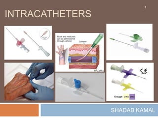
- 2. Inroduction 2 A plastic tube, usually attached to a puncturing needle, inserted into a blood vessel for infusion, injection, or pressure monitoring.
- 3. History 3 1929 when a 25 year old surgical resident named Werner Forssman inserted a plastic urethral catheter into the basilic vein in his right arm and then advanced the catheter into the right atrium of his heart. in 1956 Forssman was awarded the Nobel Prize in Medicine for performing the first right- heart catheterization in a human subject.
- 4. Types 4 Peripheral vascular catheters • Venous • Arterial Central venous catheters Peripherally inserted central venous catheters Special catheters • Hemodialysis Catheters • Introducer Sheaths • Pulmonary Artery Catheters
- 5. Catheter Material 5 Made of synthetic polymers (polyurethane and silicone) that are chemically inert, biocompatible, and resistant to chemical and thermal degradation. Peripheral vascular catheters (arterial and venous), central venous catheters, and pulmonary artery catheters are mostly made of polyurethane, peripherally- inserted central venous catheters (PICCs) are mostly made of silicone.
- 6. Catheter Size 6 • Gauge size varies inversely with OD (i.e., higher the gauge size, the smaller the OD); • however, there is no fixed relationship between gauge size and OD. Gauge Size Determined by the outside diameter (OD) of the catheter. • Superior to the gauge system because of its simplicity and uniformity. • begins at zero, and each increment of one French unit represents an increase of 1/3 (0.33) mm in OD: • French size × 0.33= OD (mm). • 3 French units in size will have an OD of 3 × 0.33 = 1.0 mm. French Size
- 8. Catheter Flow 8 Hagen-Poiseuille equation. Steady flow (Q) in a rigid tube is directly related to the fourth power of the inner radius of the tube (r4), and is inversely related to the length of the tube (L) and the viscosity of the fluid (μ).
- 9. PERIPHERAL VASCULAR CATHETERS9 Peripheral blood vessels in adults are cannulated typically 16–20 gauge catheters that are 1–2 inches in length. inserted using a catheter-over-needle device. Once flashback is evident, the catheter is advanced over the needle and into the lumen of the blood vessel.
- 10. Catheter-over-needle device for the cannulation of peripheral blood vessels. 10 The catheter fits snugly over the needle and has a tapered end to prevent fraying of the catheter tip during insertion. The needle has a clear hub to visualize the “flashback” of blood that occurs when the tip of the needle enters the lumen of a blood vessel.
- 12. 12 Safety needle guard automatically covers the needle's sharp bevel after withdrawal of needle from the hub, minimizing the risk of needle stick injuries.
- 14. 14
- 15. Indications: 15 Fluid and electrolyte replacement Administration of medicines Administration of blood/blood products Administration of Total Parenteral Nutrition Haemodynamic monitoring Blood sampling
- 16. Advantages: 16 Immediate effect Control over the rate of administration Patient cannot tolerate drugs / fluids orally Some drugs cannot be absorbed by any other route Pain and irritation is avoided compared to some substances when given SC/IM
- 17. What equipment do you need?17 Dressing Tray Non Sterile Gloves/Apron Cleaning Wipes Gauze swab IV cannula (separate slide) Tourniquet Dressing to secure cannula Alcohol wipes Saline flush and sterile syringe or fluid to be administered Sharps bin
- 18. Preperation: 18 Consult with patient Give explanation Gain consent Position the patient appropriately and identify the non-dominant hand/arm Support arm on pillow or in other suitable manner. Check for any contra-indications e.g. infection, damaged tissue, AV fistula etc.
- 19. Encourage venous filling by: 19 Correctly applying a tourniquet (applied to the patient’s upper/lower arm at a pressure which is high enough to impede venous distension but not to restrict arterial flow) Opening & closing the fist
- 20. What are the signs of a good vein?20 Bouncy Soft Above previous sites Refills when depressed Visible Has a large lumen Well supported Straight Easily palpable
- 21. Site of Choice for Veins: Identify a suitable vein
- 22. What veins should you avoid? 22 Thrombosed / sclerosed / fibrosed Inflamed / bruised Thin / Fragile Mobile Near bony prominences Areas or sites of infection, oedema or phlebitis Have undergone multiple previous punctures Do not use if patient has IV fluid in situ
- 23. Small vs. Large Veins 23 Catheters placed in small, peripheral veins have a limited life expectancy because they promote localized inflammation and thrombosis. The inflammation is prompted by mechanical injury to the blood vessel and by chemical injury to the vessel from caustic drug infusions. The thrombosis is incited by the inflammation, and is propagated by the sluggish flow in small, cannulated veins (low flow in small, cannulated veins is associated with an increase in blood viscosity, and this increases the propensity for thrombus formation).
- 24. 24 Large veins offer the advantages of a larger diameter and higher flow rates. The larger diameter allows the insertion of larger bore, multilumen catheters, which increases the efficiency of vascular access (i.e., more infusions per venipuncture). The higher flow rates reduce the damaging effects of infused fluids and thereby reduce the propensity for local thrombosis.
- 25. Procedure 25 Wash hands Prepare equipments Remove the cannula from the packaging and check all parts are operational Loosen the white cap and gently replace it Apply tourniquet Identify vein Clean the site over the vein with alcohol wipe, allow to dry
- 26. 26 Remove tourniquet if not able to proceed Put on non-sterile gloves Re-apply the tourniquet, 7-10 cm above site Remove the protective sleeve from the needle taking care not to touch it at any time Hold the cannula in your dominant hand, stretch the skin over the vein to anchor the vein with your non-dominant hand (Do not re palpate the vein)
- 27. 27 Insert the needle (bevel side up) at an angle of 10-30o to the skin (this will depend on vein depth.) Observe for blood in the flashback chamber Lower the cannula slightly to ensure it enters the lumen and does not puncture exterior wall of the vessel Gently advance the cannula over the needle whilst withdrawing the guide, noting secondary flashback along the cannula
- 28. 28 Release the tourniquet Apply gentle pressure over the vein (beyond the cannula tip) remove the white cap from the needle Remove the needle from the cannula and dispose of it into a sharps container Attach the white lock cap Secure the cannula with an appropriate dressing Flush the cannula with 2-5 mls 0.9% Sodium Chloride or attach an IV giving set and fluid
- 29. Finally: 29 Document the procedure including Date & time Site and size of cannula Any problems encountered Review date (cannula should be in situ no longer than 72 hours without appropriate risk assessment.) Note: some hospitals have pre-printed forms to record cannula events Thank the patient Clean up, dispose of rubbish
- 30. Possible Complications: 30 direct access to a patient's vascular system so provides a potential route for entry of micro organisms into that system. cause serious infection if they are allowed to enter and proliferate in the IV cannula, insertion site, or IV fluid.
- 31. IV-Site Infection: 31 Does not produce much (if any) pus or inflammation at the IV site. This is the most common cannula-related infection.
- 32. Cellulites: 32 Warm, red and often tender skin surrounding the site of cannula insertion; pus is rarely detectable.
- 33. Infiltration or tissuing 33 occurs when the infusion (fluid) leaks into the surrounding tissue. It is important to detect early as tissue necrosis could occur – re-site cannula immediately
- 34. Thrombolism / thrombophlebitis 34 occur when a small clot becomes detached from the sheath of the cannula or the vessel wall – prevention is the greatest form of defence. Flush cannula regularly and consider re-siting the cannula if in prolonged use.
- 35. Extravasation 35 accidental administration of IV drugs into the surrounding tissue, because the needle has punctured the vein and the infusion goes directly into the arm tissue. The leakage of high osmolarity solutions or chemotherapy agents can result in significant tissue destruction, and significant complications
- 36. Bruising 36 commonly results from failed IV placement - particularly in the elderly and those on anticoagulant therapy.
- 37. Air embolism 37 occurs when air enters the infusion line, although this is very rare it is best if we consider the preventive measures – Make sure all lines are well primed prior to use and connections are secure
- 38. Haematoma 38 occurs when blood leaks out of the infusion site. The common cause of this is using cannula that are not tapered at the distal end. It will also occur if on insertion the cannula has penetrated through the other side of the vessel wall – apply pressure to the site for approximately 4 minutes and elevate the limb
- 39. Phlebitis 39 It is common in IV therapy and can be caused in many ways. It is inflammation of a vein (redness and pain at the infusion site) prevention can be using aseptic insertion techniques, choosing the smallest gauge cannula possible for the prescribed treatment, secure the cannula properly to prevent movement and carry out regular checks of the infusion site.
- 40. Arterial catheter 40 a thin catheter inserted into an artery. to monitor the BP real-time (rather than by intermittent measurement), and to obtain samples for ABG measurements. not used to administer medication inserted in the wrist (radial artery); but can also be inserted into the elbow (brachial artery), groin (femoral artery), foot (dorsalis pedis artery) or the inside of the wrist (ulnar artery).
- 41. Central Venous Catheters 41 Used for cannulation of larger, more centrally placed veins (i.e., subclavian, internal jugular, and femoral veins) is often necessary for reliable vascular access in critically ill patients. They are typically 15 to 30 cm (6 to 12 inches) in length, and have single or multiple (2–4) infusion channels. Multilumen catheters are favored in the critically ill patient recquires a multitude of parenteral therapies (e.g., fluids, drugs, and nutrient mixtures). Multilumen catheters make it possible to deliver these therapies using a single venipuncture.
- 42. 42 Triple-lumen catheters are favorite for central venous access. These catheters are available in sizes of 4-9 French. Size 7 French(OD=23mm) triple lumen catheter is a popular choice in adults, which typically have one 16 gauge channel and two smaller 18 gauge channels. To prevent mixing of infusate solutions, the three outflow ports are separated.
- 43. 43
- 44. 44
- 45. Indications 45 When peripheral venous access is difficult to obtain (e.g., in obese or intravenous drug abusers) or difficult to maintain (e.g., in agitated patients). For the delivery of vasoconstrictor drugs (e.g., dopamine, norepinephrine), hypertonic solutions (e.g., parenteral nutrition formulas), or multiple parenteral medications. For prolonged parenteral drug therapy (i.e., more than a few days). For specialized tasks such as hemodialysis, transvenous cardiac pacing, or hemodynamic monitoring (e.g., with pulmonary artery catheters).
- 46. Contraindications: 46 There are no absolute contraindications except inexperienced operator is in this procedure, including the presence or severity of a coagulation disorder. Relative contraindicatios Inexperience operator with unsupervised operator, Local infection, Distorted local anatomy, Coagulopathy, Previous radiation therapy, Suspected proximal vascular injury
- 47. Insertion Technique 47 Central venous catheters are inserted by threading the catheter over a guidewire (Seldinger technique). A small bore needle (usually 20 gauge) is used to probe for the target vessel. When the tip of the needle enters the vessel, a long, thin wire with a flexible tip is passed through the needle and into the vessel lumen. The needle is then removed, and a catheter is advanced over the guidewire and into the blood vessel.
- 48. Seldinger technique 48 When cannulating deep vessels, a larger and more rigid “dilator catheter” is first threaded over the guide-wire to create a tract that facilitates insertion of the vascular catheter.
- 50. Peripherally Inserted Central Catheters50 peripherally inserted central catheters (PICCs), which are inserted in the basilic or cephalic vein in the arm (just above the antecubital fossa) and advanced into the superior vena cava. Useful in patients with concern for the adverse consequences of central venous cannulation (e.g., pneumothorax arterial puncture, poor patient acceptance) PICCs are used primarily when traditional central venous accesss are considered risky (e.g., severe thrombocytopenia) or are difficult to obtain (e.g., morbid obesity).
- 51. 51
- 52. PICC vs CVC 52 Major distinction between PICCs and central venous catheters is their length; i.e., the length of the catheters(50 cm and 70 cm) is at least double the length of the triple lumen catheters. Because of this added length there is reduction in flow capacity in PICC. Flow is particularly sluggish in the double lumen PICCs because of the smaller diameter of the infusion channels. The flow limitation of PICCs (especially the double lumen catheters) makes them ill-suited for aggressive volume therapy.
- 53. Hemodialysis Catheters 53 One of the recognized benefits of intensive care units is the ability to provide emergent hemodialysis for patients with acute renal failure, Hemodialysis catheters are the wide-body catheters of critical care, with diameters up to 16 French (5.3 mm), and they are equipped with dual 12 gauge infusion channels that can accommodate the high flow rates (200–300 mL/min) needed for effective hemodialysis. One channel carries blood from the patient to the dialysis membranes, and the other channel returns the blood to the patient.
- 54. 54
- 55. 55
- 56. Introducer Sheaths 56 Introducer sheaths are large-bore (8–9 French) catheters that serve as conduits for the insertion and removal of temporary vascular devices. They are used primarily to facilitate the placement of pulmonary artery (PA) catheters The introducer sheath is first placed in a large, central vein, and the PA catheter is then threaded through the sheath and advanced into the pulmonary artery.
- 57. 57 The placement of PA catheters often requires repeated trials of advancing and retracting the catheter to achieve the proper position in the pulmonary artery, and the introducer sheath facilitates these movements. When the PA catheter is no longer needed, the introducer sheath allows the catheter to be removed and replaced with a central venous catheter, if needed, without a new venipuncture. serve as stand-alone infusion devices-with pressurized infusion systems, flow rates of 850 mL/min have been reported
- 58. Pulmonary Artery Catheters 58 Pulmonary artery balloon-flotation catheters are highly specialized devices capable of providing various measures of cardiovascular function and systemic oxygenation.
- 59. 59
Editor's Notes
- The use of multiple infusion channels does not increase the incidence of catheter-related infections, but the larger diameter of multilumen catheters creates an increased risk of catheter-induced thrombosis.