This document discusses intestinal regeneration biology and approaches. It covers:
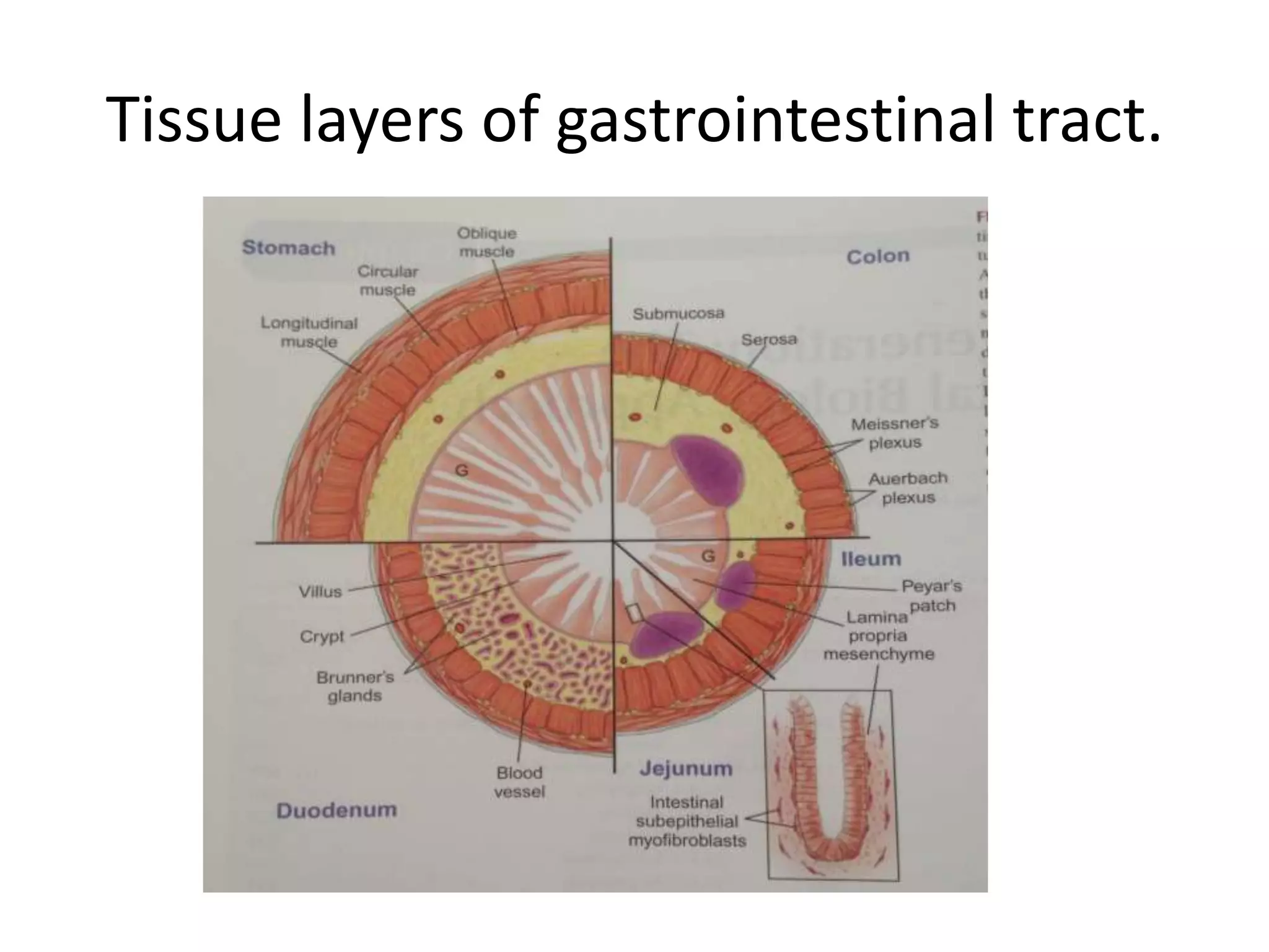
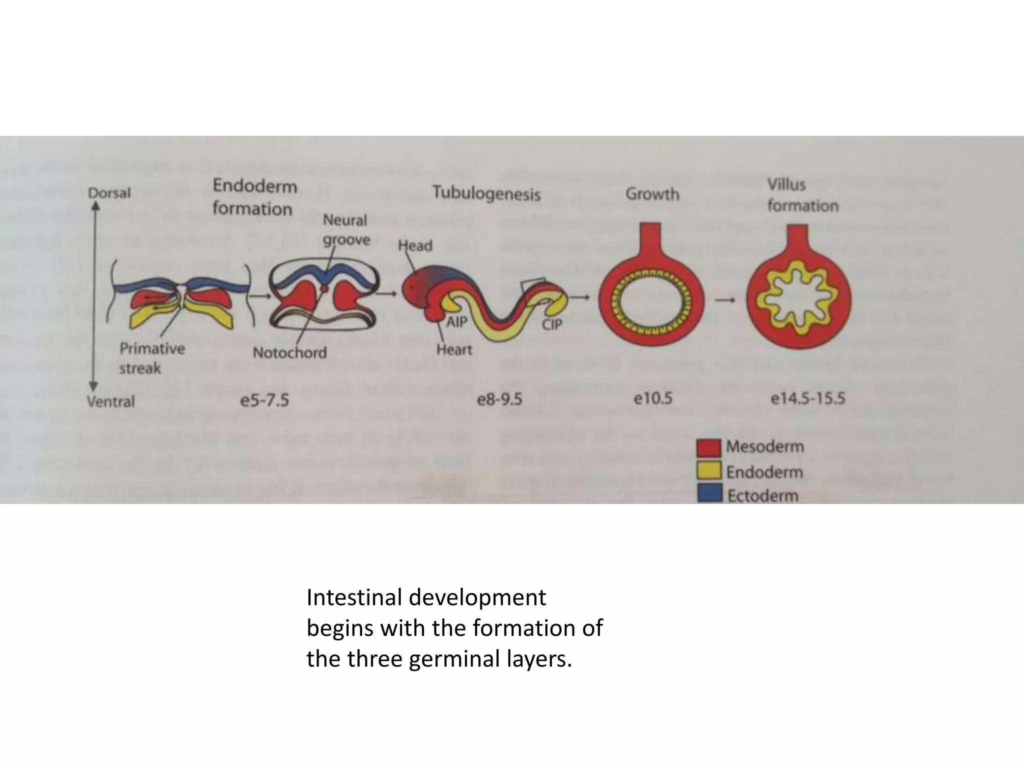
1. The tissue layers of the gastrointestinal tract and intestinal development from birth to adulthood.
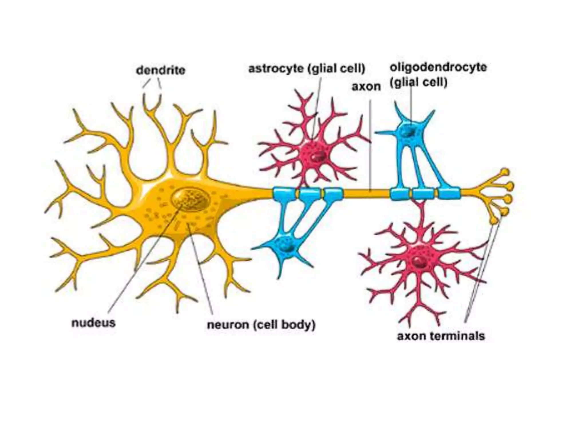
2. Sources of smooth muscle and neuronal stem cells including bone marrow, muscle tissue, and enteric nervous system.
3. Differences between stem and progenitor cells and how regenerative organogenesis occurs in the adult intestine through mucosal development and crypt differentiation.


![Inflammatory responses
Immunologic functions, which may be important in the
pathogenesis of IBD, have not been fully investigated
in SEMFs. In many tissues, inflammatory responses
are regulated in close association with the regulation
of ECM metabolism. For example, in colonic SEMFs,
coupled induction of chemokines [IL-8 and monocyte
chemoattractant protein (MCP)-1] and MMPs in response
to IL-1b and TNF-a has been observed](https://image.slidesharecdn.com/intestinalregenerationbiologyapproach-170313044644/75/Intestinal-regeneration-biology-approach-26-2048.jpg)