Downloaded 407 times


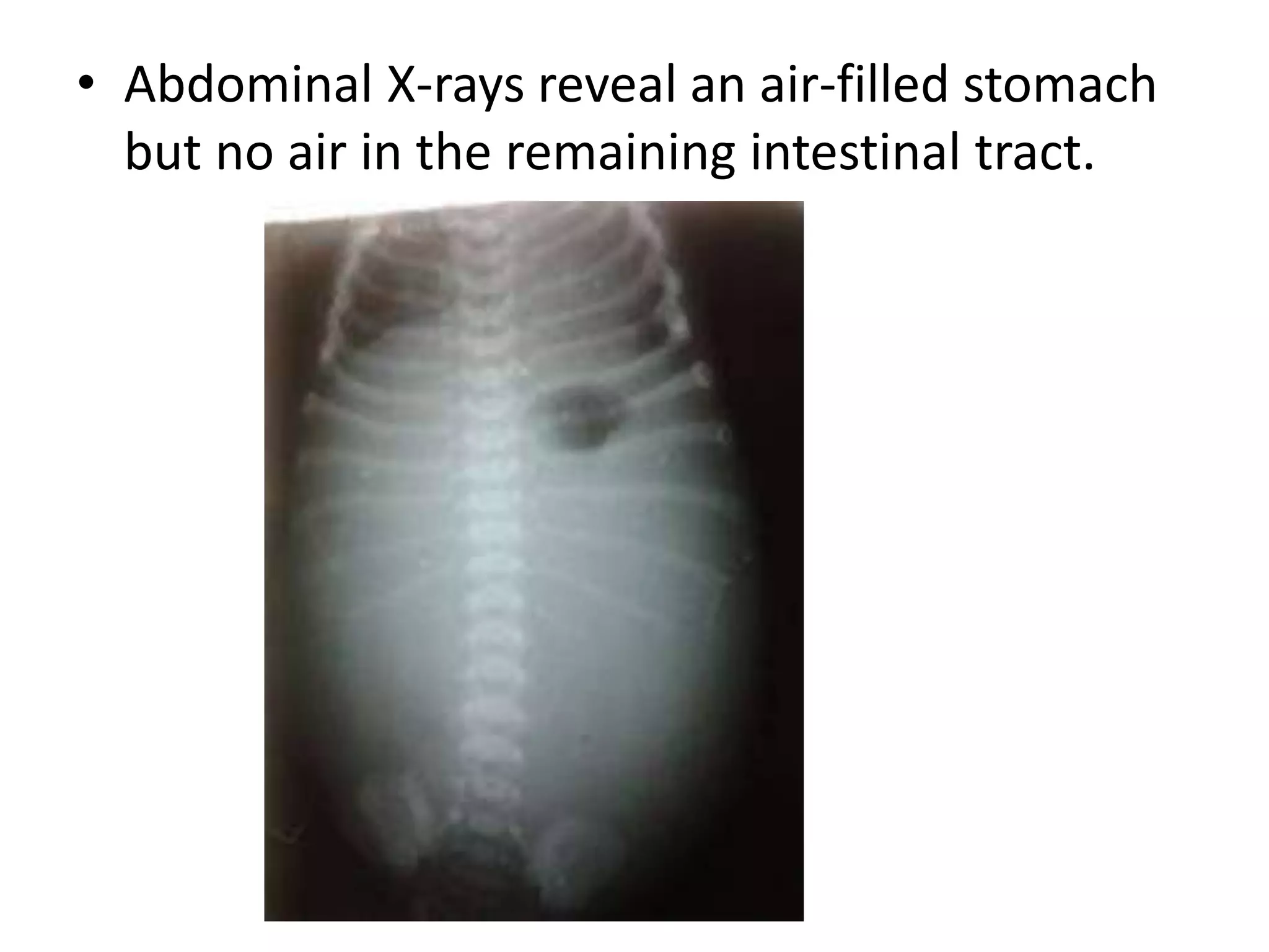
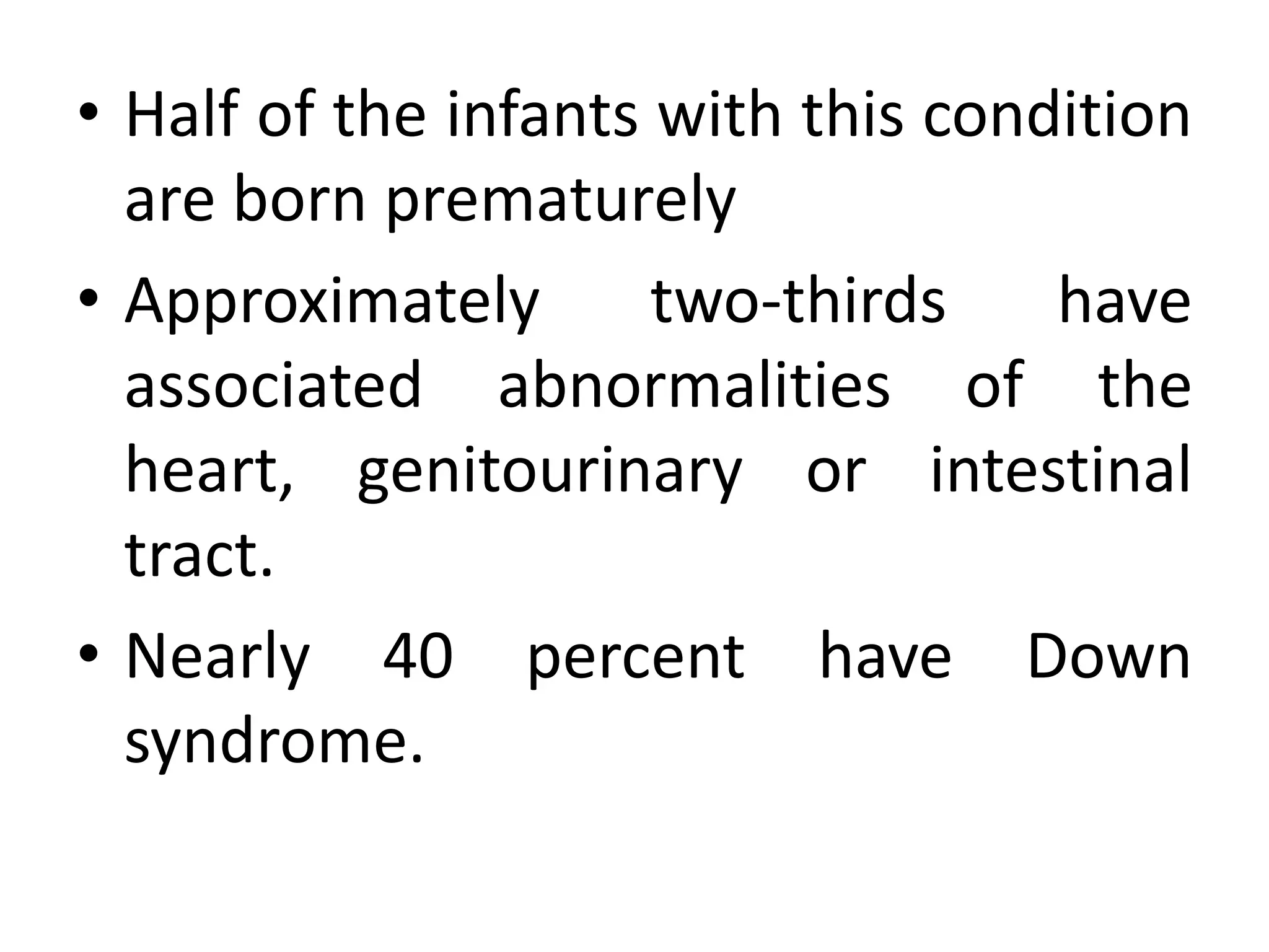


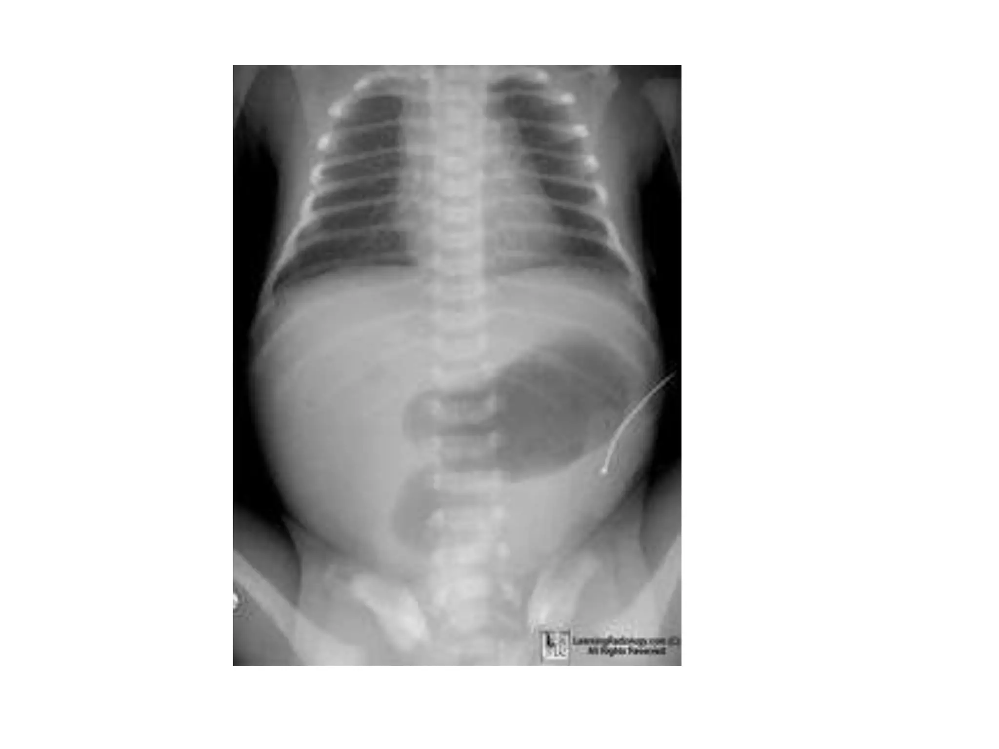


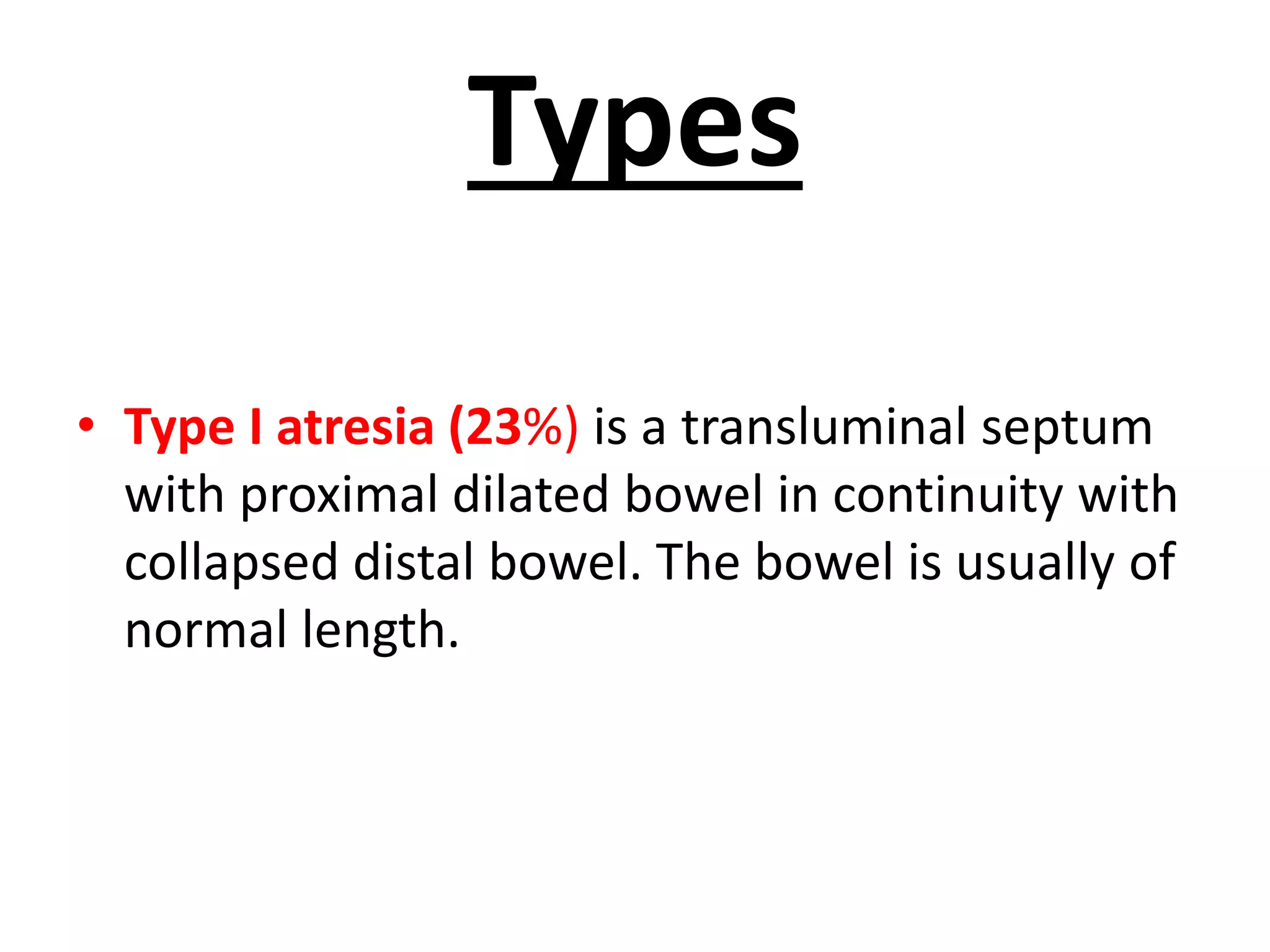

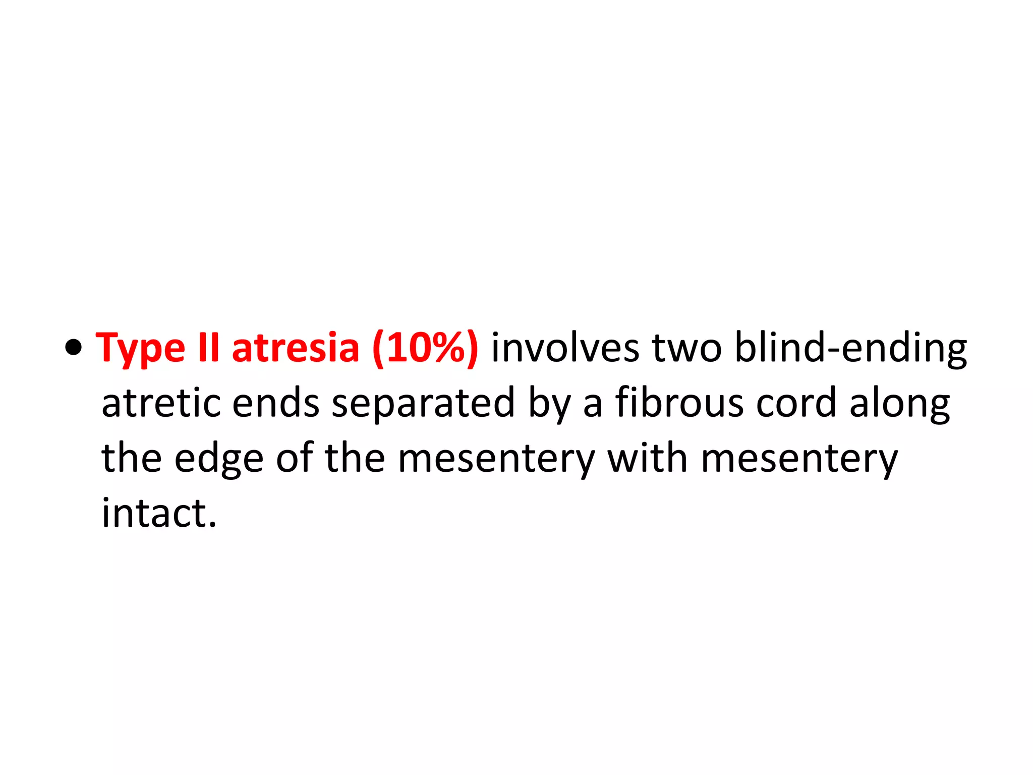
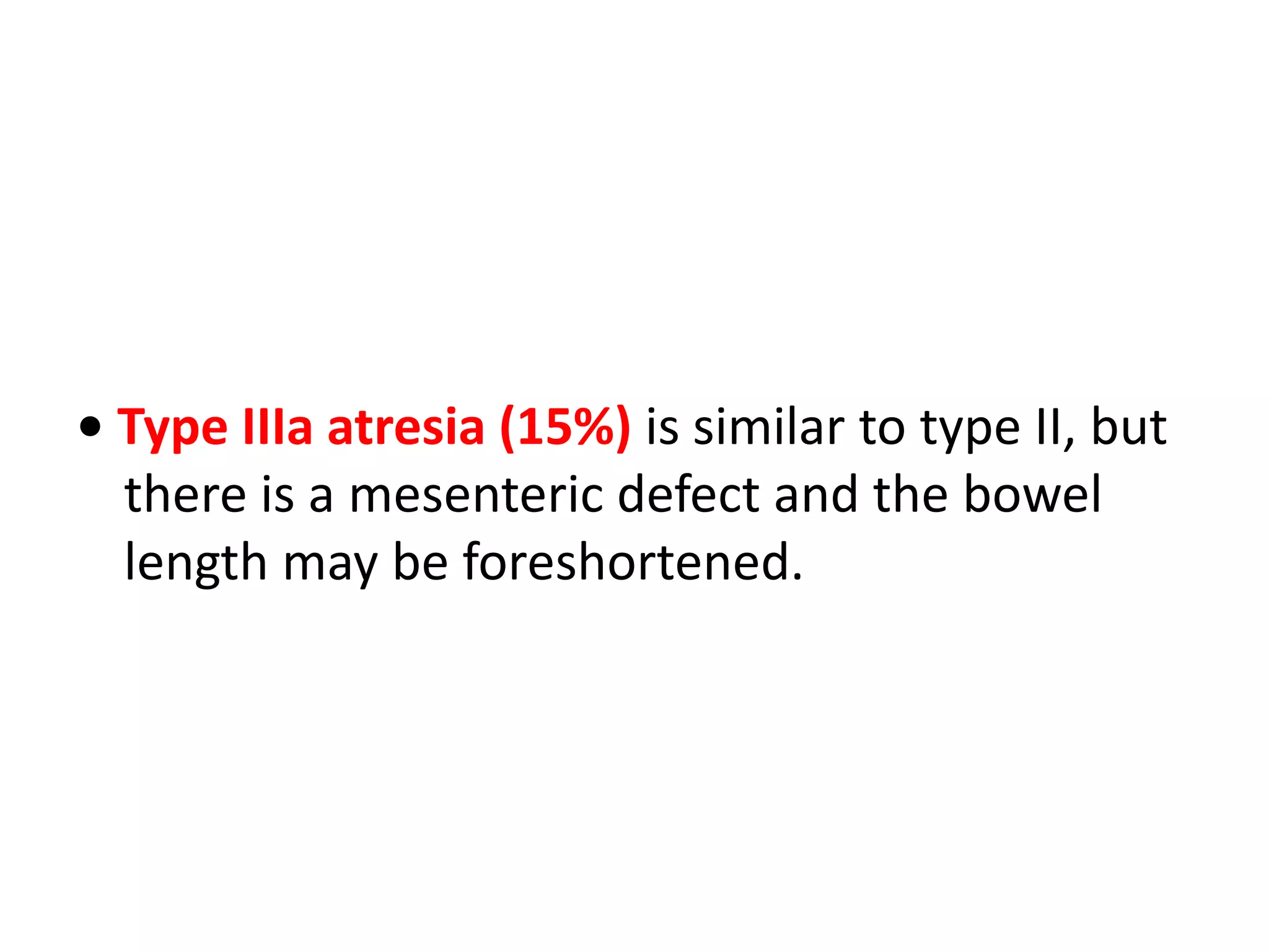


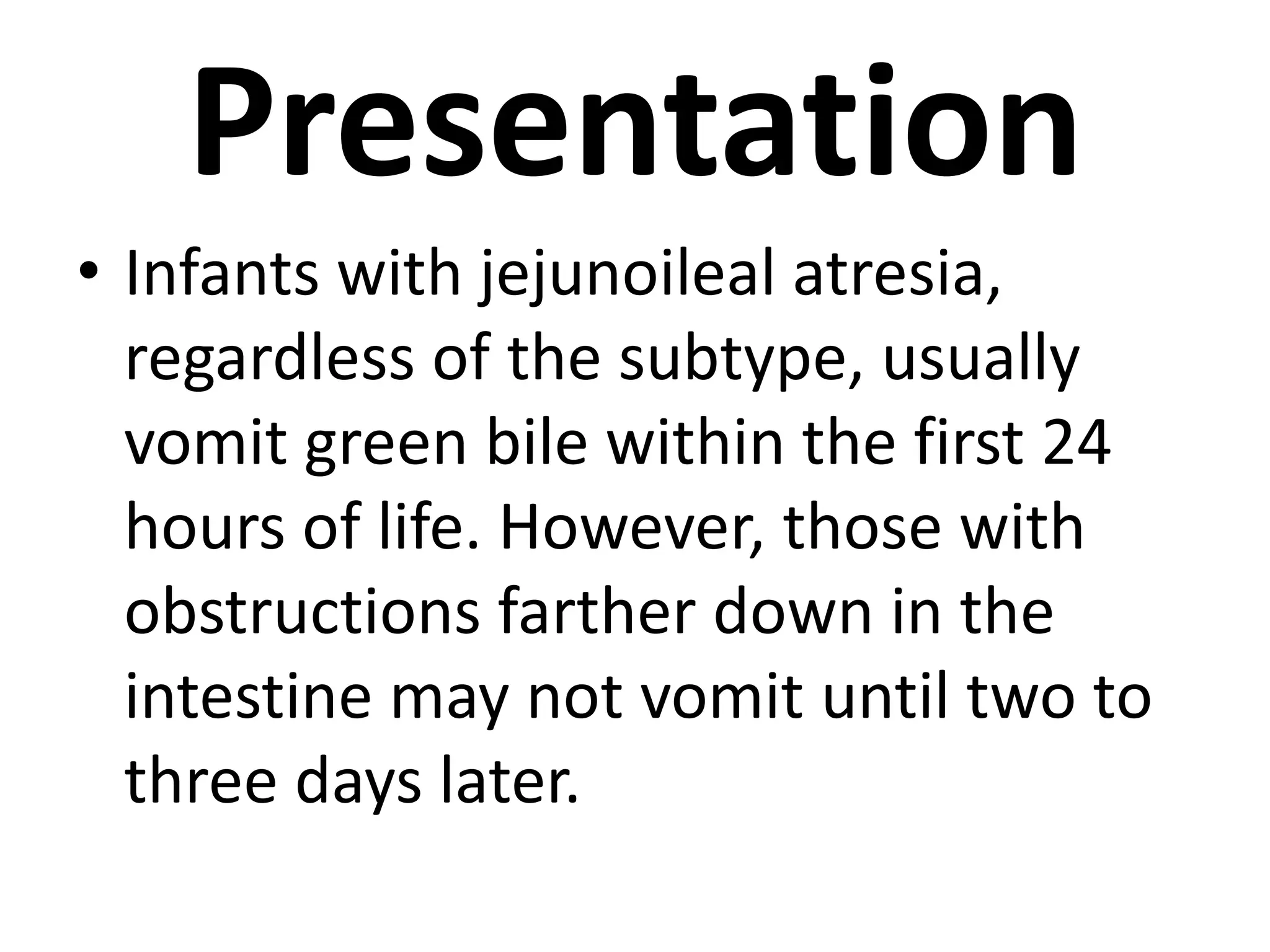


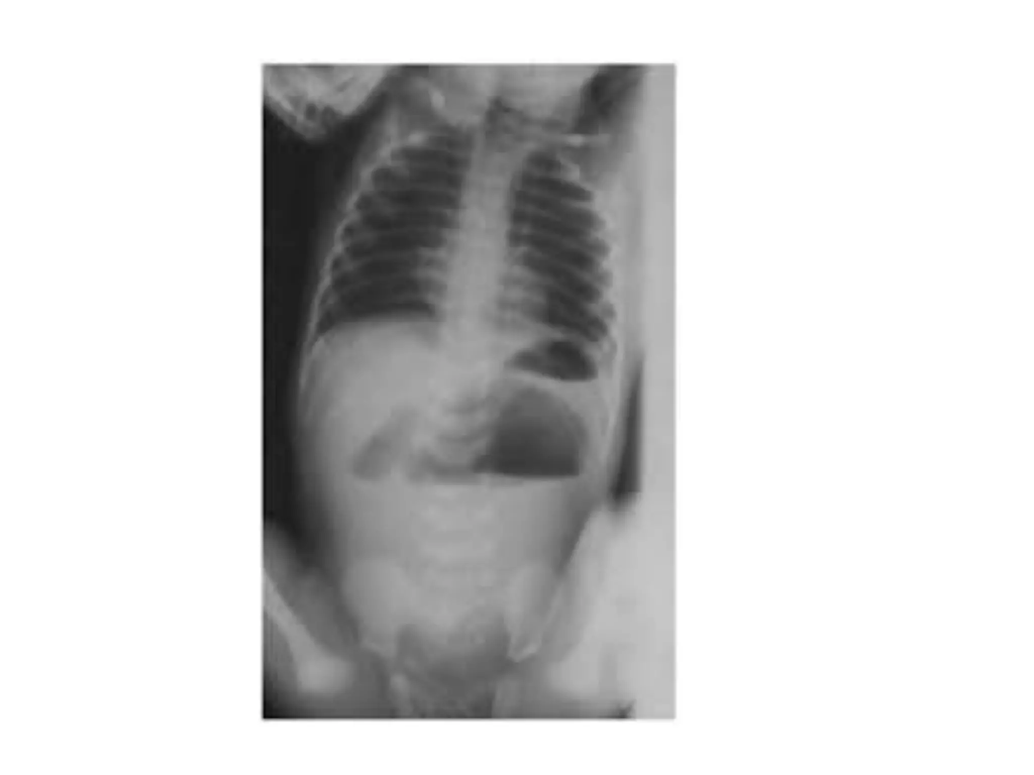


This document discusses different types of intestinal atresia, which is a complete blockage in the intestine. It describes pyloric, duodenal, jejunoileal, and colonic atresia. Pyloric atresia involves an obstruction at the stomach-duodenum junction. Duodenal atresia occurs in 1 in 2,500 births and often involves other abnormalities. Jejunoileal atresia can be classified into different types depending on the location and severity of the blockage. Colonic atresia is rare and may involve dilation of the colon. Diagnosis is often made through prenatal ultrasound or abdominal x-ray. Treatment involves surgery to remove blockages and re


