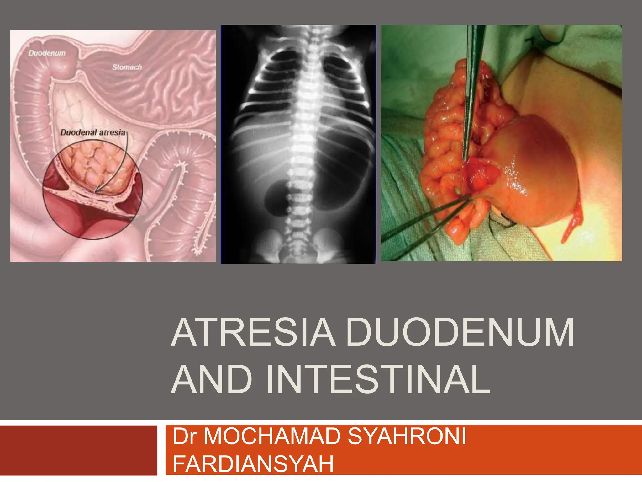
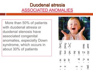
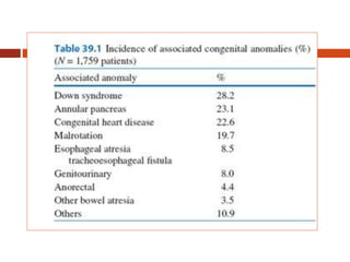
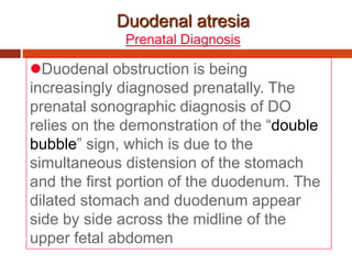
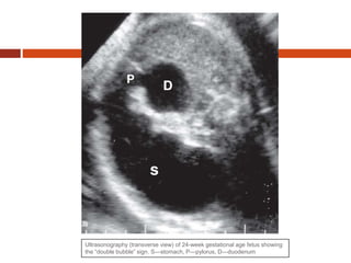
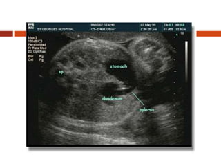
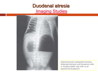
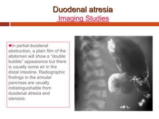
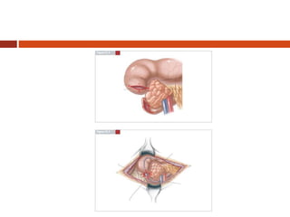
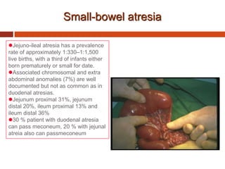
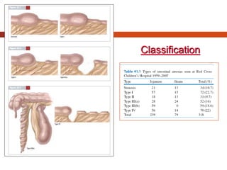
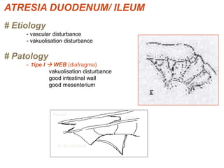
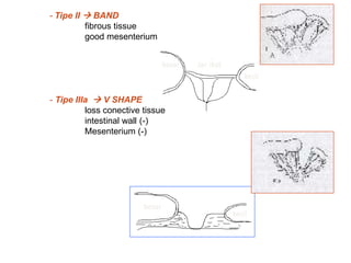
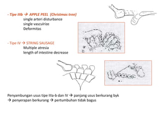
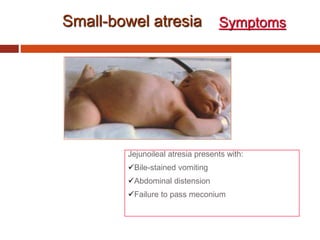
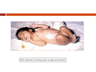
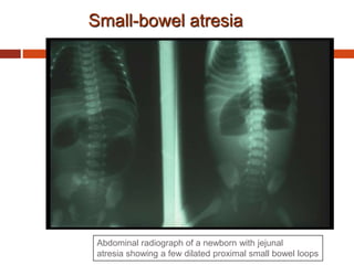
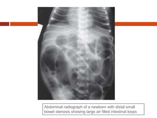
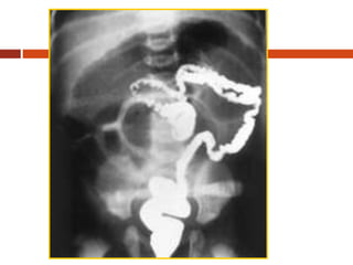
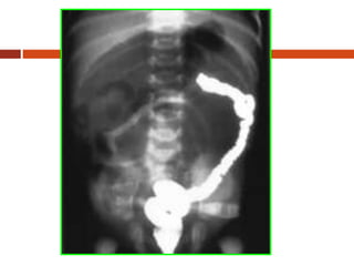
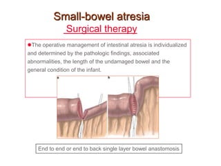
ATRESIA DUODENUM
AND INTESTINAL
Dr MOCHAMAD SYAHRONI
FARDIANSYAH
Duodenal atresia
INCIDENCE
Congenital duodenal obstruction
(DO) is a frequent cause of congenital
intestinal obstruction in the newborn,
occurring in 1 per 5,000–10,000 live
births, and affecting boys more
commonly than girls.
More than 50% of patients
with duodenal atresia or
duodenal stenosis have
associated congenital
anomalies, especially Down
syndrome, which occurs in
about 30% of patients
Duodenal atresia
ASSOCIATED ANOMALIES
Duodenal atresia
Prenatal Diagnosis
Duodenal obstruction is being
increasingly diagnosed prenatally. The
prenatal sonographic diagnosis of DO
relies on the demonstration of the “double
bubble” sign, which is due to the
simultaneous distension of the stomach
and the first portion of the duodenum. The
dilated stomach and duodenum appear
side by side across the midline of the
upper fetal abdomen
Ultrasonography (transverse view) of 24-week gestational age fetus showing
the “double bubble” sign. S—stomach, P—pylorus, D—duodenum
Duodenal atresia
Prenatal Diagnosis
Maternal polyhydramnios is also a common
ultrasonographic finding observed in 20–75% of
cases with duodenal atresia, mainly in the
second half of pregnancy.
 In all cases of combined polyhydramnios and
“double bubble” sign, a detailed evaluation for
other associated anomalies, especially cardiac
anomalies, should be undertaken.
Amniocentesis for chromosomal analysis is
helpful for counseling.
Duodenal atresia
Clinical
About half of these patients are
premature and of low birth weight.
Vomiting and intolerance of attempted
feedings are the most common symptoms
and are usually present on the first day of
life.
Since 80% of the obstructions are
located in the postampullary region of the
duodenum, vomitus in the majority of
cases is bile-stained.
Duodenal atresia
Clinical
An orogastric (OG) feeding
tube in an infant with
suspected duodenal
obstruction typically yields a
significant amount of bile-
stained fluid.
Duodenal atresia
Clinical
In supra-ampullary atresia, it is nonbilious.
There is no abdominal distention because of
the high level of obstruction.
Infants may pass some meconium in the first
24 h of life and thereafter constipation may
develop.
Dehydration with weight loss and electrolyte
imbalance soon follows if fluid and electrolyte
losses have not been adequately replaced.
Duodenal atresia
Clinical
Incomplete duodenal obstruction (e.g.,
duodenal membrane with a central
aperture) usually leads to the
delayed onset of symptoms, which may
appear a few months or years after birth.
Duodenal atresia
Imaging Studies
The diagnosis of the duodenal
atresia is confirmed on X-ray
examination. An abdominal
radiograph will show a dilated
stomach and duodenum,
giving the characteristic
appearance of a “double
bubble sign”
(the stomach and the proximal
duodenum are air filled), with
no gas beyond the duodenum
Duodenal atresia
Imaging Studies
Abdominal erect radiograph showing
distended stomach and duodenum with
a “double bubble” sign with no air
beyond the duodenum
Duodenal atresia
Imaging Studies
In partial duodenal
obstruction, a plain film of the
abdomen will show a “double
bubble” appearance but there
is usually some air in the
distal intestine. Radiographic
findings in the annular
pancreas are usually
indistinguishable from
duodenal atresia and
stenosis.
Duodenal atresia
Surgical therapy
Duodenal atresia and stenosis are
treated surgically. In patients with
duodenal obstruction, a
duodenoduodenostomy is the most
commonly performed procedure. Some
advocate duodenojejunostomy, a
procedure practiced by few because of its
higher risk of long-term complications.
Small-bowel atresia
Jejuno-ileal atresia has a prevalence
rate of approximately 1:330–1:1,500
live births, with a third of infants either
born prematurely or small for date.
Associated chromosomal and extra
abdominal anomalies (7%) are well
documented but not as common as in
duodenal atresias.
Jejunum proximal 31%, jejunum
distal 20%, ileum proximal 13% and
ileum distal 36%
30 % patient with duodenal atresia
can pass meconeum, 20 % with jejunal
atreia also can passmeconeum
Classification
ATRESIA DUODENUM/ ILEUM
# Etiology
- vascular disturbance
- vakuolisation disturbance
# Patology
- Tipe I  WEB (diafragma)
vakuolisation disturbance
good intestinal wall
good mesenterium
Parsial
Total
A. Drummond
besar Jar. ikat
kecil
besar
kecil
- Tipe II  BAND
fibrous tissue
good mesenterium
- Tipe IIIa  V SHAPE
loss conective tissue
intestinal wall (-)
Mesenterium (-)
- Tipe IIIb  APPLE PEEL (Christmas tree)
single arteri disturbance
single vasculrize
Deformitas
- Tipe IV  STRING SAUSAGE
Multiple atresia
length of intestine decrease
Penyambungan usus tipe IIIa-b dan IV  panjang usus berkurang byk
 penyerapan berkurang  pertumbuhan tidak bagus
Small-bowel atresia
Jejunoileal atresia presents with:
Bile-stained vomiting
Abdominal distension
Failure to pass meconium
Symptoms
Bile stained vomiting due to jejunal atresia
Small-bowel atresia
Abdominal radiograph of a newborn with jejunal
atresia showing a few dilated proximal small bowel loops
Abdominal radiograph of a newborn with distal small
bowel stenosis showing large air filled intestinal loops
Small-bowel atresia
Surgical therapy
The operative management of intestinal atresia is individualized
and determined by the pathologic findings, associated
abnormalities, the length of the undamaged bowel and the
general condition of the infant.
End to end or end to back single layer bowel anastomosis
THANKS FOR LEARNING

(02) Atresia Duodenum and management.ppt

  • 1.
    ATRESIA DUODENUM AND INTESTINAL DrMOCHAMAD SYAHRONI FARDIANSYAH
  • 2.
    Duodenal atresia INCIDENCE Congenital duodenalobstruction (DO) is a frequent cause of congenital intestinal obstruction in the newborn, occurring in 1 per 5,000–10,000 live births, and affecting boys more commonly than girls.
  • 3.
    More than 50%of patients with duodenal atresia or duodenal stenosis have associated congenital anomalies, especially Down syndrome, which occurs in about 30% of patients Duodenal atresia ASSOCIATED ANOMALIES
  • 5.
    Duodenal atresia Prenatal Diagnosis Duodenalobstruction is being increasingly diagnosed prenatally. The prenatal sonographic diagnosis of DO relies on the demonstration of the “double bubble” sign, which is due to the simultaneous distension of the stomach and the first portion of the duodenum. The dilated stomach and duodenum appear side by side across the midline of the upper fetal abdomen
  • 6.
    Ultrasonography (transverse view)of 24-week gestational age fetus showing the “double bubble” sign. S—stomach, P—pylorus, D—duodenum
  • 8.
    Duodenal atresia Prenatal Diagnosis Maternalpolyhydramnios is also a common ultrasonographic finding observed in 20–75% of cases with duodenal atresia, mainly in the second half of pregnancy.  In all cases of combined polyhydramnios and “double bubble” sign, a detailed evaluation for other associated anomalies, especially cardiac anomalies, should be undertaken. Amniocentesis for chromosomal analysis is helpful for counseling.
  • 9.
    Duodenal atresia Clinical About halfof these patients are premature and of low birth weight. Vomiting and intolerance of attempted feedings are the most common symptoms and are usually present on the first day of life. Since 80% of the obstructions are located in the postampullary region of the duodenum, vomitus in the majority of cases is bile-stained.
  • 10.
    Duodenal atresia Clinical An orogastric(OG) feeding tube in an infant with suspected duodenal obstruction typically yields a significant amount of bile- stained fluid.
  • 11.
    Duodenal atresia Clinical In supra-ampullaryatresia, it is nonbilious. There is no abdominal distention because of the high level of obstruction. Infants may pass some meconium in the first 24 h of life and thereafter constipation may develop. Dehydration with weight loss and electrolyte imbalance soon follows if fluid and electrolyte losses have not been adequately replaced.
  • 12.
    Duodenal atresia Clinical Incomplete duodenalobstruction (e.g., duodenal membrane with a central aperture) usually leads to the delayed onset of symptoms, which may appear a few months or years after birth.
  • 13.
    Duodenal atresia Imaging Studies Thediagnosis of the duodenal atresia is confirmed on X-ray examination. An abdominal radiograph will show a dilated stomach and duodenum, giving the characteristic appearance of a “double bubble sign” (the stomach and the proximal duodenum are air filled), with no gas beyond the duodenum
  • 14.
    Duodenal atresia Imaging Studies Abdominalerect radiograph showing distended stomach and duodenum with a “double bubble” sign with no air beyond the duodenum
  • 15.
    Duodenal atresia Imaging Studies Inpartial duodenal obstruction, a plain film of the abdomen will show a “double bubble” appearance but there is usually some air in the distal intestine. Radiographic findings in the annular pancreas are usually indistinguishable from duodenal atresia and stenosis.
  • 16.
    Duodenal atresia Surgical therapy Duodenalatresia and stenosis are treated surgically. In patients with duodenal obstruction, a duodenoduodenostomy is the most commonly performed procedure. Some advocate duodenojejunostomy, a procedure practiced by few because of its higher risk of long-term complications.
  • 18.
    Small-bowel atresia Jejuno-ileal atresiahas a prevalence rate of approximately 1:330–1:1,500 live births, with a third of infants either born prematurely or small for date. Associated chromosomal and extra abdominal anomalies (7%) are well documented but not as common as in duodenal atresias. Jejunum proximal 31%, jejunum distal 20%, ileum proximal 13% and ileum distal 36% 30 % patient with duodenal atresia can pass meconeum, 20 % with jejunal atreia also can passmeconeum
  • 19.
  • 20.
    ATRESIA DUODENUM/ ILEUM #Etiology - vascular disturbance - vakuolisation disturbance # Patology - Tipe I  WEB (diafragma) vakuolisation disturbance good intestinal wall good mesenterium Parsial Total A. Drummond
  • 21.
    besar Jar. ikat kecil besar kecil -Tipe II  BAND fibrous tissue good mesenterium - Tipe IIIa  V SHAPE loss conective tissue intestinal wall (-) Mesenterium (-)
  • 22.
    - Tipe IIIb APPLE PEEL (Christmas tree) single arteri disturbance single vasculrize Deformitas - Tipe IV  STRING SAUSAGE Multiple atresia length of intestine decrease Penyambungan usus tipe IIIa-b dan IV  panjang usus berkurang byk  penyerapan berkurang  pertumbuhan tidak bagus
  • 23.
    Small-bowel atresia Jejunoileal atresiapresents with: Bile-stained vomiting Abdominal distension Failure to pass meconium Symptoms
  • 24.
    Bile stained vomitingdue to jejunal atresia
  • 25.
    Small-bowel atresia Abdominal radiographof a newborn with jejunal atresia showing a few dilated proximal small bowel loops
  • 26.
    Abdominal radiograph ofa newborn with distal small bowel stenosis showing large air filled intestinal loops
  • 29.
    Small-bowel atresia Surgical therapy Theoperative management of intestinal atresia is individualized and determined by the pathologic findings, associated abnormalities, the length of the undamaged bowel and the general condition of the infant. End to end or end to back single layer bowel anastomosis
  • 30.