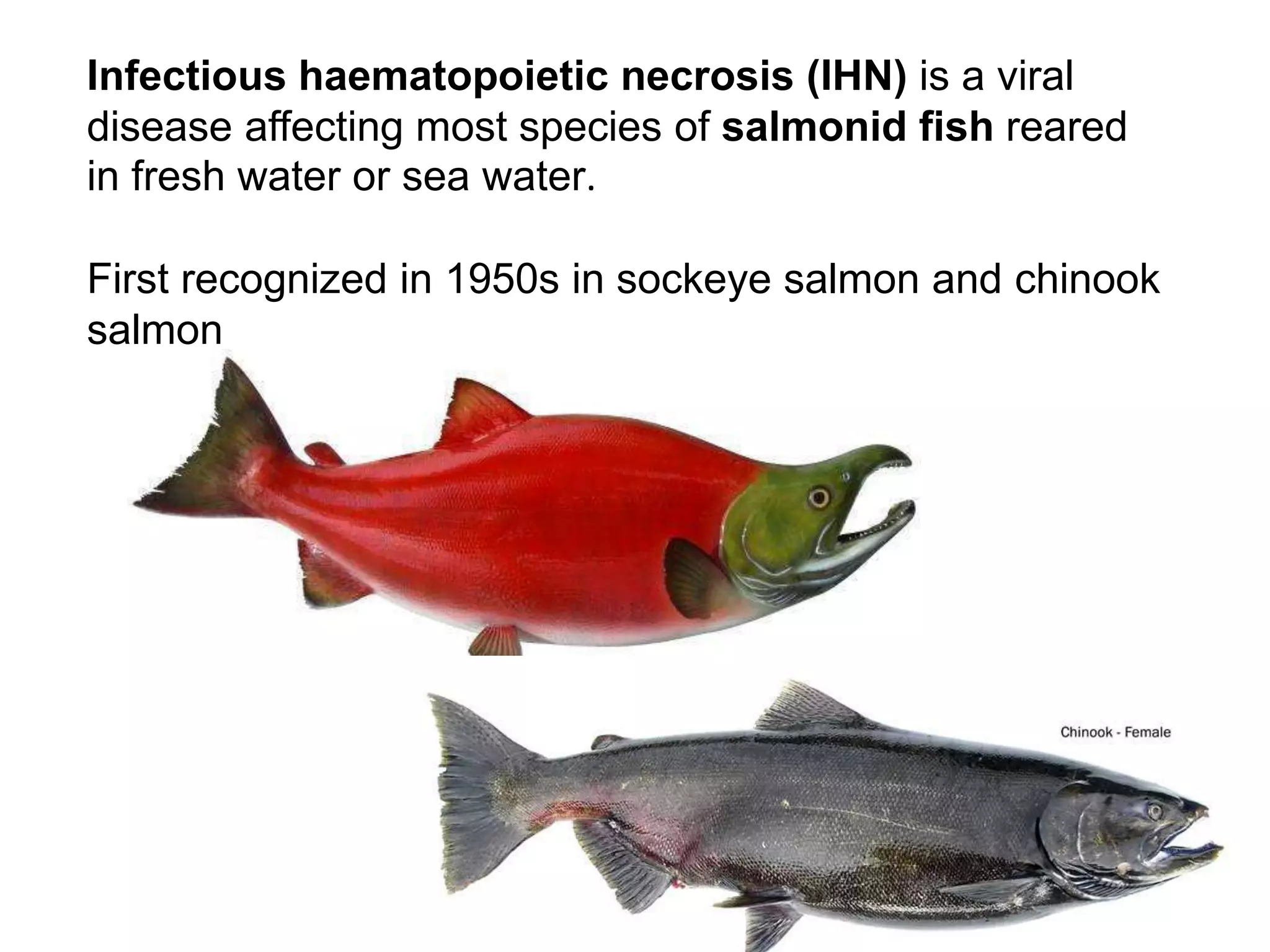
INFECTIOUS HAEMATOPOIETIC
NECROSIS
Infectious haematopoietic necrosis (IHN) is a viral
disease affecting most species of salmonid fish reared
in fresh water or sea water.
First recognized in 1950s in sockeye salmon and chinook
salmon
Caused by the rhabdovirus
the principal clinical and economic consequences of IHN
occur on farms rearing rainbow trout where acute
outbreaks can result in very high mortality.
However, both Pacific and Atlantic salmon can be
severely affected.
Aetiological agent
Agent strains
The fish rhabdovirus, IHNV, has a bullet-shaped virion containing
a non-segmented,
negative-sense,
 single-stranded RNA genome
 single-stranded RNA genome of approximately 11,000
nucleotides that encodes six proteins in the following order
a nucleoprotein (N),
 a phosphoprotein (P),
 a matrix protein (M),
a glycoprotein (G),
 a non-virion protein (NV),
 a polymerase (L).
The causative agent of IHN is a Rhabdovirus , genus
Novirhabdovirus 3 main genotypes described ( Kurath
et al., 2003)
GROUPS ORIGIN
U Isolates from Alaska, British Columbia
Washington,Oregon, California and Japan
obtained from :
Sockeye salmon ( O. nerka )
Chinook salmon ( O. tshawytscha)
M: isolates from Idaho, Washington, France and Italy
obtained from rainbow trout (O. mikiss )
L: isolates from California, Oregon and Japan obtained
from Giappone, obtained from
Survival outside the host
IHNV is heat, acid and ether labile.
The virus will survive in fresh water for at least 1 month at
cooler temperatures, especially if organic material is present.
Stability of the agent (effective inactivation methods)
IHNV is readily inactivated by common disinfectants and
drying
Life cycle
Reservoirs of IHNV are
clinically infected fish and
covert carriers among
cultured, feral or wild fish.
Virus is shed via urine,
sexual fluids and from
external mucus, whereas
kidney, spleen and other
internal organs are the sites
in which virus is most
abundant during the course
of overt infection
SUSCEPTIBLE HOSTS
rainbow or steelhead trout (O. mykiss),
Chum salmon (Oncorhynchus keta),
coho salmon (O. kisutch),
Masou salmon (O. masou),
sockeye salmon (O. nerka),
pink salmon (O. rhodurus)
Chinook salmon (O.tshawytscha),
Atlantic salmon (Salmo salar
Susceptible stages of the host
IHN occurs among several species of salmonids with fry being
the most highly susceptible stage.
Older fish are typically more
resistant to clinical disease,
but among individuals, there
is a high degree of variation
in susceptibility to IHNV.
Survivors of IHN demonstrate a
strong protective immunity with
the synthesis of circulating
antibodies to the virus
Target organs and infected tissue
Virus entry is thought to occur through
the gills and
at bases of fins
while kidney, spleen and other internal organs are the
sites in which virus is most abundant during the course of
overt infection.
Salmon affected by IHN virus, ventral congestion and pale gill
Disease pattern
Infection with IHNV often leads to mortality due to the
impairment of osmotic balance, and occurs within a
clinical context of oedema and haemorrhage.
Virus multiplication in endothelial cells of blood
capillaries, haematopoietic tissues, and cells of the
kidney underlies the clinical signs.
Transmission mechanisms
The transmission of IHNV between fish is primarily
horizontal and high levels of virus are shed from infected
juvenile fish, however, cases of vertical or egg-associated
transmission have been recorded.
Although egg-associated
transmission is
significantly reduced by
the now common practice
of surface disinfection of
eggs with an iodophor
solution, it is the only
mechanism accounting for
the appearance of IHN in
new geographical
locations among alevins
• CANADA
• USA
• DOMINICAN REP.
• JAPAN
• KOREA
• PAKISTAN
• EUROPE
• BELGIUM
• CZECH REPUBLIC
• GERMANY
• ITALY
• FRANCE
• NETHERLANDS
• POLAND
• SLOVENIA
GEOGRAPHICAL
DISTRIBUTION
officially reported from China, Iran,
Japan and the Republic of Korea.
Mortality and morbidity
Depending on the
species of fish,
rearing conditions,
temperature, and, to some extent,
the virus strain, outbreaks of IHN
may range from explosive to chronic.
Losses in acute outbreaks will exceed several per cent of the
population per day and cumulative mortality may reach 90–
95% or more .
In chronic cases, losses are protracted and fish in various
stages of disease can be observed in the pond.
IHN mortality in market size Atlantic salmon
TRASMISSION AND PATHOGENESIS
VIRUS ENTRY : Gills , skin, oral
VIRUS SHEDDING : Feces , urine , sessual fluids, mucus
TRANSMISSION : Mostly orizontally
Vertical suspected
Confirmed by vectors ( invertebrates)
TEMPERATURE : Most of the outbreaks at 8-15°C
REPLICATION : Viremia AT 5-10 days
TARGET ORGANS : haematopoietic tissues ( kidney, spleen ) ,
brain and gastro- intestinal.
MORBIDITY & MORTALITY : 90-95% in fry . Not significant in
market-size fish
BTSF
Best organs or tissues
The optimal tissue material to be examined is spleen,
anterior kidney, and either heart or encephalon.
In some cases, ovarian fluid and milt must be examined.
Infectious hematopoietic necrosis ( IHN )
in chinook salmon
Collecting-sperm
If a sample consists of whole fish with a body length between
4 cm and 6 cm, the viscera including kidney should be
collected.
If a sample consisted of whole fish less than 4 cm long,
these should be minced with sterile scissors or a scalpel,
after removal of the body behind the gut opening.
If a sample consisted of whole fish more than 6 cm
long, tissue specimens should be collected as
described above. The tissue specimens should be
minced with sterile scissors or a scalpel,
homogenised and suspended in transport medium.
Samples/tissues that are not suitable
IHNV is very sensitive to degradation, therefore
sampling tissues with high enzymatic activities or
large numbers of contaminating bacteria such as the
intestine or skin should be avoided when possible.
Muscle tissue is also less useful as it typically contains
a lower virus load.
Field diagnostic methods
Clinical signs
The disease is typically characterised by gross signs
that include
lethargy interspersed with bouts of frenzied,
 abnormal activity,
 darkening of the skin,
 pale gills,
 ascites,
distended abdomen,
exophthalmia, and
 petechial haemorrhages internally and externally.
photo of affected fry with ascites
Black body Color with
exophthalmos(a),
Abdominal enlargement (b),
Pale and weak gill filaments.
There were bleeding in
cephalosome(c), Actinost (d),
abdominal(e)
And the muscular tissue of the
dorsal fin (f).
The liver(g),
Clinical symptoms Of rainbow trout infected with IHNV
Spleen and kidney were pale,
the stomach was swollen with a milky white liquid,
And the bowel released a yellowish liquid.
There were no clinical symptoms in rainbow trout of control group(−).
Salmon affected by IHN virus exhibiting peritoneal
and caecal fat haemorrhage
Rainbow trout fry -darkening and
exopthalmia (popeye) as shown by the
fish in the lower portion of the photo.
Signs of IHNV disease in rainbow trout (Oncorhynchus mykiss)
include hemorrhage and
exophthalmia (pop-eye)
(photograph at left),
skin darkening relative to
lighter colored healthy fish
(photograph at right)
Chinook salmon fry with
infectious haematopoietic
necrosis.
Note characteristic darkening
from the tail region, swollen
stomach and haemorrhaging at
base of fins
Rainbow trout fry with (left)
and without (right)
infectious haematopoietic
necrosis.
Note the darker colour of
the infected fish
Examples of clinical signs observed in IHNV-infected
rainbow trout.
Behavioural changes
During outbreaks, fish are
typically lethargic with bouts of frenzied,
abnormal activity,
such as spiral swimming and flashing.
A trailing faecal cast is observed in some species.
 Spinal deformities are present among some of the surviving
fish.
Rainbow_trout_fingerling
Clinical methods
Gross pathology
Affected fish exhibit
darkening of the skin,
pale gills,
 ascites,
distended abdomen,
exophthalmia, and
 petechial haemorrhages internally and externally.
Internally, fish appear anaemic and lack food in the gut.
The liver, kidney and spleen are pale.
Ascitic fluid is present and petechiae are observed in the
organs of the body cavity.
Larger, more robust individuals die first.
Fry are lethargic (swim feebly and avoid current by moving to
the edge of the raceway) with sporadic hyperactivity.
A long, thick, off - white fecal pseudocast trailing from the
rectum is diagnostic.
Other clinical signs include darkening, abdominal distension,
exophthalmos, and hemorrhage at the base of the
fins.
Gills are pale, and internally, there is visceral pallor, caused
by anemia.
There is no food in the gastrointestinal tract, which is
distended with an off - white, translucent, mucoid, fluid.
There may be petechiation of the visceral fat, mesenteries,
peritoneum, swim bladder, meninges, and pericardium.
In sockeye salmon, 5% or more
of surviving fish may have
spinal deformities.
Clinical signs are less severe in
older fish and may be absent or
simply appear as lateral
compression because of
anorexia .
Microscopic pathology
Histopathological findings reveal degenerative necrosis
in haematopoietic tissues, kidney, spleen, liver,
pancreas, and digestive tract.
Necrosis of eosinophilic granular cells in the
intestinal wall is pathognomonic of IHNV infection
CLINICAL PATHOLOGY
IHN causes profound changes in cellular and chemical blood
constituents, primarily because of renal damage.
The most diagnostic change is the presence of remnants
of necrotic cells (“ necrobiotic bodies ” ), probably
erythrocytes, in kidney smears.
These cells are less frequent in peripheral blood.
Fish are anemic and leukopenic, and there is evidence of
osmotic imbalance (hypoosmolality).
7-8 months old sockeye salmon naturally infected with IHN.
Fig 1-Kidney imprint. Note necrobiotic body (arrow).
Fig. 2. Kidney imprint. Note necrobiotic bodies (arrows).
HISTOPATHOLOGY
In affected fry, major changes are necrosis of
 the kidney,
hematopoetic tissue,
pancreas,
gastrointestinal tract, and
interrenal tissue (adrenal cortex).
.
Splenic and renal
hematopoetic
tissues are usually
affected first and
most severely
Fig. 3. Kidney section, showing area of focal degeneration
and necrosis (arrow).
Fig. 4. Spleen section, showing area of focal necrosis
involving few cells (arrows)
Interrenal tissue may eventually
be involved, as well as glomeruli
and tubules.
Pancreatic necrosis is
common.
Pleiomorphic
intracytoplasmic and
intranuclear inclusions are
present in the pancreatic
acinar and islet cells.
Hepatic necrosis has been reported in some cases.
Necrosis of the eosinophilic granule cells of the intestinal
submucosa (Fig) is highly diagnostic but is only evident in fish at
least 3 – 4 months old
In older fingerlings, lesions are similar (splenic and renal
hematopoetic necrosis, moderate sloughing of intestinal
mucosa, degeneration of pancreas) but more subtle.
One distinguishing feature may be the presence of gill lesions
(branchial hyperplasia and fusion)
Peripheral blood smear.
Note large cell in the center,
probably a monocyte, showing
foamy cytoplasm.
10-13 month old sockeye salmon
experimentally infected with IHN
10-13 month old sockeye salmon experimentally infected with IHN.
Fig. 7. Spleen section. Note some degeneration and necrosis and the
lack of lymphoid cells.
Fig. 8. Intestine just posterior to the caeca. Sloughing of the epithelial
layer of the intestine is clearly evident.
Histopathology should be supported by at least confirmation
with immunological or molecular probes, when possible.
Virus - infected cells can be immunologically identified in
histological sections or tissue smears from target organs or
blood
Kidney section.
Note the hyaline droplets in
the epithelium of tubules
(arrows).
(A) Arrows showed that the
liver cells hemorrhaged
and necrosed, the cell
nucleus was prominent, the
chromatin was condensed,
and the liver cytoplasm
exhibited extensive vacuole
formation.
Histological analysis of rainbow trout experimentally
infected with IHNV
(C) Kidney tissue hemorrhaged and necrosed extensively
with characteristics similar to those of the liver.
Renal tubular necrosis led to red blood cells and cell debris
appearing in the lumen.
The interspace was blocked between the necrotic epithelial
cells of the glomerulus and the renal corpusculum
(E) The myocardium showed
bleeding, and there were
erythrocytes between muscle
fibers
(G) Some back muscle fibers were broken, and the fiber
streak dis-appeared. (H) The muscle fibers showed
bleeding, and red blood cell clusters were visible. (I)
normal muscles
Histopathology and immunohistochemistry of Atlantic
salmon infected with the high virulence IHNV
(a) shows kidney necrosis H&E, and panels
show
immunohistochemical
staining (red) of viral
nucleocapsid antigen in
necrotic lesions of exocrine
pancreas cells,
immunohistochemic
al staining (red) of
viral nucleocapsid
antigen in necrotic
lesions of kidney
hematopoietic tissue
immunohistochemical staining (red) of viral nucleocapsid
antigen in necrotic lesions of(d) skin – subepidermally below
the basement membrane, (e) granular layer of pyloric cecae
IHNV is a relatively weak immunogen.
However, there is little antigenic variation among various
isolates, making serological identification of the virus
relatively simple.
SUBCLINICAL CARRIERS OF IHNV
Adult carriers are asymptomatic.
In female carriers the most sensitive tissues for virus
isolation are ovarian fluid, gills, pyloric ceca, and
kidney.
Postspawning examination of a carrier’s ovarian fluid
is best, since no virus may be detectable for as little as 2
weeks before spawning.
Diagnosis
Wet mounts
Wet mounts have limited
diagnostic value.
Tissue imprints and smears
Necrobiotic bodies and foamy
macrophages, indicative of a
clinical manifestation of IHN,
can be best observed using
tissue imprints obtained from
the kidney and spleen rather
than smears.
Electron microscopy/cytopathology
Electron microscopy of virus-infected cells reveals bullet-
shaped virions of approximately 150–190 nm in length and
65–75 nm in width.
The virions are visible at the
cell surface or within vacuoles
or intracellular spaces after
budding through cellular
membranes.
The virion possesses an outer
envelope containing host
lipids and the viral
glycoprotein spikes that react
with immunogold staining to
decorate the virion surface.
Electron micrograph of IHN virus
(the rod-like particles)
Identification of IHN viruses under transmission
electron microscopy (TEM).
After inoculation with viruses, the EPC cells were craped off.
The virions were cut into slices and observed under TEM.
The virions of rIHNV-EGFP were showed in
A (scale bar 2000 nm), B(scale bar 500 nm)
C(scale bar 1000 nm) and D(scale bar 1000 nm).
The virions of IHNV
were showed in
E(scale bar 500 nm),
F(scale bar 100 nm),
G(scale bar 300 nm)
and
H(scale bar 100 nm).
IHNV immunofluorescence pattern in EPC cells.
DIAGNOSIS
The suspicion of IHN may be confirmed by virus
isolation and identification of the causative
agent
Select at least 10 symptomatic specimen and
test virologically according to the following
methods :
virus isolation on
epc/bf-2 or fhm/rtg-2
followed by identification
virus isolation and
serological identification
by ifat or elisa or
pcr
PREVENTION AND CONTROL
In addition to the disinfection of eggs , the control of IHN
may be obtained by
ERADICATION METHODS
– harvest and eliminate all
the fish population
– dry all the basins simultaneously
(6 weeks)
– disinfect
– restoke with free fish
VACCINATION
– a dna vaccine has been registerd in canada to be used
in salmon industry ( salmo salar) .
BTSF
Infectious Heamopoietic Necrosis Virus

Infectious Heamopoietic Necrosis Virus

  • 1.
  • 2.
    Infectious haematopoietic necrosis(IHN) is a viral disease affecting most species of salmonid fish reared in fresh water or sea water. First recognized in 1950s in sockeye salmon and chinook salmon
  • 3.
    Caused by therhabdovirus the principal clinical and economic consequences of IHN occur on farms rearing rainbow trout where acute outbreaks can result in very high mortality. However, both Pacific and Atlantic salmon can be severely affected.
  • 4.
    Aetiological agent Agent strains Thefish rhabdovirus, IHNV, has a bullet-shaped virion containing a non-segmented, negative-sense,  single-stranded RNA genome
  • 5.
     single-stranded RNAgenome of approximately 11,000 nucleotides that encodes six proteins in the following order a nucleoprotein (N),  a phosphoprotein (P),  a matrix protein (M), a glycoprotein (G),  a non-virion protein (NV),  a polymerase (L).
  • 6.
    The causative agentof IHN is a Rhabdovirus , genus Novirhabdovirus 3 main genotypes described ( Kurath et al., 2003) GROUPS ORIGIN U Isolates from Alaska, British Columbia Washington,Oregon, California and Japan obtained from : Sockeye salmon ( O. nerka ) Chinook salmon ( O. tshawytscha) M: isolates from Idaho, Washington, France and Italy obtained from rainbow trout (O. mikiss ) L: isolates from California, Oregon and Japan obtained from Giappone, obtained from
  • 8.
    Survival outside thehost IHNV is heat, acid and ether labile. The virus will survive in fresh water for at least 1 month at cooler temperatures, especially if organic material is present. Stability of the agent (effective inactivation methods) IHNV is readily inactivated by common disinfectants and drying
  • 9.
    Life cycle Reservoirs ofIHNV are clinically infected fish and covert carriers among cultured, feral or wild fish. Virus is shed via urine, sexual fluids and from external mucus, whereas kidney, spleen and other internal organs are the sites in which virus is most abundant during the course of overt infection
  • 10.
    SUSCEPTIBLE HOSTS rainbow orsteelhead trout (O. mykiss), Chum salmon (Oncorhynchus keta), coho salmon (O. kisutch), Masou salmon (O. masou), sockeye salmon (O. nerka), pink salmon (O. rhodurus) Chinook salmon (O.tshawytscha), Atlantic salmon (Salmo salar
  • 11.
    Susceptible stages ofthe host IHN occurs among several species of salmonids with fry being the most highly susceptible stage. Older fish are typically more resistant to clinical disease, but among individuals, there is a high degree of variation in susceptibility to IHNV. Survivors of IHN demonstrate a strong protective immunity with the synthesis of circulating antibodies to the virus
  • 12.
    Target organs andinfected tissue Virus entry is thought to occur through the gills and at bases of fins while kidney, spleen and other internal organs are the sites in which virus is most abundant during the course of overt infection. Salmon affected by IHN virus, ventral congestion and pale gill
  • 13.
    Disease pattern Infection withIHNV often leads to mortality due to the impairment of osmotic balance, and occurs within a clinical context of oedema and haemorrhage. Virus multiplication in endothelial cells of blood capillaries, haematopoietic tissues, and cells of the kidney underlies the clinical signs.
  • 14.
    Transmission mechanisms The transmissionof IHNV between fish is primarily horizontal and high levels of virus are shed from infected juvenile fish, however, cases of vertical or egg-associated transmission have been recorded.
  • 15.
    Although egg-associated transmission is significantlyreduced by the now common practice of surface disinfection of eggs with an iodophor solution, it is the only mechanism accounting for the appearance of IHN in new geographical locations among alevins
  • 16.
    • CANADA • USA •DOMINICAN REP. • JAPAN • KOREA • PAKISTAN • EUROPE • BELGIUM • CZECH REPUBLIC • GERMANY • ITALY • FRANCE • NETHERLANDS • POLAND • SLOVENIA GEOGRAPHICAL DISTRIBUTION officially reported from China, Iran, Japan and the Republic of Korea.
  • 17.
    Mortality and morbidity Dependingon the species of fish, rearing conditions, temperature, and, to some extent, the virus strain, outbreaks of IHN may range from explosive to chronic. Losses in acute outbreaks will exceed several per cent of the population per day and cumulative mortality may reach 90– 95% or more . In chronic cases, losses are protracted and fish in various stages of disease can be observed in the pond. IHN mortality in market size Atlantic salmon
  • 18.
    TRASMISSION AND PATHOGENESIS VIRUSENTRY : Gills , skin, oral VIRUS SHEDDING : Feces , urine , sessual fluids, mucus TRANSMISSION : Mostly orizontally Vertical suspected Confirmed by vectors ( invertebrates) TEMPERATURE : Most of the outbreaks at 8-15°C REPLICATION : Viremia AT 5-10 days TARGET ORGANS : haematopoietic tissues ( kidney, spleen ) , brain and gastro- intestinal. MORBIDITY & MORTALITY : 90-95% in fry . Not significant in market-size fish BTSF
  • 19.
    Best organs ortissues The optimal tissue material to be examined is spleen, anterior kidney, and either heart or encephalon. In some cases, ovarian fluid and milt must be examined. Infectious hematopoietic necrosis ( IHN ) in chinook salmon Collecting-sperm
  • 20.
    If a sampleconsists of whole fish with a body length between 4 cm and 6 cm, the viscera including kidney should be collected. If a sample consisted of whole fish less than 4 cm long, these should be minced with sterile scissors or a scalpel, after removal of the body behind the gut opening.
  • 21.
    If a sampleconsisted of whole fish more than 6 cm long, tissue specimens should be collected as described above. The tissue specimens should be minced with sterile scissors or a scalpel, homogenised and suspended in transport medium.
  • 22.
    Samples/tissues that arenot suitable IHNV is very sensitive to degradation, therefore sampling tissues with high enzymatic activities or large numbers of contaminating bacteria such as the intestine or skin should be avoided when possible. Muscle tissue is also less useful as it typically contains a lower virus load.
  • 23.
    Field diagnostic methods Clinicalsigns The disease is typically characterised by gross signs that include lethargy interspersed with bouts of frenzied,  abnormal activity,  darkening of the skin,  pale gills,  ascites, distended abdomen, exophthalmia, and  petechial haemorrhages internally and externally. photo of affected fry with ascites
  • 24.
    Black body Colorwith exophthalmos(a), Abdominal enlargement (b), Pale and weak gill filaments. There were bleeding in cephalosome(c), Actinost (d), abdominal(e) And the muscular tissue of the dorsal fin (f). The liver(g), Clinical symptoms Of rainbow trout infected with IHNV Spleen and kidney were pale, the stomach was swollen with a milky white liquid, And the bowel released a yellowish liquid. There were no clinical symptoms in rainbow trout of control group(−).
  • 25.
    Salmon affected byIHN virus exhibiting peritoneal and caecal fat haemorrhage
  • 26.
    Rainbow trout fry-darkening and exopthalmia (popeye) as shown by the fish in the lower portion of the photo. Signs of IHNV disease in rainbow trout (Oncorhynchus mykiss) include hemorrhage and exophthalmia (pop-eye) (photograph at left), skin darkening relative to lighter colored healthy fish (photograph at right)
  • 27.
    Chinook salmon frywith infectious haematopoietic necrosis. Note characteristic darkening from the tail region, swollen stomach and haemorrhaging at base of fins Rainbow trout fry with (left) and without (right) infectious haematopoietic necrosis. Note the darker colour of the infected fish
  • 28.
    Examples of clinicalsigns observed in IHNV-infected rainbow trout.
  • 29.
    Behavioural changes During outbreaks,fish are typically lethargic with bouts of frenzied, abnormal activity, such as spiral swimming and flashing. A trailing faecal cast is observed in some species.  Spinal deformities are present among some of the surviving fish. Rainbow_trout_fingerling
  • 30.
    Clinical methods Gross pathology Affectedfish exhibit darkening of the skin, pale gills,  ascites, distended abdomen, exophthalmia, and  petechial haemorrhages internally and externally. Internally, fish appear anaemic and lack food in the gut. The liver, kidney and spleen are pale. Ascitic fluid is present and petechiae are observed in the organs of the body cavity.
  • 31.
    Larger, more robustindividuals die first. Fry are lethargic (swim feebly and avoid current by moving to the edge of the raceway) with sporadic hyperactivity. A long, thick, off - white fecal pseudocast trailing from the rectum is diagnostic.
  • 32.
    Other clinical signsinclude darkening, abdominal distension, exophthalmos, and hemorrhage at the base of the fins. Gills are pale, and internally, there is visceral pallor, caused by anemia. There is no food in the gastrointestinal tract, which is distended with an off - white, translucent, mucoid, fluid. There may be petechiation of the visceral fat, mesenteries, peritoneum, swim bladder, meninges, and pericardium.
  • 33.
    In sockeye salmon,5% or more of surviving fish may have spinal deformities. Clinical signs are less severe in older fish and may be absent or simply appear as lateral compression because of anorexia .
  • 34.
    Microscopic pathology Histopathological findingsreveal degenerative necrosis in haematopoietic tissues, kidney, spleen, liver, pancreas, and digestive tract. Necrosis of eosinophilic granular cells in the intestinal wall is pathognomonic of IHNV infection
  • 35.
    CLINICAL PATHOLOGY IHN causesprofound changes in cellular and chemical blood constituents, primarily because of renal damage. The most diagnostic change is the presence of remnants of necrotic cells (“ necrobiotic bodies ” ), probably erythrocytes, in kidney smears. These cells are less frequent in peripheral blood. Fish are anemic and leukopenic, and there is evidence of osmotic imbalance (hypoosmolality).
  • 36.
    7-8 months oldsockeye salmon naturally infected with IHN. Fig 1-Kidney imprint. Note necrobiotic body (arrow). Fig. 2. Kidney imprint. Note necrobiotic bodies (arrows).
  • 37.
    HISTOPATHOLOGY In affected fry,major changes are necrosis of  the kidney, hematopoetic tissue, pancreas, gastrointestinal tract, and interrenal tissue (adrenal cortex). . Splenic and renal hematopoetic tissues are usually affected first and most severely
  • 38.
    Fig. 3. Kidneysection, showing area of focal degeneration and necrosis (arrow). Fig. 4. Spleen section, showing area of focal necrosis involving few cells (arrows)
  • 39.
    Interrenal tissue mayeventually be involved, as well as glomeruli and tubules. Pancreatic necrosis is common. Pleiomorphic intracytoplasmic and intranuclear inclusions are present in the pancreatic acinar and islet cells. Hepatic necrosis has been reported in some cases. Necrosis of the eosinophilic granule cells of the intestinal submucosa (Fig) is highly diagnostic but is only evident in fish at least 3 – 4 months old
  • 40.
    In older fingerlings,lesions are similar (splenic and renal hematopoetic necrosis, moderate sloughing of intestinal mucosa, degeneration of pancreas) but more subtle. One distinguishing feature may be the presence of gill lesions (branchial hyperplasia and fusion) Peripheral blood smear. Note large cell in the center, probably a monocyte, showing foamy cytoplasm. 10-13 month old sockeye salmon experimentally infected with IHN
  • 41.
    10-13 month oldsockeye salmon experimentally infected with IHN. Fig. 7. Spleen section. Note some degeneration and necrosis and the lack of lymphoid cells. Fig. 8. Intestine just posterior to the caeca. Sloughing of the epithelial layer of the intestine is clearly evident.
  • 42.
    Histopathology should besupported by at least confirmation with immunological or molecular probes, when possible. Virus - infected cells can be immunologically identified in histological sections or tissue smears from target organs or blood Kidney section. Note the hyaline droplets in the epithelium of tubules (arrows).
  • 43.
    (A) Arrows showedthat the liver cells hemorrhaged and necrosed, the cell nucleus was prominent, the chromatin was condensed, and the liver cytoplasm exhibited extensive vacuole formation. Histological analysis of rainbow trout experimentally infected with IHNV
  • 45.
    (C) Kidney tissuehemorrhaged and necrosed extensively with characteristics similar to those of the liver. Renal tubular necrosis led to red blood cells and cell debris appearing in the lumen. The interspace was blocked between the necrotic epithelial cells of the glomerulus and the renal corpusculum
  • 46.
    (E) The myocardiumshowed bleeding, and there were erythrocytes between muscle fibers
  • 47.
    (G) Some backmuscle fibers were broken, and the fiber streak dis-appeared. (H) The muscle fibers showed bleeding, and red blood cell clusters were visible. (I) normal muscles
  • 48.
    Histopathology and immunohistochemistryof Atlantic salmon infected with the high virulence IHNV (a) shows kidney necrosis H&E, and panels
  • 49.
    show immunohistochemical staining (red) ofviral nucleocapsid antigen in necrotic lesions of exocrine pancreas cells, immunohistochemic al staining (red) of viral nucleocapsid antigen in necrotic lesions of kidney hematopoietic tissue
  • 50.
    immunohistochemical staining (red)of viral nucleocapsid antigen in necrotic lesions of(d) skin – subepidermally below the basement membrane, (e) granular layer of pyloric cecae
  • 51.
    IHNV is arelatively weak immunogen. However, there is little antigenic variation among various isolates, making serological identification of the virus relatively simple. SUBCLINICAL CARRIERS OF IHNV Adult carriers are asymptomatic. In female carriers the most sensitive tissues for virus isolation are ovarian fluid, gills, pyloric ceca, and kidney. Postspawning examination of a carrier’s ovarian fluid is best, since no virus may be detectable for as little as 2 weeks before spawning.
  • 52.
    Diagnosis Wet mounts Wet mountshave limited diagnostic value. Tissue imprints and smears Necrobiotic bodies and foamy macrophages, indicative of a clinical manifestation of IHN, can be best observed using tissue imprints obtained from the kidney and spleen rather than smears.
  • 53.
    Electron microscopy/cytopathology Electron microscopyof virus-infected cells reveals bullet- shaped virions of approximately 150–190 nm in length and 65–75 nm in width. The virions are visible at the cell surface or within vacuoles or intracellular spaces after budding through cellular membranes. The virion possesses an outer envelope containing host lipids and the viral glycoprotein spikes that react with immunogold staining to decorate the virion surface. Electron micrograph of IHN virus (the rod-like particles)
  • 54.
    Identification of IHNviruses under transmission electron microscopy (TEM). After inoculation with viruses, the EPC cells were craped off. The virions were cut into slices and observed under TEM. The virions of rIHNV-EGFP were showed in A (scale bar 2000 nm), B(scale bar 500 nm)
  • 55.
    C(scale bar 1000nm) and D(scale bar 1000 nm).
  • 56.
    The virions ofIHNV were showed in E(scale bar 500 nm), F(scale bar 100 nm), G(scale bar 300 nm) and H(scale bar 100 nm).
  • 57.
  • 59.
    DIAGNOSIS The suspicion ofIHN may be confirmed by virus isolation and identification of the causative agent Select at least 10 symptomatic specimen and test virologically according to the following methods : virus isolation on epc/bf-2 or fhm/rtg-2 followed by identification virus isolation and serological identification by ifat or elisa or pcr
  • 60.
    PREVENTION AND CONTROL Inaddition to the disinfection of eggs , the control of IHN may be obtained by ERADICATION METHODS – harvest and eliminate all the fish population – dry all the basins simultaneously (6 weeks) – disinfect – restoke with free fish VACCINATION – a dna vaccine has been registerd in canada to be used in salmon industry ( salmo salar) . BTSF

Editor's Notes

  • #15 Horizontal transmission of IHNV is typically by direct exposure, but invertebrate vectors have been proposed to play a role in some cases
  • #29 Examples of clinical signs observed in IHNV-infected rainbow trout.
  • #58 IHNV immunofluorescence pattern in EPC cells.