SlideShare a Scribd company logo
1 of 31
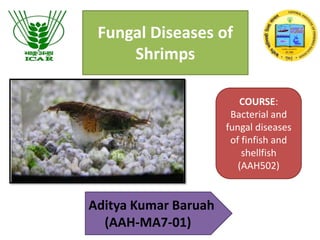
Fungal Diseases of
Shrimps
Aditya Kumar Baruah
(AAH-MA7-01)
COURSE:
Bacterial and
fungal diseases
of finfish and
shellfish
(AAH502)
Introduction
• Fungal infections are common in the fish culture, but it possible for shrimp to get
fungal infections as well.
• It's unavoidable, since fungal spores are everywhere, in the air and water.
• Fungi are plant like organisms but unlike plants are not capable of photosynthesis.
• All fungal diseases are called Mycosis (plural: mycoses).
• Fungi are usually fought off by a healthy immune system, so we only see this in
weakened or injured shrimp or just after a moult.
• Spores attach themselves to weakened sites on the shrimp and break out as a
cottony white growth.
• If not treated quickly, the spores will invade any dead tissue cells and in the process
infect more tissue causing a greater infection.
• At times, if the infection is only on the surface of the shrimp's shell, a moult can get
rid of the fungus. It is only by timeliness/chance that such a situation could rectify
itself. At other times, treatment is required.
MAJOR FUNGAL DISEASES IN
SHRIMPS
• Larval mycosis- Lagenidium sp.
Sirolpidium sp.
• Black gill disease- Fusarium sp.
• Red disease- Alfatoxicosis
Larval Mycosis
CAUSATIVE AGENTS:
• Lagenidium spp., Sirolpidium spp., Haliphthoros spp.
SPECIES AFFECTED:
• All Penaeus species, crabs (e.g. Scylla serrata)
GROSS SIGNS:
• Sudden onset of mortalities in larval stages of shrimps and crabs.
• Crab eggs are also susceptible for mycotic infection.
• The commonly affected larval stages among shrimp species are
the protozoeal and mysis stages.
• Infected larvae become immobile and will settle to the bottom of
the tank if aeration/circulation is interrupted.
• Presence of excessive mycelial network visible through the
exoskeleton of moribund and dead larvae.
EFFECTS OF HOSTS:
• Progressive systemic mycosis that is accompanied by
little or no host inflammatory response can be
observed.
• Infection is apparently lethal, accumulating mortality of
20-100% within 48-72 h after onset of infection.
Causative agents:
Lagenidium sp.
Phylum: Heterokonta
Class: Oomycota
Order: Lagenidiales
Family: Lagenidiaceae
Genus: Lagenidium
(Schenk, 1859)
Lagenidium sp.
• In wild crustacean population, it is known as an
egg parasite, while in aquaculture it affects both
eggs and larvae of crustaceans.
• Lagenidium callinectes (Couch, 1942) is a marine
phycomycetous parasitic fungus in eggs and
larvae of marine crustaceans.
• First report in larvae of penaeid white shrimp
Penaeus setiferus (Lightner & Fontaine,1973)
• The hyphae of Lagenidium callinectes are irregularly branched, septate, and
contain a cell wall and membrane, vacuoles, mitochondria, ribosomes, small
and large vesicles.
• The spores occur singly or in pairs.
• The fungal mycelium may either invade and embed itself in the tissues, or
alternatively, replace all the muscle tissues of the infected larval P. monodon.
• Fungus infected, untreated populations of nauplii, zoea and mysis exhibited
heavy mortalities
• Lagenidium sp. exhibited growth in potato
dextrose agar medium and in Sabouraud's agar
at 28 °C.
• The pathology of Lagenidium in all host species
is apparently similar.
• Lagenidium is particularly troublesome in
aquaculture of marine decapod crustaceans and
it is reported to be responsible for major
epizootics of the larvae of most species of
penaeid shrimp throughout the world (Lightner
1977, 1981).
Species affecting crustaceans
Lagenidium infection in crustacean larvae.
Larva of Penaeus monodon heavily infested with the fungus (note
the mycelia [arrows] completely replacing the body tissues of the
larva; and the vesicles [arrowheads] ready to release the
zoospores).
Filaments of Lagenidium in the tail of Penaeus
monodon larva (arrows). Zoospores in a vesicle
(arrowhead)
Zoosporangial development of
Haliphthoros sp. by hyphal
fragmentation
Sirolpidium sp.
Phylum: Oomycota
Class: Peronosporea
Order: Myzocytiopsidales
Family: Sirolpidiaceae
Genus: Sirolpidium
Species: Sirolpidium zoophthorum
A larval shrimp showing
hyphae of Sirolpidium inside
abdominal appendages.
Pathogenesis
• Affects larvae ranging in stage from early veliger
to post-metamorphic juveniles up to 400 µm in
diameter.
• Fungus spreads throughout the soft-tissues
causing them to disintegrate.
• The sporangia produce tubes which protrude
outside the shell and release motile zoospores.
• In heavily infected larval cultures over 90% of
larvae can be killed within two days.
DIAGNOSIS FOR LARVAL MYCOSIS
• Microscopic examination of affected larvae will reveal extensive, non-septate,
highly branched fungal mycelia throughout the body and appendages
• Specialized hyphae or discharge tubes, with or without terminal vesicles, may
be present, and could be the basis for identification of the causative agent.
• Motile zoospores may be observed being released from the discharge tubes in
the case of some species.
• Classification of the type of organism causing particular epizootic of larval
mycosis is dependent upon the microscopic examination as follows:
– Lagenidium – zoospores are released from terminal vesicle
– Haliphthoros – absence of terminal vesicles; zoospores are released through
discharge tubes formed by the zoosporangia
– Sirolpidium- Larvae contain looped, sparsely branched mycelia, with
constrictions at intervals between segments which may be swollen and
frequently lobed.
Molecular diagnosis
• PCR-based detection of Lagenidium DNA in frozen and
ethanol-fixed tissues
 First-round PCR, which utilized universal fungal primers ITS1
and ITS2P,
 Second-round PCR using the Lagenidium-specific primers
LAG1 and LAG2 (Nadine et.al., 2002)
• Research has been going on for developing DNA-based
diagnostics, PCR primers and DNA probes, specific for
Sirolpidium zoophthorum that allowsdetection of early
infections, so that control measures can be implemented
within the hatchery.
PREVENTION AND CONTROL
• Disinfection of contaminated larval rearing tanks and chlorination and/or
filtration of the incoming water can prevent outbreaks. Different
antimycotic compounds have been tested in vitro.
• Use of calcium hypochloride @500ppm for 24 hours in hatcheries is found
to be mycocidal.
• Malachite green oxalate @ 6-10ppb in prawn/shrimp hatcheries.
• 1-10 ppm formalin
• egg disinfection with 20 ppm detergent followed by thorough rinsing before
hatching.
• 0.5 ppm treatment with Trifluralin significantly reduced the mortality of
infected larval populations (i.e. 1-1% nauplii, 3-28% zoea and 5-21% mysis
mortality).
• For Sirolpidium: No known treatment.
Batches containing infected individuals should be destroyed in an
approved manner; disinfect all containers and equipment in contact with
the infected stock.
Black Gill Disease
CAUSATIVE AGENT: Fusarium solani
SPECIES AFFECTED: All Penaeus species
Fusarium sp.
Kingdom: Fungi
Phylum: Ascomycota
Class: Sordariomycetes
Subclass: Hypocreomycetidae
Order: Hypocreales
Family: Nectriaceae
Genus: Fusarium
Species: F. solani
Some spp.- Fusarium graminearum, Fusarium asiaticum, Fusarium
culmorum, and Fusarium avenaceumFusarium oxysporum
• Fusarium solani produces colonies that are white and cottony.
• However, instead of developing a pink or violet centre like
most Fusarium species, F. solani becomes blue-green or bluish
brown.
• On the underside, they may be pale, tea-with-milk-brown, or
red-brown.
• However, some clinical isolates have been blue-green or ink-blue
on the underside.
• F. solani colonies are low-floccose, loose, slimy, and sporadical.
• When grown on Potato Dextrose Agar (PDA), this fungus grows
rapidly, colonies reach a diameter of 64–70 mm in 7 days.
• Fusarium solani has aerial hyphae that give rise to conidiophores laterally.
• The conidiophores branch into thin, elongated monophialides that
produce conidia.
• Phialides that produce macroconidia are shorter than those that produce
microconidia.
• The macroconidia produced by F. solani are slightly curved, hyaline, and
broad, often aggregating in fascicles.
• Typically the macroconidia of this species have 3 septa but may have as many
as 4–5.
• Microconidia have thickened basal cells and tapered, rounded apical cells.
• Fusarium solani also forms chlamydospores most commonly under suboptimal
growth conditions
GROSS SIGNS:
Appearance of “black spots” that
preceded mortalities in juvenile shrimps grown in ponds.
EFFECTS ON HOSTS:
• Infection usually starts on damaged tissues such as wounds, gills
damaged from chemical treatments or pollutants, and lesions
resulting from other disease processes.
• Once infection is established, it is usually progressive with 30%
remission rate.
•Lesions may also serve as a route of entry for other opportunistic
pathogens.
DIAGNOSIS
• Microscopic examination of wet mounts of infected tissues
will reveal the presence of canoe-shaped macroconidia.
Infection may begin at different loci and spread slowly.
• Nested PCR: EF1/EF2 primer pair.
• A nested PCR with Fa/Ra in both steps was tested but
produced only a smear or no product at all.
• Therefore, new primers Fa+7 and Ra+6 were designed by
extending primers Fa and Ra at their 5′ ends.
• The TEF1 sequences of several Fusarium species are of
concern.
Aflatoxicosis (Red Disease)
CAUSATIVE AGENT:
• Aflatoxin produced by Aspergillus flavus and other Aspergillus
spp. which are common contaminants of not-properly stored or
expired feeds.
SPECIES AFFECTED: Penaeus monodon, other Penaeus spp.
.
GROSS SIGNS:
• Yellowish, and eventually reddish discoloration of the shrimp
body and appendages can be observed among pond-cultured
shrimp juveniles.
• Affected animals become lethargic with weak swimming
activity near pond dikes. Soft shelling can also be observed
DIAGNOSIS:
• Affected shrimps will not survive for more than 30 seconds
when collected from the feeding trays. There will also be loss
of appetite.
• Confirmation is by chemical analysis for the presence of
aflatoxin in the suspected feed/ingredient.
EFFECTS ON HOSTS:
• Histopathologically, necrosis in the tubule
epithelium that proceeds from proximal
portion of the tubules to peripheral tubule
tips in the hepatopancreas can be observed.
• Growth will be retarded.
Pathological changes in the hepatopancreas and heamatopoetic organs of shrimp
having red disease
Conclusion
• Fungal diseases arise due to bad pond hygiene.
• Keeping the organic load less in the system avoids majority of
fungal diseases.
• The diseases caused by fungi are mostly secondary.
• Very few works has been done on these shell fish fungus and
not much works has been done on the histopathological
effects of these fungus and diagnostic techniques are also not
well studied.
• Proper management strategies are to be taken in shrimp
culture to avoid loses due to these opportunistic pathogens.
References
• Davis, H.C. and V.L. Loosanoff. 1955. A fungus disease in bivalve larvae.
Proceedings of the National Shellfisheries Association 45: 151-156.
• Davis, H.C., V.L. Loosanoff, W.H. Weston and C. Martin. 1954. A fungus
disease in clam and oyster larvae. Science 120: 36-38.
• NADINE R. ZNAJDA, AMY M. GROOTERS‡ and ROSANNA MARSELLA,2002.
PCR-based detection of Pythium and Lagenidium DNA in frozenand
ethanol-fixed animal tissues. Veterinary Dermatology, 13: 187–194
ackwellScience,Ltd.
• Woo, P.T.K, 2006, Fish Diseases and Disorders, volume3, pp.205-229
• Muraosa,Y., Lawhavinit, O., Hatai, K., Lagenidium thermophilum isolated
from eggs and larvae of black tiger shrimp Penaeus monodon in Thailand,
Fish Pathology.41(1), 35-40, 2006.3
Fungal diseases of shrimp

More Related Content

What's hot

Viral diseases in crustaceans
Viral diseases in crustaceansViral diseases in crustaceans
Viral diseases in crustaceansgopika v
 
Diagnosis of fish diseases
Diagnosis of fish diseasesDiagnosis of fish diseases
Diagnosis of fish diseasesEngy Tarek
 
vaccination in fish
vaccination in fishvaccination in fish
vaccination in fish40024
 
Parasitic diseases of fish
Parasitic diseases of fishParasitic diseases of fish
Parasitic diseases of fishAvijit Pramanik
 
Bacterial haemorraghic septicemia
Bacterial haemorraghic septicemiaBacterial haemorraghic septicemia
Bacterial haemorraghic septicemiaBijayananda Seth
 
fungal disease in fishes
 fungal  disease  in fishes fungal  disease  in fishes
fungal disease in fishessanchu yadav
 
bacterial disease of fishes
 bacterial disease of fishes bacterial disease of fishes
bacterial disease of fishessanchu yadav
 
Biosecurity in aquaculture
Biosecurity in aquacultureBiosecurity in aquaculture
Biosecurity in aquacultureVikasUjjania
 
Epizootic Ulcerative Syndrome
Epizootic Ulcerative SyndromeEpizootic Ulcerative Syndrome
Epizootic Ulcerative SyndromeAMITJADHAV83080
 
White spot disease in fish
White spot disease in fishWhite spot disease in fish
White spot disease in fishharapriya behera
 
Fish disease and health management
Fish disease and health managementFish disease and health management
Fish disease and health managementNazmul Ahmed Oli
 
Hybridization in fishes
Hybridization in fishesHybridization in fishes
Hybridization in fishesAbhishek Giri
 
Aquarium fish breeding
Aquarium fish breedingAquarium fish breeding
Aquarium fish breedingSameer Chebbi
 

What's hot (20)

Pathogens of fish
Pathogens of fishPathogens of fish
Pathogens of fish
 
Viral diseases in crustaceans
Viral diseases in crustaceansViral diseases in crustaceans
Viral diseases in crustaceans
 
Diagnosis of fish diseases
Diagnosis of fish diseasesDiagnosis of fish diseases
Diagnosis of fish diseases
 
vaccination in fish
vaccination in fishvaccination in fish
vaccination in fish
 
Fish Health Management
Fish Health Management Fish Health Management
Fish Health Management
 
Spf and spr
Spf and sprSpf and spr
Spf and spr
 
Parasitic diseases of fish
Parasitic diseases of fishParasitic diseases of fish
Parasitic diseases of fish
 
viral disease
 viral disease viral disease
viral disease
 
Protozoan disease
Protozoan diseaseProtozoan disease
Protozoan disease
 
Bacterial haemorraghic septicemia
Bacterial haemorraghic septicemiaBacterial haemorraghic septicemia
Bacterial haemorraghic septicemia
 
fungal disease in fishes
 fungal  disease  in fishes fungal  disease  in fishes
fungal disease in fishes
 
bacterial disease of fishes
 bacterial disease of fishes bacterial disease of fishes
bacterial disease of fishes
 
Biosecurity in aquaculture
Biosecurity in aquacultureBiosecurity in aquaculture
Biosecurity in aquaculture
 
Epizootic Ulcerative Syndrome
Epizootic Ulcerative SyndromeEpizootic Ulcerative Syndrome
Epizootic Ulcerative Syndrome
 
White spot disease in fish
White spot disease in fishWhite spot disease in fish
White spot disease in fish
 
Fish disease and health management
Fish disease and health managementFish disease and health management
Fish disease and health management
 
Hybridization in fishes
Hybridization in fishesHybridization in fishes
Hybridization in fishes
 
Fish parasite
Fish parasite Fish parasite
Fish parasite
 
Aquarium fish breeding
Aquarium fish breedingAquarium fish breeding
Aquarium fish breeding
 
Identification of fish seed
Identification of fish seedIdentification of fish seed
Identification of fish seed
 

Similar to Fungal diseases of shrimp

Class Trematoda. Tapeworms as parasites of human being
Class Trematoda. Tapeworms as parasites of human beingClass Trematoda. Tapeworms as parasites of human being
Class Trematoda. Tapeworms as parasites of human beingEneutron
 
bacillusgp-170124093136.pdf
bacillusgp-170124093136.pdfbacillusgp-170124093136.pdf
bacillusgp-170124093136.pdfOsmanAli92
 
Zoonotic Infections. Yersinia. Agents of Plague, Enteric yersiniosis & Pdeudo...
Zoonotic Infections. Yersinia. Agents of Plague, Enteric yersiniosis & Pdeudo...Zoonotic Infections. Yersinia. Agents of Plague, Enteric yersiniosis & Pdeudo...
Zoonotic Infections. Yersinia. Agents of Plague, Enteric yersiniosis & Pdeudo...Eneutron
 
CONTAMINANTS IN CELL CULTURE & PRECAUTIONS.pptx
CONTAMINANTS IN CELL CULTURE & PRECAUTIONS.pptxCONTAMINANTS IN CELL CULTURE & PRECAUTIONS.pptx
CONTAMINANTS IN CELL CULTURE & PRECAUTIONS.pptxPriyansha Singh
 
Strongyloides_stercoralis.ppt
Strongyloides_stercoralis.pptStrongyloides_stercoralis.ppt
Strongyloides_stercoralis.pptJoshuaKalunda
 
Fungal infections part I
Fungal infections part IFungal infections part I
Fungal infections part IIbrahim Farag
 
Gram positive bacteria in ocular pathology
Gram positive bacteria in ocular pathologyGram positive bacteria in ocular pathology
Gram positive bacteria in ocular pathologySudheer Kumar
 
bacillusgp-170124093136 (1).pdf
bacillusgp-170124093136 (1).pdfbacillusgp-170124093136 (1).pdf
bacillusgp-170124093136 (1).pdfStephenNjoroge22
 
Nematodes and their Management
Nematodes and their Management Nematodes and their Management
Nematodes and their Management Amit Pundir
 
Introduction to Microbiology
Introduction to MicrobiologyIntroduction to Microbiology
Introduction to MicrobiologyDerar ALJarrah
 

Similar to Fungal diseases of shrimp (20)

Class Trematoda. Tapeworms as parasites of human being
Class Trematoda. Tapeworms as parasites of human beingClass Trematoda. Tapeworms as parasites of human being
Class Trematoda. Tapeworms as parasites of human being
 
Cryptosporadium
CryptosporadiumCryptosporadium
Cryptosporadium
 
Bacillus
Bacillus Bacillus
Bacillus
 
bacillusgp-170124093136.pdf
bacillusgp-170124093136.pdfbacillusgp-170124093136.pdf
bacillusgp-170124093136.pdf
 
Zoonotic Infections. Yersinia. Agents of Plague, Enteric yersiniosis & Pdeudo...
Zoonotic Infections. Yersinia. Agents of Plague, Enteric yersiniosis & Pdeudo...Zoonotic Infections. Yersinia. Agents of Plague, Enteric yersiniosis & Pdeudo...
Zoonotic Infections. Yersinia. Agents of Plague, Enteric yersiniosis & Pdeudo...
 
CONTAMINANTS IN CELL CULTURE & PRECAUTIONS.pptx
CONTAMINANTS IN CELL CULTURE & PRECAUTIONS.pptxCONTAMINANTS IN CELL CULTURE & PRECAUTIONS.pptx
CONTAMINANTS IN CELL CULTURE & PRECAUTIONS.pptx
 
Chapter5 microbiology farid
Chapter5 microbiology faridChapter5 microbiology farid
Chapter5 microbiology farid
 
Strongyloides_stercoralis.ppt
Strongyloides_stercoralis.pptStrongyloides_stercoralis.ppt
Strongyloides_stercoralis.ppt
 
Bacillus1
Bacillus1Bacillus1
Bacillus1
 
Fungal infections part I
Fungal infections part IFungal infections part I
Fungal infections part I
 
Maleria
MaleriaMaleria
Maleria
 
Gram positive bacteria in ocular pathology
Gram positive bacteria in ocular pathologyGram positive bacteria in ocular pathology
Gram positive bacteria in ocular pathology
 
Protozoan disease of fishes
Protozoan disease of fishesProtozoan disease of fishes
Protozoan disease of fishes
 
bacillusgp-170124093136 (1).pdf
bacillusgp-170124093136 (1).pdfbacillusgp-170124093136 (1).pdf
bacillusgp-170124093136 (1).pdf
 
Papillomatosis
PapillomatosisPapillomatosis
Papillomatosis
 
Ascariasis
AscariasisAscariasis
Ascariasis
 
Nematodes and their Management
Nematodes and their Management Nematodes and their Management
Nematodes and their Management
 
Introduction to Microbiology
Introduction to MicrobiologyIntroduction to Microbiology
Introduction to Microbiology
 
Health considerations done
Health considerations doneHealth considerations done
Health considerations done
 
Cryptosporidium
CryptosporidiumCryptosporidium
Cryptosporidium
 

Recently uploaded

Proudly South Africa powerpoint Thorisha.pptx
Proudly South Africa powerpoint Thorisha.pptxProudly South Africa powerpoint Thorisha.pptx
Proudly South Africa powerpoint Thorisha.pptxthorishapillay1
 
Framing an Appropriate Research Question 6b9b26d93da94caf993c038d9efcdedb.pdf
Framing an Appropriate Research Question 6b9b26d93da94caf993c038d9efcdedb.pdfFraming an Appropriate Research Question 6b9b26d93da94caf993c038d9efcdedb.pdf
Framing an Appropriate Research Question 6b9b26d93da94caf993c038d9efcdedb.pdfUjwalaBharambe
 
Final demo Grade 9 for demo Plan dessert.pptx
Final demo Grade 9 for demo Plan dessert.pptxFinal demo Grade 9 for demo Plan dessert.pptx
Final demo Grade 9 for demo Plan dessert.pptxAvyJaneVismanos
 
Enzyme, Pharmaceutical Aids, Miscellaneous Last Part of Chapter no 5th.pdf
Enzyme, Pharmaceutical Aids, Miscellaneous Last Part of Chapter no 5th.pdfEnzyme, Pharmaceutical Aids, Miscellaneous Last Part of Chapter no 5th.pdf
Enzyme, Pharmaceutical Aids, Miscellaneous Last Part of Chapter no 5th.pdfSumit Tiwari
 
Alper Gobel In Media Res Media Component
Alper Gobel In Media Res Media ComponentAlper Gobel In Media Res Media Component
Alper Gobel In Media Res Media ComponentInMediaRes1
 
How to Configure Email Server in Odoo 17
How to Configure Email Server in Odoo 17How to Configure Email Server in Odoo 17
How to Configure Email Server in Odoo 17Celine George
 
Historical philosophical, theoretical, and legal foundations of special and i...
Historical philosophical, theoretical, and legal foundations of special and i...Historical philosophical, theoretical, and legal foundations of special and i...
Historical philosophical, theoretical, and legal foundations of special and i...jaredbarbolino94
 
Software Engineering Methodologies (overview)
Software Engineering Methodologies (overview)Software Engineering Methodologies (overview)
Software Engineering Methodologies (overview)eniolaolutunde
 
Full Stack Web Development Course for Beginners
Full Stack Web Development Course  for BeginnersFull Stack Web Development Course  for Beginners
Full Stack Web Development Course for BeginnersSabitha Banu
 
Organic Name Reactions for the students and aspirants of Chemistry12th.pptx
Organic Name Reactions  for the students and aspirants of Chemistry12th.pptxOrganic Name Reactions  for the students and aspirants of Chemistry12th.pptx
Organic Name Reactions for the students and aspirants of Chemistry12th.pptxVS Mahajan Coaching Centre
 
Biting mechanism of poisonous snakes.pdf
Biting mechanism of poisonous snakes.pdfBiting mechanism of poisonous snakes.pdf
Biting mechanism of poisonous snakes.pdfadityarao40181
 
Introduction to ArtificiaI Intelligence in Higher Education
Introduction to ArtificiaI Intelligence in Higher EducationIntroduction to ArtificiaI Intelligence in Higher Education
Introduction to ArtificiaI Intelligence in Higher Educationpboyjonauth
 
Roles & Responsibilities in Pharmacovigilance
Roles & Responsibilities in PharmacovigilanceRoles & Responsibilities in Pharmacovigilance
Roles & Responsibilities in PharmacovigilanceSamikshaHamane
 
Computed Fields and api Depends in the Odoo 17
Computed Fields and api Depends in the Odoo 17Computed Fields and api Depends in the Odoo 17
Computed Fields and api Depends in the Odoo 17Celine George
 
Crayon Activity Handout For the Crayon A
Crayon Activity Handout For the Crayon ACrayon Activity Handout For the Crayon A
Crayon Activity Handout For the Crayon AUnboundStockton
 
POINT- BIOCHEMISTRY SEM 2 ENZYMES UNIT 5.pptx
POINT- BIOCHEMISTRY SEM 2 ENZYMES UNIT 5.pptxPOINT- BIOCHEMISTRY SEM 2 ENZYMES UNIT 5.pptx
POINT- BIOCHEMISTRY SEM 2 ENZYMES UNIT 5.pptxSayali Powar
 
History Class XII Ch. 3 Kinship, Caste and Class (1).pptx
History Class XII Ch. 3 Kinship, Caste and Class (1).pptxHistory Class XII Ch. 3 Kinship, Caste and Class (1).pptx
History Class XII Ch. 3 Kinship, Caste and Class (1).pptxsocialsciencegdgrohi
 
Solving Puzzles Benefits Everyone (English).pptx
Solving Puzzles Benefits Everyone (English).pptxSolving Puzzles Benefits Everyone (English).pptx
Solving Puzzles Benefits Everyone (English).pptxOH TEIK BIN
 
Incoming and Outgoing Shipments in 1 STEP Using Odoo 17
Incoming and Outgoing Shipments in 1 STEP Using Odoo 17Incoming and Outgoing Shipments in 1 STEP Using Odoo 17
Incoming and Outgoing Shipments in 1 STEP Using Odoo 17Celine George
 

Recently uploaded (20)

Proudly South Africa powerpoint Thorisha.pptx
Proudly South Africa powerpoint Thorisha.pptxProudly South Africa powerpoint Thorisha.pptx
Proudly South Africa powerpoint Thorisha.pptx
 
Framing an Appropriate Research Question 6b9b26d93da94caf993c038d9efcdedb.pdf
Framing an Appropriate Research Question 6b9b26d93da94caf993c038d9efcdedb.pdfFraming an Appropriate Research Question 6b9b26d93da94caf993c038d9efcdedb.pdf
Framing an Appropriate Research Question 6b9b26d93da94caf993c038d9efcdedb.pdf
 
Final demo Grade 9 for demo Plan dessert.pptx
Final demo Grade 9 for demo Plan dessert.pptxFinal demo Grade 9 for demo Plan dessert.pptx
Final demo Grade 9 for demo Plan dessert.pptx
 
Enzyme, Pharmaceutical Aids, Miscellaneous Last Part of Chapter no 5th.pdf
Enzyme, Pharmaceutical Aids, Miscellaneous Last Part of Chapter no 5th.pdfEnzyme, Pharmaceutical Aids, Miscellaneous Last Part of Chapter no 5th.pdf
Enzyme, Pharmaceutical Aids, Miscellaneous Last Part of Chapter no 5th.pdf
 
Alper Gobel In Media Res Media Component
Alper Gobel In Media Res Media ComponentAlper Gobel In Media Res Media Component
Alper Gobel In Media Res Media Component
 
How to Configure Email Server in Odoo 17
How to Configure Email Server in Odoo 17How to Configure Email Server in Odoo 17
How to Configure Email Server in Odoo 17
 
Historical philosophical, theoretical, and legal foundations of special and i...
Historical philosophical, theoretical, and legal foundations of special and i...Historical philosophical, theoretical, and legal foundations of special and i...
Historical philosophical, theoretical, and legal foundations of special and i...
 
Software Engineering Methodologies (overview)
Software Engineering Methodologies (overview)Software Engineering Methodologies (overview)
Software Engineering Methodologies (overview)
 
Full Stack Web Development Course for Beginners
Full Stack Web Development Course  for BeginnersFull Stack Web Development Course  for Beginners
Full Stack Web Development Course for Beginners
 
Organic Name Reactions for the students and aspirants of Chemistry12th.pptx
Organic Name Reactions  for the students and aspirants of Chemistry12th.pptxOrganic Name Reactions  for the students and aspirants of Chemistry12th.pptx
Organic Name Reactions for the students and aspirants of Chemistry12th.pptx
 
Biting mechanism of poisonous snakes.pdf
Biting mechanism of poisonous snakes.pdfBiting mechanism of poisonous snakes.pdf
Biting mechanism of poisonous snakes.pdf
 
Introduction to ArtificiaI Intelligence in Higher Education
Introduction to ArtificiaI Intelligence in Higher EducationIntroduction to ArtificiaI Intelligence in Higher Education
Introduction to ArtificiaI Intelligence in Higher Education
 
Roles & Responsibilities in Pharmacovigilance
Roles & Responsibilities in PharmacovigilanceRoles & Responsibilities in Pharmacovigilance
Roles & Responsibilities in Pharmacovigilance
 
Computed Fields and api Depends in the Odoo 17
Computed Fields and api Depends in the Odoo 17Computed Fields and api Depends in the Odoo 17
Computed Fields and api Depends in the Odoo 17
 
Crayon Activity Handout For the Crayon A
Crayon Activity Handout For the Crayon ACrayon Activity Handout For the Crayon A
Crayon Activity Handout For the Crayon A
 
POINT- BIOCHEMISTRY SEM 2 ENZYMES UNIT 5.pptx
POINT- BIOCHEMISTRY SEM 2 ENZYMES UNIT 5.pptxPOINT- BIOCHEMISTRY SEM 2 ENZYMES UNIT 5.pptx
POINT- BIOCHEMISTRY SEM 2 ENZYMES UNIT 5.pptx
 
History Class XII Ch. 3 Kinship, Caste and Class (1).pptx
History Class XII Ch. 3 Kinship, Caste and Class (1).pptxHistory Class XII Ch. 3 Kinship, Caste and Class (1).pptx
History Class XII Ch. 3 Kinship, Caste and Class (1).pptx
 
TataKelola dan KamSiber Kecerdasan Buatan v022.pdf
TataKelola dan KamSiber Kecerdasan Buatan v022.pdfTataKelola dan KamSiber Kecerdasan Buatan v022.pdf
TataKelola dan KamSiber Kecerdasan Buatan v022.pdf
 
Solving Puzzles Benefits Everyone (English).pptx
Solving Puzzles Benefits Everyone (English).pptxSolving Puzzles Benefits Everyone (English).pptx
Solving Puzzles Benefits Everyone (English).pptx
 
Incoming and Outgoing Shipments in 1 STEP Using Odoo 17
Incoming and Outgoing Shipments in 1 STEP Using Odoo 17Incoming and Outgoing Shipments in 1 STEP Using Odoo 17
Incoming and Outgoing Shipments in 1 STEP Using Odoo 17
 

Fungal diseases of shrimp

  • 1. Fungal Diseases of Shrimps Aditya Kumar Baruah (AAH-MA7-01) COURSE: Bacterial and fungal diseases of finfish and shellfish (AAH502)
  • 2. Introduction • Fungal infections are common in the fish culture, but it possible for shrimp to get fungal infections as well. • It's unavoidable, since fungal spores are everywhere, in the air and water. • Fungi are plant like organisms but unlike plants are not capable of photosynthesis. • All fungal diseases are called Mycosis (plural: mycoses). • Fungi are usually fought off by a healthy immune system, so we only see this in weakened or injured shrimp or just after a moult. • Spores attach themselves to weakened sites on the shrimp and break out as a cottony white growth. • If not treated quickly, the spores will invade any dead tissue cells and in the process infect more tissue causing a greater infection. • At times, if the infection is only on the surface of the shrimp's shell, a moult can get rid of the fungus. It is only by timeliness/chance that such a situation could rectify itself. At other times, treatment is required.
  • 3. MAJOR FUNGAL DISEASES IN SHRIMPS • Larval mycosis- Lagenidium sp. Sirolpidium sp. • Black gill disease- Fusarium sp. • Red disease- Alfatoxicosis
  • 4. Larval Mycosis CAUSATIVE AGENTS: • Lagenidium spp., Sirolpidium spp., Haliphthoros spp. SPECIES AFFECTED: • All Penaeus species, crabs (e.g. Scylla serrata)
  • 5. GROSS SIGNS: • Sudden onset of mortalities in larval stages of shrimps and crabs. • Crab eggs are also susceptible for mycotic infection. • The commonly affected larval stages among shrimp species are the protozoeal and mysis stages. • Infected larvae become immobile and will settle to the bottom of the tank if aeration/circulation is interrupted. • Presence of excessive mycelial network visible through the exoskeleton of moribund and dead larvae.
  • 6. EFFECTS OF HOSTS: • Progressive systemic mycosis that is accompanied by little or no host inflammatory response can be observed. • Infection is apparently lethal, accumulating mortality of 20-100% within 48-72 h after onset of infection.
  • 7. Causative agents: Lagenidium sp. Phylum: Heterokonta Class: Oomycota Order: Lagenidiales Family: Lagenidiaceae Genus: Lagenidium (Schenk, 1859)
  • 8. Lagenidium sp. • In wild crustacean population, it is known as an egg parasite, while in aquaculture it affects both eggs and larvae of crustaceans. • Lagenidium callinectes (Couch, 1942) is a marine phycomycetous parasitic fungus in eggs and larvae of marine crustaceans. • First report in larvae of penaeid white shrimp Penaeus setiferus (Lightner & Fontaine,1973)
  • 9. • The hyphae of Lagenidium callinectes are irregularly branched, septate, and contain a cell wall and membrane, vacuoles, mitochondria, ribosomes, small and large vesicles. • The spores occur singly or in pairs. • The fungal mycelium may either invade and embed itself in the tissues, or alternatively, replace all the muscle tissues of the infected larval P. monodon. • Fungus infected, untreated populations of nauplii, zoea and mysis exhibited heavy mortalities
  • 10. • Lagenidium sp. exhibited growth in potato dextrose agar medium and in Sabouraud's agar at 28 °C. • The pathology of Lagenidium in all host species is apparently similar. • Lagenidium is particularly troublesome in aquaculture of marine decapod crustaceans and it is reported to be responsible for major epizootics of the larvae of most species of penaeid shrimp throughout the world (Lightner 1977, 1981).
  • 12. Lagenidium infection in crustacean larvae. Larva of Penaeus monodon heavily infested with the fungus (note the mycelia [arrows] completely replacing the body tissues of the larva; and the vesicles [arrowheads] ready to release the zoospores).
  • 13. Filaments of Lagenidium in the tail of Penaeus monodon larva (arrows). Zoospores in a vesicle (arrowhead) Zoosporangial development of Haliphthoros sp. by hyphal fragmentation
  • 14. Sirolpidium sp. Phylum: Oomycota Class: Peronosporea Order: Myzocytiopsidales Family: Sirolpidiaceae Genus: Sirolpidium Species: Sirolpidium zoophthorum A larval shrimp showing hyphae of Sirolpidium inside abdominal appendages.
  • 15. Pathogenesis • Affects larvae ranging in stage from early veliger to post-metamorphic juveniles up to 400 µm in diameter. • Fungus spreads throughout the soft-tissues causing them to disintegrate. • The sporangia produce tubes which protrude outside the shell and release motile zoospores. • In heavily infected larval cultures over 90% of larvae can be killed within two days.
  • 16. DIAGNOSIS FOR LARVAL MYCOSIS • Microscopic examination of affected larvae will reveal extensive, non-septate, highly branched fungal mycelia throughout the body and appendages • Specialized hyphae or discharge tubes, with or without terminal vesicles, may be present, and could be the basis for identification of the causative agent. • Motile zoospores may be observed being released from the discharge tubes in the case of some species. • Classification of the type of organism causing particular epizootic of larval mycosis is dependent upon the microscopic examination as follows: – Lagenidium – zoospores are released from terminal vesicle – Haliphthoros – absence of terminal vesicles; zoospores are released through discharge tubes formed by the zoosporangia – Sirolpidium- Larvae contain looped, sparsely branched mycelia, with constrictions at intervals between segments which may be swollen and frequently lobed.
  • 17. Molecular diagnosis • PCR-based detection of Lagenidium DNA in frozen and ethanol-fixed tissues  First-round PCR, which utilized universal fungal primers ITS1 and ITS2P,  Second-round PCR using the Lagenidium-specific primers LAG1 and LAG2 (Nadine et.al., 2002) • Research has been going on for developing DNA-based diagnostics, PCR primers and DNA probes, specific for Sirolpidium zoophthorum that allowsdetection of early infections, so that control measures can be implemented within the hatchery.
  • 18. PREVENTION AND CONTROL • Disinfection of contaminated larval rearing tanks and chlorination and/or filtration of the incoming water can prevent outbreaks. Different antimycotic compounds have been tested in vitro. • Use of calcium hypochloride @500ppm for 24 hours in hatcheries is found to be mycocidal. • Malachite green oxalate @ 6-10ppb in prawn/shrimp hatcheries. • 1-10 ppm formalin • egg disinfection with 20 ppm detergent followed by thorough rinsing before hatching. • 0.5 ppm treatment with Trifluralin significantly reduced the mortality of infected larval populations (i.e. 1-1% nauplii, 3-28% zoea and 5-21% mysis mortality). • For Sirolpidium: No known treatment. Batches containing infected individuals should be destroyed in an approved manner; disinfect all containers and equipment in contact with the infected stock.
  • 19. Black Gill Disease CAUSATIVE AGENT: Fusarium solani SPECIES AFFECTED: All Penaeus species
  • 20. Fusarium sp. Kingdom: Fungi Phylum: Ascomycota Class: Sordariomycetes Subclass: Hypocreomycetidae Order: Hypocreales Family: Nectriaceae Genus: Fusarium Species: F. solani
  • 21. Some spp.- Fusarium graminearum, Fusarium asiaticum, Fusarium culmorum, and Fusarium avenaceumFusarium oxysporum • Fusarium solani produces colonies that are white and cottony. • However, instead of developing a pink or violet centre like most Fusarium species, F. solani becomes blue-green or bluish brown. • On the underside, they may be pale, tea-with-milk-brown, or red-brown. • However, some clinical isolates have been blue-green or ink-blue on the underside. • F. solani colonies are low-floccose, loose, slimy, and sporadical. • When grown on Potato Dextrose Agar (PDA), this fungus grows rapidly, colonies reach a diameter of 64–70 mm in 7 days.
  • 22. • Fusarium solani has aerial hyphae that give rise to conidiophores laterally. • The conidiophores branch into thin, elongated monophialides that produce conidia. • Phialides that produce macroconidia are shorter than those that produce microconidia. • The macroconidia produced by F. solani are slightly curved, hyaline, and broad, often aggregating in fascicles. • Typically the macroconidia of this species have 3 septa but may have as many as 4–5. • Microconidia have thickened basal cells and tapered, rounded apical cells. • Fusarium solani also forms chlamydospores most commonly under suboptimal growth conditions
  • 23. GROSS SIGNS: Appearance of “black spots” that preceded mortalities in juvenile shrimps grown in ponds. EFFECTS ON HOSTS: • Infection usually starts on damaged tissues such as wounds, gills damaged from chemical treatments or pollutants, and lesions resulting from other disease processes. • Once infection is established, it is usually progressive with 30% remission rate. •Lesions may also serve as a route of entry for other opportunistic pathogens.
  • 24. DIAGNOSIS • Microscopic examination of wet mounts of infected tissues will reveal the presence of canoe-shaped macroconidia. Infection may begin at different loci and spread slowly. • Nested PCR: EF1/EF2 primer pair. • A nested PCR with Fa/Ra in both steps was tested but produced only a smear or no product at all. • Therefore, new primers Fa+7 and Ra+6 were designed by extending primers Fa and Ra at their 5′ ends. • The TEF1 sequences of several Fusarium species are of concern.
  • 25. Aflatoxicosis (Red Disease) CAUSATIVE AGENT: • Aflatoxin produced by Aspergillus flavus and other Aspergillus spp. which are common contaminants of not-properly stored or expired feeds. SPECIES AFFECTED: Penaeus monodon, other Penaeus spp. .
  • 26. GROSS SIGNS: • Yellowish, and eventually reddish discoloration of the shrimp body and appendages can be observed among pond-cultured shrimp juveniles. • Affected animals become lethargic with weak swimming activity near pond dikes. Soft shelling can also be observed DIAGNOSIS: • Affected shrimps will not survive for more than 30 seconds when collected from the feeding trays. There will also be loss of appetite. • Confirmation is by chemical analysis for the presence of aflatoxin in the suspected feed/ingredient.
  • 27. EFFECTS ON HOSTS: • Histopathologically, necrosis in the tubule epithelium that proceeds from proximal portion of the tubules to peripheral tubule tips in the hepatopancreas can be observed. • Growth will be retarded.
  • 28. Pathological changes in the hepatopancreas and heamatopoetic organs of shrimp having red disease
  • 29. Conclusion • Fungal diseases arise due to bad pond hygiene. • Keeping the organic load less in the system avoids majority of fungal diseases. • The diseases caused by fungi are mostly secondary. • Very few works has been done on these shell fish fungus and not much works has been done on the histopathological effects of these fungus and diagnostic techniques are also not well studied. • Proper management strategies are to be taken in shrimp culture to avoid loses due to these opportunistic pathogens.
  • 30. References • Davis, H.C. and V.L. Loosanoff. 1955. A fungus disease in bivalve larvae. Proceedings of the National Shellfisheries Association 45: 151-156. • Davis, H.C., V.L. Loosanoff, W.H. Weston and C. Martin. 1954. A fungus disease in clam and oyster larvae. Science 120: 36-38. • NADINE R. ZNAJDA, AMY M. GROOTERS‡ and ROSANNA MARSELLA,2002. PCR-based detection of Pythium and Lagenidium DNA in frozenand ethanol-fixed animal tissues. Veterinary Dermatology, 13: 187–194 ackwellScience,Ltd. • Woo, P.T.K, 2006, Fish Diseases and Disorders, volume3, pp.205-229 • Muraosa,Y., Lawhavinit, O., Hatai, K., Lagenidium thermophilum isolated from eggs and larvae of black tiger shrimp Penaeus monodon in Thailand, Fish Pathology.41(1), 35-40, 2006.3