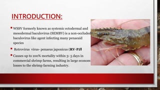
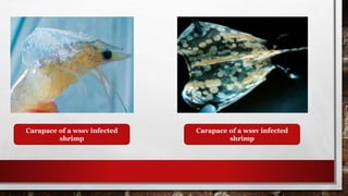
INTRODUCTION:
WSSV formerly known as systemic ectodermal and
mesodermal baculovirus (SEMBV) is a non-occluded
baculovirus like agent infecting many penaeaid
species
 Retrovirus virus- penaeus japonicus (RV-PJ)
Causes up to 100% mortality within 3- 5 days in
commercial shrimp farms, resulting in large economic
losses to the shrimp farming industry.
OUTBREAK OF DISEASE:
1. WSSV mostly attack to post larvae.
2. But disease occur in seen on growing juveniles shrimp of all ages and size but mostly
from 1-3 months after stocking in the grow out ponds.
3. WSSV outbreak can occur irrespective of farming system, stocking density, water
quality and salinity.
TRANSMISSION OF DISEASE:
1. Dead, infected shrimp are considered to be the source of subsequent infection for
WSSV this is mainly due to a higher opportunity of horizontal transmission of the virus
through cannibalism and the water borne route.
2. The major source of infection for shrimp farms is from infected spawners and
postlarvae.
3. The vertical transmission of WSSV by (trans- ovum)
VECTORS:
1. Rotifers,
2. marine molluscs,
3. polychaeteworms and
4. Artemia salina
5. sea slaters(isopoda) and
6. euphydradaeinsect larvae
EXTERNAL PATHOLOGY:
1. Rapid mortality accompanied by gross signs in moribound shrimp.
2. Lethargy, dirty gills, anorexia, broken antennae.
3. Swollen branchiostegites.
4. White deposits of calcium of 0.5-2.0mm dia on the cuticle of the shrimp
cephalothorax or carapace.
5. Display expanded chromatophores cephalothorax cuticle.
Carapace of a wssv infected
shrimp
Carapace of a wssv infected
shrimp
TARGET TISSUES:
White spot syndrome virus can infect cells of mesodermal and ectodermal origin,
1. Cuticular Epithelium
2. the lymphoid organ
3. Heart
4. Haematopoietic tissue
5. Stomach cuticular epidermis
6. Sub cuticular connective tissue
7. Antennal gland and hind gut
8. Nervous system and compound eyes are infected in very late stage.
Target organs for WSSV disease
INTERNAL PATHOLOGY:
1. The lymphoid organ of diseased shrimp may be distended, and haemolymph
infiltration in the enlarged haemal sinuses and interstitial spaces may result in a
hypertrophied yellowish hepatopancreas.
2. Sometimes empty gut also indicate the disease occurance.
3. Cuticular epiboint fouling
4. Lymphoid organ swelling.
(a) WSSV-infected
nucleus in gill (arrow).
(b) Low-power view of
hindgut showing
numerous infected
nuclei (arrow).
(c,d) Midgut epithelium
nuclei with inclusions
and emarginated
chromatin clearly
visible
HISTOPATHOLOGY:
1. Moribund shrimp show tissue destruction, accompanied by low to high basophilic
(H&E stained) central inclusion bodies within the hypertrophied nuclei of the cuticular
epithelial and connective tissue cells.
2. Presence of intranuclear basophilic inclusion bodies in the ectodermal and
mesodermal tissue found in cuticle, foregut and hindgut. But they are not found in the
tissues of endodermal origin. i.e midgut and hepatopancreas.
3. Cowdry type A inclusion bodies are intranuclear, eosinophilic, amorphous and
surrounded by a clear halo beneath the nuclear membrane but later, inclusions
become lightly to deeply basophilic and fill the entire nucleus.
Necrosis in different tissue of penaeids
PATHOGENESIS:
DISEASE OCCUR IN 3 STEPS
1. Latent stage
2. Transition stage
3. Patent stage
LATENT PHASE :
1. Initially they act active.
2. No presence of virus.
3. No mortality.
4. No inclusion bodies.
5. Pcr analysis –ve for 1st step.
6. But +ve for 2nd step.
TRANSISTION PHASE :
1. Shrimps are active.
2. They may or may not have WSSV.
3. In few cases there is appearance of inclusion bodies.
4. PCR analysis +ve by 1st step.
5. Severity index is medium.
PATENT STAGE:
1. Appear weak.
2. Stop feeding
3. Prominent white spot upto 2mm dia.
4. Basophilic intranuclear inclusion bodies.
5. Pcr 1st step +ve.
6. Shigh mortality.
AETIOLOGY:
1. Member of the genus whispovirus within the nimaviridae family.
2. Virions of WSSV are rod or ellipsoid to bacilliform in shape, have a regular symmetry,
and measure 80–120 nm in diameter and 250–380 nm in length. Most notable is the
thread- or flagella-like extension (appendage) at one end of the virion with 39
structural protein.
3. This virus has a large circular double-stranded DNA genome with diffrent size(292.9-
307.2 kb), where the diverse isolates show differences in virulence.
Shape of whispovirus
GEOGRAPHICAL DISTRIBUTION:
White spot syndrome virus was first detected in Southeast Asia
1. Central and south Americas
2.Malaysia
3.Thailand
4.Mexico
5.China
6.Japanvc
7. Korea
8.Indian subcontinent
HOST RANGE:
White spot syndrome virus is highly infective for marine penaeid shrimp including marine,
brackish and freshwater prawns, crabs, crayfish and lobsters
1. Macrobrachium rosenbergii
2. P. monodon
3. L. vannamei, L. stylirostris,
4. Marsupenaeus japonicus,
5. Fenneropenaeus chinensis
6. Procambarus clarkii
7. Cray fish Pacifastacus leniusculus
Control and prevention:
1. No effective vaccination methods for WSSV have been developed.
2.Chemotherapy no scientifically confirmed reports.
3.Low temperatures (12 ± 2˚ C) affect the WSSV pathogenicity and inhibit mortality in crayfish
and shrimp.
4.Other elements such as sulphated polysaccharides, fucoidan or microalgae cell walls have
been used as immunostimulants for shrimp.
5.These products have been somewhat successful against pathogens like WSSV around the
world.
6.Declairing crop holidays by the farmers.
PROPHYLACTIC:
1. As avoiding stocking in the cold season SPF free larvae or PCR tested larvae in the culture
system.
2. B-1,3-glucan, whose addition to the shrimp feed effectively improves the immunity and
survival of P.monodon
3. Some products such as Dunaliella extract and probiotics show a positive effect on resistance
of shrimp to WSSV infection.
4. Finally, the development of good management practices, the control of environmental
variables, genetic improvement in the penaeid species, understanding of virus physiology,
modulation of the Shrimp immune system, supported by functional Genomics and
proteomics of this crustacean, can reduce the epizootics.
REFERENCE:
1. OIE aquatic manual.
2. E course.
3. A review article. WSSV in cultured shrimp.
4. Web source.
• THANK YOU…

White spot syndrome_disease[1]

  • 2.
    INTRODUCTION: WSSV formerly knownas systemic ectodermal and mesodermal baculovirus (SEMBV) is a non-occluded baculovirus like agent infecting many penaeaid species  Retrovirus virus- penaeus japonicus (RV-PJ) Causes up to 100% mortality within 3- 5 days in commercial shrimp farms, resulting in large economic losses to the shrimp farming industry.
  • 3.
    OUTBREAK OF DISEASE: 1.WSSV mostly attack to post larvae. 2. But disease occur in seen on growing juveniles shrimp of all ages and size but mostly from 1-3 months after stocking in the grow out ponds. 3. WSSV outbreak can occur irrespective of farming system, stocking density, water quality and salinity.
  • 4.
    TRANSMISSION OF DISEASE: 1.Dead, infected shrimp are considered to be the source of subsequent infection for WSSV this is mainly due to a higher opportunity of horizontal transmission of the virus through cannibalism and the water borne route. 2. The major source of infection for shrimp farms is from infected spawners and postlarvae. 3. The vertical transmission of WSSV by (trans- ovum)
  • 5.
    VECTORS: 1. Rotifers, 2. marinemolluscs, 3. polychaeteworms and 4. Artemia salina 5. sea slaters(isopoda) and 6. euphydradaeinsect larvae
  • 6.
    EXTERNAL PATHOLOGY: 1. Rapidmortality accompanied by gross signs in moribound shrimp. 2. Lethargy, dirty gills, anorexia, broken antennae. 3. Swollen branchiostegites. 4. White deposits of calcium of 0.5-2.0mm dia on the cuticle of the shrimp cephalothorax or carapace. 5. Display expanded chromatophores cephalothorax cuticle.
  • 7.
    Carapace of awssv infected shrimp Carapace of a wssv infected shrimp
  • 8.
    TARGET TISSUES: White spotsyndrome virus can infect cells of mesodermal and ectodermal origin, 1. Cuticular Epithelium 2. the lymphoid organ 3. Heart 4. Haematopoietic tissue 5. Stomach cuticular epidermis 6. Sub cuticular connective tissue 7. Antennal gland and hind gut 8. Nervous system and compound eyes are infected in very late stage.
  • 9.
    Target organs forWSSV disease
  • 10.
    INTERNAL PATHOLOGY: 1. Thelymphoid organ of diseased shrimp may be distended, and haemolymph infiltration in the enlarged haemal sinuses and interstitial spaces may result in a hypertrophied yellowish hepatopancreas. 2. Sometimes empty gut also indicate the disease occurance. 3. Cuticular epiboint fouling 4. Lymphoid organ swelling.
  • 11.
    (a) WSSV-infected nucleus ingill (arrow). (b) Low-power view of hindgut showing numerous infected nuclei (arrow). (c,d) Midgut epithelium nuclei with inclusions and emarginated chromatin clearly visible
  • 12.
    HISTOPATHOLOGY: 1. Moribund shrimpshow tissue destruction, accompanied by low to high basophilic (H&E stained) central inclusion bodies within the hypertrophied nuclei of the cuticular epithelial and connective tissue cells. 2. Presence of intranuclear basophilic inclusion bodies in the ectodermal and mesodermal tissue found in cuticle, foregut and hindgut. But they are not found in the tissues of endodermal origin. i.e midgut and hepatopancreas. 3. Cowdry type A inclusion bodies are intranuclear, eosinophilic, amorphous and surrounded by a clear halo beneath the nuclear membrane but later, inclusions become lightly to deeply basophilic and fill the entire nucleus.
  • 13.
    Necrosis in differenttissue of penaeids
  • 14.
    PATHOGENESIS: DISEASE OCCUR IN3 STEPS 1. Latent stage 2. Transition stage 3. Patent stage
  • 15.
    LATENT PHASE : 1.Initially they act active. 2. No presence of virus. 3. No mortality. 4. No inclusion bodies. 5. Pcr analysis –ve for 1st step. 6. But +ve for 2nd step.
  • 16.
    TRANSISTION PHASE : 1.Shrimps are active. 2. They may or may not have WSSV. 3. In few cases there is appearance of inclusion bodies. 4. PCR analysis +ve by 1st step. 5. Severity index is medium.
  • 17.
    PATENT STAGE: 1. Appearweak. 2. Stop feeding 3. Prominent white spot upto 2mm dia. 4. Basophilic intranuclear inclusion bodies. 5. Pcr 1st step +ve. 6. Shigh mortality.
  • 18.
    AETIOLOGY: 1. Member ofthe genus whispovirus within the nimaviridae family. 2. Virions of WSSV are rod or ellipsoid to bacilliform in shape, have a regular symmetry, and measure 80–120 nm in diameter and 250–380 nm in length. Most notable is the thread- or flagella-like extension (appendage) at one end of the virion with 39 structural protein. 3. This virus has a large circular double-stranded DNA genome with diffrent size(292.9- 307.2 kb), where the diverse isolates show differences in virulence.
  • 19.
  • 20.
    GEOGRAPHICAL DISTRIBUTION: White spotsyndrome virus was first detected in Southeast Asia 1. Central and south Americas 2.Malaysia 3.Thailand 4.Mexico 5.China 6.Japanvc 7. Korea 8.Indian subcontinent
  • 21.
    HOST RANGE: White spotsyndrome virus is highly infective for marine penaeid shrimp including marine, brackish and freshwater prawns, crabs, crayfish and lobsters 1. Macrobrachium rosenbergii 2. P. monodon 3. L. vannamei, L. stylirostris, 4. Marsupenaeus japonicus, 5. Fenneropenaeus chinensis 6. Procambarus clarkii 7. Cray fish Pacifastacus leniusculus
  • 22.
    Control and prevention: 1.No effective vaccination methods for WSSV have been developed. 2.Chemotherapy no scientifically confirmed reports. 3.Low temperatures (12 ± 2˚ C) affect the WSSV pathogenicity and inhibit mortality in crayfish and shrimp. 4.Other elements such as sulphated polysaccharides, fucoidan or microalgae cell walls have been used as immunostimulants for shrimp. 5.These products have been somewhat successful against pathogens like WSSV around the world. 6.Declairing crop holidays by the farmers.
  • 23.
    PROPHYLACTIC: 1. As avoidingstocking in the cold season SPF free larvae or PCR tested larvae in the culture system. 2. B-1,3-glucan, whose addition to the shrimp feed effectively improves the immunity and survival of P.monodon 3. Some products such as Dunaliella extract and probiotics show a positive effect on resistance of shrimp to WSSV infection. 4. Finally, the development of good management practices, the control of environmental variables, genetic improvement in the penaeid species, understanding of virus physiology, modulation of the Shrimp immune system, supported by functional Genomics and proteomics of this crustacean, can reduce the epizootics.
  • 24.
    REFERENCE: 1. OIE aquaticmanual. 2. E course. 3. A review article. WSSV in cultured shrimp. 4. Web source.
  • 25.