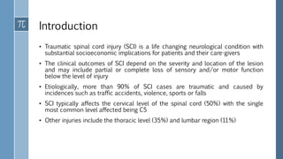
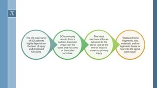
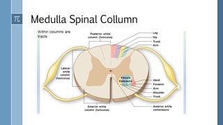
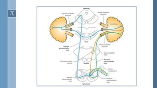
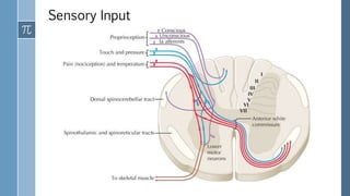
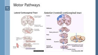
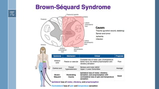
The document provides an overview of incomplete spinal cord injuries. Some key points:
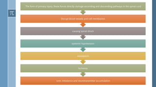
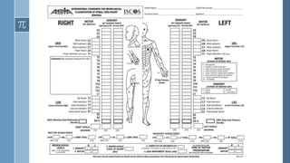
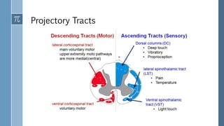
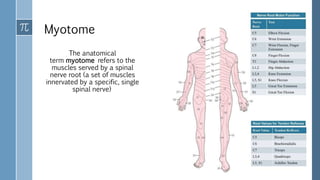
- Spinal cord injuries can range from complete to incomplete, depending on the severity and location of the lesion in the spinal cord. Incomplete injuries result in partial preservation of motor or sensory function below the injury level.
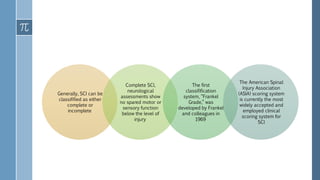
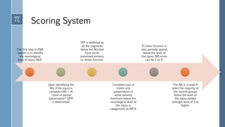
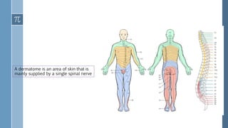
- The American Spinal Injury Association (ASIA) scoring system is used to clinically classify spinal cord injuries based on motor and sensory function. Injuries are classified on a scale from A (complete injury) to D (near normal function).
- Recovery from incomplete injuries is possible, though most occurs within the first year as spontaneous recovery plateaus. Sensory preservation is a predictor of potential motor recovery.
Brachial plexus injury (BPI) is a severe peripheral nerve injury affecting upper extremities, causing functional damage and physical disability
Root injury is defined as root avulsion from the spinal cord and rupture in the preganglionic root zone or dorsal ganglion at the vertebral foramen
Post-ganglionic injury are injuries distal to the ganglion, which divided into supra and infra-clavicular injury
This document provides information about the American Spinal Injury Association (ASIA) scale for classifying spinal cord injuries. It outlines the 5 steps for determining the ASIA grade: 1) determine sensory levels, 2) determine motor levels, 3) determine the neurological level of injury, 4) determine if the injury is complete or incomplete, and 5) determine the ASIA Impairment Scale grade (A-E). A 30-year-old man who fell 8 feet and had no motor function or voluntary anal contraction below his inguinal region is presented as a case example to demonstrate how to apply the ASIA scale.
Peripheral nerve injuries can occur through various mechanisms and be classified in different ways. The median, radial, and ulnar nerves are commonly injured in the upper limb. Examination of specific muscles innervated by each nerve helps to localize the level and severity of injury. Precise history taking and clinical assessment including motor, sensory and trophic changes are needed to diagnose peripheral nerve lesions.
This document discusses carpal tunnel syndrome, which is caused by compression of the median nerve as it passes through the carpal tunnel of the wrist. It can cause numbness, tingling, and weakness in the hand. The presentation outlines the causes, clinical features, diagnosis, and treatment options for carpal tunnel syndrome, which include wrist splints, oral anti-inflammatory medications, local steroid injections, and carpal tunnel release surgery if conservative measures fail. The document provides details on physical exam findings and special tests like Tinel's and Phalen's maneuvers used to diagnose carpal tunnel syndrome.
This document discusses spinal dysraphism, which refers to congenital anomalies resulting from failed fusion of the dorsal spinal elements. It describes different types including spina bifida occulta, meningocele, myelomeningocele, myelocystocele, and lipomeningocele. Meningocele involves a mass composed of CSF, meninges, and skin, but no neural elements. Myelomeningocele contains neural tissue and is usually associated with neurological deficits. Myelocystocele and lipomeningocele involve fatty tissue herniating through spinal defects. Radiological imaging plays an important role in diagnosis, with ultrasound, CT, MRI, and myelography discussed.
The Colles' fracture is a break of the radius bone close to the wrist that results in an upward displacement of the bone. It is typically caused by falling on an outstretched hand. Treatment depends on the severity but may include casting, closed reduction, open reduction and internal fixation. Physiotherapy focuses on regaining range of motion and strength through exercises over several phases of rehabilitation. Prevention includes proper nutrition, exercise, and wrist protection.
This document provides an overview of syringomyelia, including its pathogenesis, pathology, classification, clinical features, and natural history. Syringomyelia is a condition characterized by fluid-filled cavities within the spinal cord. It most commonly affects the cervical and thoracic regions. Clinical features include pain and sensory loss. The natural history varies, but symptoms typically progress slowly over years, with some patients experiencing stabilization or spontaneous resolution in rare cases.
Traumatic brain injury (TBI) is caused by an external force to the head that can lead to temporary or permanent impairment. It is a leading cause of death and disability, especially in young people. A TBI can be closed, without skull fracture, or open, with skull penetration. Initial management involves assessing severity with CT or MRI scans and monitoring for complications like increased intracranial pressure. Rehabilitation therapies like physiotherapy and occupational therapy aim to restore functions and prevent issues like spasticity or contractures. Outcomes depend on the severity of injury but long-term disabilities can impact cognition, movement, speech, and behavior.
This document discusses the anatomy, landmarks, measurements, common anomalies, syndromes, and injuries of the craniovertebral junction. It begins with a brief description of the craniovertebral junction's development and components. It then outlines several key anatomical landmarks and measurements used to evaluate the region on imaging. The remainder of the document details various congenital anomalies, developmental abnormalities, syndromes, and acquired conditions that can affect the craniovertebral junction.
This document discusses the anatomy, types of injuries, clinical presentation, investigations and classification of brachial plexus injuries.
It describes the formation of the brachial plexus from the cervical nerve roots and its divisions. Injuries can be preganglionic or postganglionic, and include traction injuries, avulsions or lacerations. Clinical exam focuses on assessing motor and sensory deficits. Investigations include imaging like MRI/CT, myelography and EMG/NCV to localize the lesion. Seddon's classification is used to describe the severity of injury.
The document discusses various syndromes of the spinal cord including complete and incomplete cord syndromes. It describes Brown-Sequard syndrome, central cord syndrome, anterior cord syndrome, posterior cord syndrome, and conus medullaris syndrome. Complete cord transection results in paralysis, sensory loss, and autonomic dysfunction below the level of injury. Incomplete syndromes can cause varying degrees of motor weakness, sensory loss, and autonomic dysfunction depending on the areas of the spinal cord affected. Specific vascular, inflammatory, and traumatic causes are discussed for each syndrome.
This document classifies spinal cord syndromes as either complete or incomplete. Complete cord syndromes result from total damage across the spinal cord and cause paralysis and loss of sensation below the level of injury. Incomplete cord syndromes result from partial damage and cause more localized neurological deficits. Several types of incomplete cord syndromes are described, including Brown-Sequard syndrome, central cord syndrome, and anterior cord syndrome. The causes, clinical features, and management of these conditions are outlined.
This document provides an overview of syringomyelia, including:
- It is a spinal cord cavity filled with cerebrospinal fluid, with a prevalence of 9 per 100,000 people.
- It can be caused by traumatic injury, Chiari malformation, or other craniovertebral junction anomalies.
- Symptoms depend on the location and extent of the syrinx and can include sensory loss, weakness, pain, and autonomic dysfunction.
- Magnetic resonance imaging is the best way to diagnose and assess syringomyelia.
- Treatment may involve surgery to decompress the craniovertebral junction, open the syrinx, or place a shunt
Syringomyelia is a condition where a cyst, called a syrinx, develops in the spinal cord. It most commonly affects the lower cervical spine. It is often associated with abnormalities of the skull or spinal column. The majority of cases are linked to Chiari malformation type 1, where the cerebellar tonsils are displaced into the spinal canal. Symptoms vary depending on the location of the syrinx but can include pain, loss of sensation, muscle weakness or atrophy, and autonomic dysfunction. Diagnosis is made using imaging like MRI. Treatment involves surgery to decompress pressure on the spinal cord like laminectomy with the goal of resolving the syrinx.
Spinal cord injuries can be either traumatic, from events like car accidents or falls, or non-traumatic, from conditions that damage the spinal cord. They are classified as either tetraplegia or paraplegia depending on whether the arms or legs are affected. Physiotherapy focuses on managing symptoms, preventing complications, and improving function through exercises for mobility, transfers, wheelchair skills, and more. The goal is to maximize independence and allow patients to safely perform daily living activities. Prognosis depends on the completeness of the injury and potential for recovery decreases over time as improvement plateaus.
Parkinson's disease is a chronic, progressive neurological disorder characterized by rigidity, bradykinesia, tremor, and postural instability. It is caused by the loss of dopamine-producing neurons in the substantia nigra. Symptoms worsen over time and can include impaired motor skills and coordination, speech and swallowing difficulties, sensory changes, and cognitive impairment. Physiotherapy aims to improve mobility, balance, and function through exercises targeting flexibility, strength, posture, gait, and functional skills.
The document summarizes information about clavicle fractures, including their epidemiology, anatomy, mechanisms of injury, classification systems, treatment options, and evidence for management approaches. Specifically, it finds that:
1) Clavicle fractures are among the most common fractures and midshaft fractures account for 69-80% of cases.
2) They are often caused by a direct blow to the shoulder and result in characteristic deformities from surrounding muscle and ligament forces.
3) Nonoperative treatment is usually indicated for nondisplaced fractures but recent evidence shows displaced or shortened fractures have higher risks of poor outcomes like pain, weakness and nonunion.
The document discusses the sensory system and how it processes and transmits sensory information from peripheral receptors to the sensory cortex. It describes how different sensory modalities like pain, temperature, vibration and proprioception are carried by different nerve fiber types and pathways in the body. It provides details on testing various sensory modalities and how the patterns of sensory loss can help localize lesions to different parts of the central or peripheral nervous system.
This document discusses cervical spine injuries, their classification, mechanisms of injury, diagnosis, and management. Some key points:
1. Cervical injuries can be caused by traction, direct impact, or indirect forces like flexion, compression, or rotation. Imaging helps classify injuries and assess stability.
2. Unstable injuries with neurological deficits or multiple injuries may require urgent surgical stabilization. Otherwise, initial treatment focuses on immobilization using rigid collars, braces, halo traction, or halo vests.
3. Common injuries include fractures of C1-C2 and the odontoid process. Type II odontoid fractures are prone to displacement and non-union, so may need open reduction and fusion
This document discusses arachnoiditis, an inflammatory disease of the arachnoid membrane surrounding the spinal cord. It develops in three stages: initial nerve inflammation, scar tissue formation causing nerve adhesion, and complete nerve root encapsulation. Causes include trauma from surgery, chemical exposure, and infection. Symptoms include pain, paresthesia, sensory loss, and muscle weakness. Diagnosis involves MRI or CT myelography. Treatment includes corticosteroids, NSAIDs, rhizotomy for pain, and decompression surgery to remove cysts. Physical therapy techniques like exercises, heat, TENS, and neural mobilization can help manage symptoms.
Waddling gait- definition|role of muscle|gait analysis|kinematic and spatiote...jasna ok
This document discusses waddling gait, which is an abnormal gait pattern seen when there is bilateral weakness of the gluteus medius muscles, the primary hip abductors. During walking, individuals with waddling gait are unable to stabilize the pelvis and it drops on both sides, causing the trunk to laterally bend and the person to walk with a wide base like a duck. Treatment focuses on strengthening exercises for the hip abductors and gluteal muscles, gait training, and balance exercises. Physiotherapy aims to improve muscle strength, correct posture, and retrain a normal walking pattern.
This document discusses the anatomy and physiology of the thoracolumbar spine and classifies different types of thoracolumbar spine injuries. It describes the anatomy of the spinal cord, blood supply, and biomechanics of the thoracic and lumbar regions. Various injury mechanisms are outlined including compression fractures, burst fractures, and chance fractures. Imaging techniques like x-rays, CT, and MRI are discussed. The Denis three-column theory and TLICS classification system are introduced to classify injuries as stable or unstable. Non-operative and surgical treatment options are provided based on the injury classification.
This document provides an overview of spinal cord injury (SCI) management. It describes a case of a 47-year-old male who suffered a cervical spine injury in a motor vehicle accident 4 months ago and is now presenting with paraplegia and paraparesis. The document then outlines key topics related to SCI, including anatomy, causes, types, pathophysiology, clinical syndromes, diagnosis, neurological assessment classification, and management. Tables and diagrams are provided to illustrate spinal cord anatomy and tracts, dermatomes, myotomes, and the American Spinal Injury Association classification system.
This document provides information on orthopaedic spinal injuries from Zagazig University in Egypt. It discusses several topics in 3 paragraphs or less:
Spinal injuries are less common than extremity injuries but have worse functional outcomes. They involve the cervical, thoracic, and lumbar spine. Neurological involvement is common in high-energy trauma or polytrauma patients.
Cervical spine injuries account for one-third of spinal injuries. The C2 vertebrae and lower C6-C7 vertebrae are most commonly injured. A neurological injury occurs in 15% of spine trauma patients. Exam of the peripheral nervous system is important to fully assess injuries.
Initial management follows ATLS protocols - stabilize
Spinal cord injury (SCI) results in paralysis and sensory deficits depending on the level and completeness of injury. SCI commonly results from traumatic injuries like motor vehicle accidents or falls in young adults. Injuries are classified by neurological level and completeness. Individuals with incomplete injuries have better outcomes than those with complete injuries. Physical therapy for SCI focuses on restoring function and independence through interventions like mobility training, respiratory management, and prevention of secondary complications. Proper home and access modifications can also facilitate community reintegration.
The document provides an overview of the anatomy and functional organization of the spinal cord. It describes:
1) The spinal cord extends from the medulla to the L1 vertebra and has cervical and lumbar enlargements. It is protected by vertebrae, discs, meninges, cerebrospinal fluid, and ligaments.
2) The spinal cord has 31 pairs of spinal nerves that contain both motor and sensory fibers. Each spinal segment innervates a specific dermatome and myotome.
3) Major ascending and descending tracts in the spinal cord include the corticospinal, spinothalamic, and dorsal column tracts which are involved in motor function and different types of
1. The document discusses various types of spinal cord injuries including complete injuries which involve a complete loss of motor and sensory function below the level of injury, and incomplete injuries which partially compromise spinal cord function with some sensation and muscle movement retained below the injury site.
2. It provides details on specific spinal cord syndromes like anterior cord syndrome, Brown-Séquard syndrome, and central cord syndrome which are characterized by variable patterns of motor and sensory loss.
3. The management of spinal cord injuries involves stabilizing the spine, treating shock, addressing airway and breathing issues, screening for associated injuries, and preventing complications like pressure sores through regular turning of immobilized patients.
The document provides information on spinal cord and cervical spine anatomy, mechanisms of spinal cord injury, clinical assessment of spinal cord injury patients, imaging for spinal cord injuries, classification of spinal cord injuries, and management principles for spinal cord injuries. Key points covered include the incidence of spinal cord injuries, common mechanisms and levels of injury, assessment of motor and sensory function, classification systems for incomplete versus complete injuries, and guidelines for cervical spine clearance in trauma patients.
This document provides an overview of thoracolumbar fractures, including epidemiology, clinical evaluation, classification systems, radiographic evaluation, treatment approaches, and specific surgical techniques. It discusses the anatomy of the thoracolumbar region, mechanisms of injury, neurological assessment tools, radiographic indicators of instability, and non-operative and operative treatment options depending on the fracture classification.
Evaluation of Spinal Injury & Emergency ManagementAtif Shahzad
This document provides information on spinal injuries, including:
- Traumatic spinal cord injuries result in 12,000 new cases per year in the US. Most injuries occur in men aged 16-30 from vehicle crashes, falls, or sports.
- Injuries are categorized by location (cervical, thoracic, lumbar), stability (stable or unstable), and neurological status (complete or incomplete paralysis).
- Initial treatment follows ATLS protocols to stabilize the spine and assess airway, breathing, circulation, disability, and exposure. Advanced imaging can further evaluate bone and neurological injuries.
Spinal trauma management involves immobilization, intravenous fluids, medications, and prompt referral. Anatomy and mechanisms of injury vary by spinal region. Evaluation assesses neurological function using dermatomes, myotomes, and reflexes to localize injury level. Injuries may cause hypovolaemic or neurogenic shock. Corticosteroids within 8 hours may improve outcomes but evidence is limited. Prompt management aims to prevent secondary spinal cord injury.
A spinal cord injury refers to any injury to the spinal cord that is caused by trauma instead of diseases resulting in a change either temporary or permanent, in its normal motor, sensory or autonomic function.
This document discusses spinal cord disorders and provides information on spinal cord anatomy and different types of spinal cord injuries and conditions. It outlines 15 questions regarding the anatomy of the spinal cord and characteristics of various spinal cord disorders including central cord syndrome, Brown-Sequard syndrome, anterior cord syndrome, transverse myelitis, syringomyelia, spinal epidural hematoma, spinal epidural abscess, diskitis, and spinal cord neoplasms. The document seeks to test the reader's knowledge on the topics covered through true or false questions.
The spinal cord extends from the brainstem and down the back, containing bundles of nerve fibers that connect the brain to the peripheral nervous system. It carries sensory information from the body to the brain and motor commands from the brain to the body. Damage to the spinal cord can occur through trauma, compression, or reduced blood flow, resulting in paralysis and loss of sensation depending on the level and completeness of injury. Management involves pharmacological treatment, physical therapy, and sometimes surgery to decompress the spinal cord.
Traumatic spinal cord injuries can cause tetraplegia or paraplegia depending on the level of the lesion in the spinal cord. Injuries are often classified based on whether they are complete or incomplete. Common clinical manifestations include motor and sensory impairments, autonomic dysreflexia, respiratory issues, spasticity, and bladder and bowel dysfunction. Indirect impairments from SCI can also occur and include respiratory complications, pressure sores, deep vein thrombosis, and other issues.
This document presents a case report of a 53-year-old female who sustained a traumatic brachial plexus injury after falling down a flight of stairs. On examination, she had sensory and motor deficits in her right upper limb consistent with an infraclavicular brachial plexus palsy. She underwent conservative therapy including physiotherapy and splinting. Over an 18-month period, her function gradually improved but she was left with some residual weakness. The document discusses the anatomy of the brachial plexus, mechanisms of injury, clinical examination findings, and prognosis factors for peripheral nerve injuries.
This document provides an overview of acute trauma to the spine. It discusses the epidemiology of spine injuries, noting they most commonly occur in males during their productive years, often from motor vehicle accidents or falls. The presentation outlines evaluation of spine trauma patients, including maintaining cervical spine immobilization, assessing the airway, breathing, and circulation during the primary survey. It also details the physical exam, neurological exam including ASIA scoring, and imaging used in diagnosis. Common injury mechanisms and classifications of complete versus incomplete injuries are presented.
The term Spinal Cord Injury is used to refer to neurological damage of the spinal cord
Any lesion involving the spinal cord result a syndrome called a “myelopathy”
Spinal cord injuries are defined as complete or incomplete according to the International Standards for the Neurological Classifification of SCI and the American Spinal Injuries Association Impairment Scale (AIS)
Complete lesions are defifined as AIS A, and incomplete lesions are defifined as AIS B, AIS C, AIS D or AIS E (Harvey, 2016)
Final case presentation sci (kimberly walsh)Kimberly Walsh
This document provides an overview of cervical myelopathy and spinal cord injury, including:
- Definitions of spinal cord injury and cervical myelopathy.
- Descriptions of anatomy including the spine, cervical spine, intervertebral discs, and ligaments.
- Causes, pathophysiology, and clinical manifestations of both cervical myelopathy and spinal cord injury.
- Details on epidemiology, diagnosis, complications and management of spinal cord injury.
Fibromyalgia adalah gangguan nyeri kronis yang menyebar pada otot dan jaringan lunak. Gejalanya meliputi rasa kaku dan nyeri otot serta kelelahan. Patofisiologinya belum jelas tetapi terkait dengan gangguan sistem saraf pusat dan periferal serta inflamasi. Terapi fisik seperti aerobic, peregangan, dan pijatan dapat membantu mengurangi gejala pasien.
Central Post Stroke Pain (CPSP) adalah sindroma nyeri neuropatik yang terjadi setelah serangan stroke akibat lesi pada thalamus dan posterior limb internal capsule. Nyeri CPSP berupa rasa terbakar, tertusuk, dan kesetrum yang dapat muncul secara spontan atau dengan stimulus dan sulit dihilangkan. Diagnosa CPSP didasarkan pada karakteristik klinis seperti nyeri, allodynia, dan hipersensitivitas terhadap stimulus dingin. Terapi utama CPS
Russian Current adalah stimulasi arus bolak-balik berfrekuensi tinggi 2500 Hz yang diberikan dalam bentuk pulsa selama 10 detik diikuti istirahat 50 detik, untuk meningkatkan kekuatan otot secara progresif tanpa menimbulkan kelelahan. Arus ini ditemukan oleh ilmuwan Rusia Yakov Kotz pada tahun 1977 dan digunakan dalam latihan kekuatan dan rehabilitasi otot.
Dokumen tersebut membahas tentang konsep dasar elektroterapi. Secara singkat, dokumen menjelaskan bahwa elektroterapi melibatkan stimulasi jaringan tubuh menggunakan arus listrik dengan berbagai karakteristik seperti voltase, arus, frekuensi, dan bentuk gelombang. Stimulasi dapat menimbulkan respon sensoris, motoris, atau nyeri tergantung tingkatannya.
Lecture 6 -- Memory 2015.pptlearning occurs when a stimulus (unconditioned st...AyushGadhvi1
learning occurs when a stimulus (unconditioned stimulus) eliciting a response (unconditioned response) • is paired with another stimulus (conditioned stimulus)
Travel Clinic Cardiff: Health Advice for International TravelersNX Healthcare
Travel Clinic Cardiff offers comprehensive travel health services, including vaccinations, travel advice, and preventive care for international travelers. Our expert team ensures you are well-prepared and protected for your journey, providing personalized consultations tailored to your destination. Conveniently located in Cardiff, we help you travel with confidence and peace of mind. Visit us: www.nxhealthcare.co.uk
These lecture slides, by Dr Sidra Arshad, offer a simplified look into the mechanisms involved in the regulation of respiration:
Learning objectives:
1. Describe the organisation of respiratory center
2. Describe the nervous control of inspiration and respiratory rhythm
3. Describe the functions of the dorsal and respiratory groups of neurons
4. Describe the influences of the Pneumotaxic and Apneustic centers
5. Explain the role of Hering-Breur inflation reflex in regulation of inspiration
6. Explain the role of central chemoreceptors in regulation of respiration
7. Explain the role of peripheral chemoreceptors in regulation of respiration
8. Explain the regulation of respiration during exercise
9. Integrate the respiratory regulatory mechanisms
10. Describe the Cheyne-Stokes breathing
Study Resources:
1. Chapter 42, Guyton and Hall Textbook of Medical Physiology, 14th edition
2. Chapter 36, Ganong’s Review of Medical Physiology, 26th edition
3. Chapter 13, Human Physiology by Lauralee Sherwood, 9th edition
Promoting Wellbeing - Applied Social Psychology - Psychology SuperNotesPsychoTech Services
A proprietary approach developed by bringing together the best of learning theories from Psychology, design principles from the world of visualization, and pedagogical methods from over a decade of training experience, that enables you to: Learn better, faster!
5-hydroxytryptamine or 5-HT or Serotonin is a neurotransmitter that serves a range of roles in the human body. It is sometimes referred to as the happy chemical since it promotes overall well-being and happiness.
It is mostly found in the brain, intestines, and blood platelets.
5-HT is utilised to transport messages between nerve cells, is known to be involved in smooth muscle contraction, and adds to overall well-being and pleasure, among other benefits. 5-HT regulates the body's sleep-wake cycles and internal clock by acting as a precursor to melatonin.
It is hypothesised to regulate hunger, emotions, motor, cognitive, and autonomic processes.
NAVIGATING THE HORIZONS OF TIME LAPSE EMBRYO MONITORING.pdfRahul Sen
Time-lapse embryo monitoring is an advanced imaging technique used in IVF to continuously observe embryo development. It captures high-resolution images at regular intervals, allowing embryologists to select the most viable embryos for transfer based on detailed growth patterns. This technology enhances embryo selection, potentially increasing pregnancy success rates.
The skin is the largest organ and its health plays a vital role among the other sense organs. The skin concerns like acne breakout, psoriasis, or anything similar along the lines, finding a qualified and experienced dermatologist becomes paramount.
“Psychiatry and the Humanities”: An Innovative Course at the University of Mo...Université de Montréal
“Psychiatry and the Humanities”: An Innovative Course at the University of Montreal Expanding the medical model to embrace the humanities. Link: https://www.psychiatrictimes.com/view/-psychiatry-and-the-humanities-an-innovative-course-at-the-university-of-montreal
Cell Therapy Expansion and Challenges in Autoimmune DiseaseHealth Advances
There is increasing confidence that cell therapies will soon play a role in the treatment of autoimmune disorders, but the extent of this impact remains to be seen. Early readouts on autologous CAR-Ts in lupus are encouraging, but manufacturing and cost limitations are likely to restrict access to highly refractory patients. Allogeneic CAR-Ts have the potential to broaden access to earlier lines of treatment due to their inherent cost benefits, however they will need to demonstrate comparable or improved efficacy to established modalities.
In addition to infrastructure and capacity constraints, CAR-Ts face a very different risk-benefit dynamic in autoimmune compared to oncology, highlighting the need for tolerable therapies with low adverse event risk. CAR-NK and Treg-based therapies are also being developed in certain autoimmune disorders and may demonstrate favorable safety profiles. Several novel non-cell therapies such as bispecific antibodies, nanobodies, and RNAi drugs, may also offer future alternative competitive solutions with variable value propositions.
Widespread adoption of cell therapies will not only require strong efficacy and safety data, but also adapted pricing and access strategies. At oncology-based price points, CAR-Ts are unlikely to achieve broad market access in autoimmune disorders, with eligible patient populations that are potentially orders of magnitude greater than the number of currently addressable cancer patients. Developers have made strides towards reducing cell therapy COGS while improving manufacturing efficiency, but payors will inevitably restrict access until more sustainable pricing is achieved.
Despite these headwinds, industry leaders and investors remain confident that cell therapies are poised to address significant unmet need in patients suffering from autoimmune disorders. However, the extent of this impact on the treatment landscape remains to be seen, as the industry rapidly approaches an inflection point.
Test bank for karp s cell and molecular biology 9th edition by gerald karp.pdfrightmanforbloodline
Test bank for karp s cell and molecular biology 9th edition by gerald karp.pdf
Nano-gold for Cancer Therapy chemistry investigatory projectSIVAVINAYAKPK
chemistry investigatory project
The development of nanogold-based cancer therapy could revolutionize oncology by providing a more targeted, less invasive treatment option. This project contributes to the growing body of research aimed at harnessing nanotechnology for medical applications, paving the way for future clinical trials and potential commercial applications.
Cancer remains one of the leading causes of death worldwide, prompting the need for innovative treatment methods. Nanotechnology offers promising new approaches, including the use of gold nanoparticles (nanogold) for targeted cancer therapy. Nanogold particles possess unique physical and chemical properties that make them suitable for drug delivery, imaging, and photothermal therapy.
2. Introduction
• Traumatic spinal cord injury (SCI) is a life changing neurological condition with
substantial socioeconomic implications for patients and their care-givers
• The clinical outcomes of SCI depend on the severity and location of the lesion
and may include partial or complete loss of sensory and/or motor function
below the level of injury
• Etiologically, more than 90% of SCI cases are traumatic and caused by
incidences such as traffic accidents, violence, sports or falls
• SCI typically affects the cervical level of the spinal cord (50%) with the single
most common level affected being C5
• Other injuries include the thoracic level (35%) and lumbar region (11%)
3. “displaced bone
fragments, disc
materials, and/or
ligaments bruise or
tear into the spinal
cord tissue”
The initial
mechanical forces
delivered to the
spinal cord at the
time of injury is
known as primary
injury
SCI commonly
results from a
sudden, traumatic
impact on the
spine that fractures
or dislocates
vertebrae
The life expectancy
of SCI patients
highly depends on
the level of injury
and preserved
functions
5. The most common form of
primary injury is impact plus
persistent compression, which
typically occurs through burst
fractures with bone fragments
compressing the spinal cord
Impact alone with transient
compression is observed less
frequently but most
commonly in hyperextension
injuries
Distraction injuries occur
when two adjacent vertebrae
are pulled apart causing the
spinal column to stretch and
tear in the axial plane
Lastly, laceration and
transection injuries can occur
through missile injuries,
severe dislocations, or sharp
bone fragment dislocations
6. ionic imbalance and neurotransmitter accumulation
Ischemia
vasospasm,
systemic hypotension
causing spinal shock
Disrupt blood vessels and cell membranes
The form of primary injury, these forces directly damage ascending and descending pathways in the spinal cord
7. To date, the most
effffective clinical
treatment to limit tissue
damage following
primary injury is the
early surgical
decompression (<24 h
post-injury) of the
injured spinal cord
8. Generally, SCI can be
classifified as either
complete or
incomplete
Complete SCI,
neurological
assessments show
no spared motor or
sensory function
below the level of
injury
The first
classifification
system, “Frankel
Grade,” was
developed by Frankel
and colleagues in
1969
The American Spinal
Injury Association
(ASIA) scoring system
is currently the most
widely accepted and
employed clinical
scoring system for
SCI
9. American Spinal Injury Association (ASIA) Scoring System
In this system, sensory function
is scored rom 0 to 2
(0 = absent, 1 = altered, 2=
normal)
And motor function from 0
to 5
10. Scoring System
The first step in ASIA
system is to identify
the neurological
level of injury (NLI)
Upon identifying the
NLI, if the injury is
complete (AIS = A),
“zone of partial
preservation” (ZPP)
is determined
ZPP is defifined as
all the segments
below the NLI that
have some
preserved sensory
or motor function
Complete loss of
motor and
preservation of
some sensory
functions below the
neurological level of
the injury is
categorized as AIS B
If motor function is
also partially spared
below the level of
the injury, AIS score
can be C or D
The AIS is scored D
when the majority of
the muscle groups
below the level of
the injury exhibit
strength level of 3 or
higher
11.
12. As time passes, SCI patients experience some spontaneous recovery of motor and sensory functions
Most of the functional recovery occurs during the first 3 months and in most cases reaches a plateau by
9 months after injury
However, additional recovery may occur up to 12–18 months post-injury
Patients with incomplete paraplegia generally have a good prognosis in regaining locomotor ability
(∼76% of patients) within a year
Complete paraplegic patients, however, experience limited recovery of lower limb function if their NLI is
above T9
An NLI below T9 is associated with 38% chance of regaining some lower extremity function
In patients with complete paraplegia, the chance of recovery to an incomplete status is only 4% with
only half of these patients regaining bladder and bowel control
13. One proposed reason for
this association is that
pinprick fibers in lateral
spinothalamic tract travel in
proximity of motor fifibers in
the lateral corticospinal
tract, and thus, preservation
of sensory fifibers can be an
indicator of the integrity of
motor fifiber
Maintenance of pinprick
sensation at the zone of
partial preservation or in
sacral segments has been
shown as a reliable
predictor of motor recovery
An association between
sensory and motor recovery
has been demonstrated in
SCI where spontaneous
sensory recovery usually
follows the pattern of motor
recovery
14. Hypovolemia and hemodynamic shock in SCI patients due to excessive bleeding and
neurogenic shock result in compromised spinal cord perfusion and ischemia
Increased tissue pressure in edematous injured spinal cord and hemorrhage-induced
vasospasm in intact vessels further disrupts blood flow to the spinal cord
Vascular insult, hemorrhage and ischemia ultimately lead to cell death and tissue destruction
through multiple mechanisms, including oxygen deprivation, loss of adenosine triphosphate
(ATP), excitotoxicity, ionic imbalance, free radical formation, and necrotic cell death
Cellular necrosis and release of cytoplasmic content increase the extracellular level of
glutamate causing glutamate excitotoxicity (the main excitatory neurotransmitter in the CNS)
15.
16. Severe acute spinal cord lesion there are two
clinical stages:
Spinal shock : loss of all reflex activity
below the level of lesion, flaccid limbs,
atonic bladder with incontinence, atonic
bowel, loss of genital reflexes and
vasomotor control
Heightened reflex activity : after 1-2 weeks
associated with spasticity of the limbs,
brisk reflexes and extensor plantar
response, spastic bladder and hyperactive
autonomic function (sweating)
17. The clinical feature
Below L1 level, the cauda equina will be affected and the patient will have LMN syndrome affecting the leg and
bladder
when it occurs above the level L1 causes spastic paraparesis or tetraparesis at high cervical level
Lesion above C5 cause UMN signs in the arms and legs
Lesion between C5 and T1 cause LMN and sometime UMN signs in the arm and UMN signs in the legs
Lesion affecting the cord below T1 will not involve the arms
18. The spinal cord extends from the top of the C1 vertebra to the bottom of the body of the L1
27. Myotome
The anatomical
term myotome refers to the
muscles served by a spinal
nerve root (a set of muscles
innervated by a specific, single
spinal nerve)
30. The Clinical Syndrome of Spinal Cord Disease
Paresis is the correct term to describe incomplete paralysis, Plegia means complete paralysis and Palsy used when the paralysis affects cranial motor
nerve (bell’s palsy, pseudobulbar palsy) or a static weakness (cerebral palsy)
The words “plegia” “palsy” and “paresis” are sometimes used interchangeably to describe weakness
Paralysis is the complete loss of voluntary movement
Brown-Sequard syndrome (Unilateral lesion causing UMN involvement of one side)
Tetraparesis (UMN involvement of all four limbs)
Paraparesis (UMN involvement of legs only)
There are tree main motor syndromes associated with spinal cord disease
31. PARAPARESIS (SPASTIC PARAPARESIS OR PARAPLEGIA)
Paraparesis
indicates bilateral
UMN damage
involving the
axons that
innervate the legs
from both
corticospinal
tracts
The clinical signs
include :
Increased tone
with spasticity
Increased
reflexes with
clonus
Extensor plantar
response
(Babinski sign)
Sphincter
dysfunction
32. TETRAPARESIS (SPASTIC TETRAPARESIS, TETRAPLEGIA, QUADRIPARESIS,
QUADRIPLEGIA)
It is usually caused
by lesion in the
high cervical cord,
occasionally due to
brainstem
Spastic tetraparesis
produces the same
clinical picture as
paraparesis but
involves both arms
and legs
33.
34. BROWN SEQUARD SYNDROME (UNILATERAL CORD LESION)
Brown Sequard syndrome
is rare in its pure form but
partial forms are more
common
Ipsilateral spastic leg and
sometimes arm if the
lesion above C5, with brisk
reflexes and an extensor
plantar response
Ipsilateral loss of joint
position sense and
vibration or touch (dorsal
column), contralateral loss
of pain and temperature
o Pain fibers ascend a few spinal segments before entering the dorsal horn
to synapse
o If the lesion damages the anterior horn cell as well as the white matter
tracts then the patient will have UMN signs below the level of the lesion
and LMN signs at and about the level of the lesion
35.
36. A framework of Physiotherapy
Management
The overall purpose of
Physiotherapy for patients with
spinal cord injury is to improve
health related quality of life
By improving patient’s ability to
participate in activities of daily
life
The International Classification
of Functioning Disability and
Helath (ICF) can be used to
describe the process involved in
formulating a physiotherapy
programs
37. Step one : Assessing impairments, activity limitation and participation retrictions
Step two : Setting goal with respect to activity limitation and participation retrictions
Step three : Identifying key impairments
Step four : Indentifying and administering
treatments
Step five : Measuring
outcomes
38. Assessing impairements, functional limitations and
participation restrictions
Various sources need
to be used
Age, cause of injury,
time since injury,
neurological status,
orthopaedic status,
other injuries and
complications,
medical and surgical
management etc
Well accepted
assessment tool used
to measure functional
limitation and
parcitipation restriction
including :
Functional
Independence Measure
(FIM)
Spinal Cord
Independence
Measure
Quadriplegic Index of
Function
Specific assessment i.e
ability to walk 6
minutes walk test, TUG,
10m walk test
39. These goals should
be expressed in
term of
participation
restrictions (i.e
return to work or
school)
Sub domains of mobility,
self care and domestic
life
Goals should be
SMART (Specific,
Measurable,
Attainable,
Realistic and
Timebound)
41. Once goals of treatment
are defined in term of
activity limitation and
participation restriction, it
is necessary to determine
which impairements
prevent the attainment of
each goal
Key impairement need to be linked to
specific activity limitation and
participation restriction
Anatomical and functional
limitation are different
42. C6 - tetraplegia sitting
Passive tension in the paralysed
hamstring muscle helps maintain
the trunk in upright position
Hamstring cannot prevent a
forward fall if they are highly
extensible