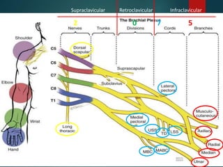
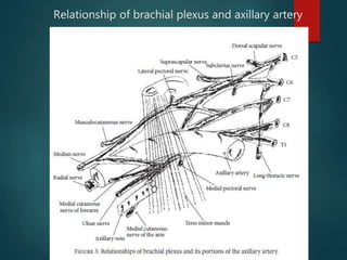
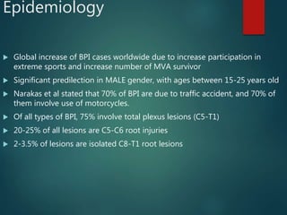
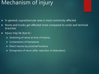
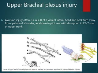
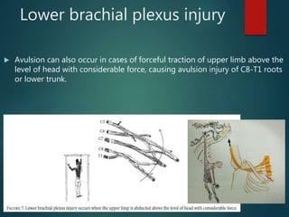
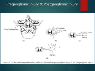
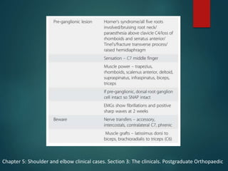
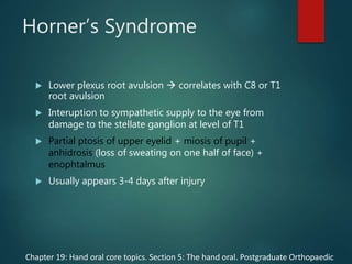
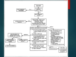
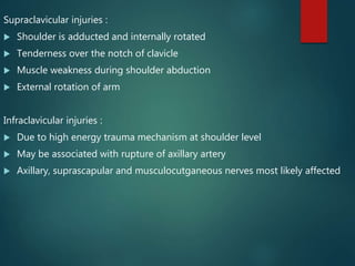
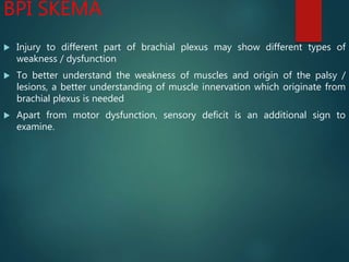
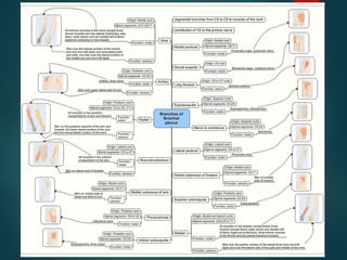
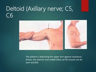
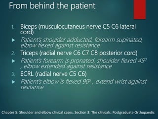
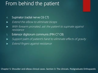
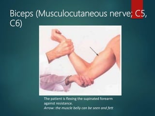
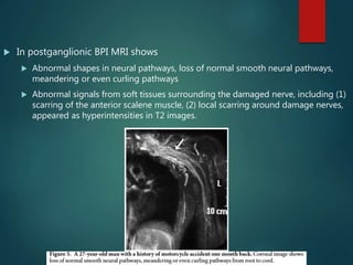
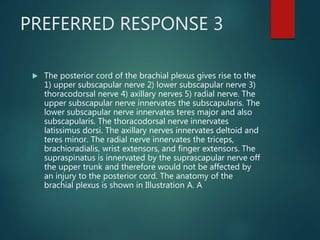
This document presents a case report of a 53-year-old female who sustained a traumatic brachial plexus injury after falling down a flight of stairs. On examination, she had sensory and motor deficits in her right upper limb consistent with an infraclavicular brachial plexus palsy. She underwent conservative therapy including physiotherapy and splinting. Over an 18-month period, her function gradually improved but she was left with some residual weakness. The document discusses the anatomy of the brachial plexus, mechanisms of injury, clinical examination findings, and prognosis factors for peripheral nerve injuries.
Brachial plexus injury (BPI) is a severe peripheral nerve injury affecting upper extremities, causing functional damage and physical disability
Root injury is defined as root avulsion from the spinal cord and rupture in the preganglionic root zone or dorsal ganglion at the vertebral foramen
Post-ganglionic injury are injuries distal to the ganglion, which divided into supra and infra-clavicular injury
1.Anatomy
a.Course
b.Motor distribution
c.Sensory distribution
2.Common sites affected
3.Level of median nerve injury
4.Clinical feature with various test performed
5.Various syndromes related to median nerve
6.Treatment
7.Summary
anatomy of median nerve,course in arm and struthers ligament, branches in the forearm, carpal tunnel and course in hand, high and low median nerve injuries, principles of surgical management, pronator teres syndrome, anterior interosseous nerve syndrome, open and endoscopic carpal tunnel release
4th year medical student's seminar presentation under supervision of orthopedic lecturer. Reference is from Dr. Sameh Doss Textbook of upper and lower limb, and also other multiple websites.
Peripheral nerve damage affecting the upper extremities can vary widely in cause and extent.
Many disorders, ranging from mild carpal tunnel syndrome to severe brachial plexopathy, need to be considered in a patient presenting with pain, sensory loss, or weakness involving the shoulder, arm, or hand.
These lecture slides, by Dr Sidra Arshad, offer a quick overview of physiological basis of a normal electrocardiogram.
Learning objectives:
1. Define an electrocardiogram (ECG) and electrocardiography
2. Describe how dipoles generated by the heart produce the waveforms of the ECG
3. Describe the components of a normal electrocardiogram of a typical bipolar leads (limb II)
4. Differentiate between intervals and segments
5. Enlist some common indications for obtaining an ECG
Study Resources:
1. Chapter 11, Guyton and Hall Textbook of Medical Physiology, 14th edition
2. Chapter 9, Human Physiology - From Cells to Systems, Lauralee Sherwood, 9th edition
3. Chapter 29, Ganong’s Review of Medical Physiology, 26th edition
4. Electrocardiogram, StatPearls - https://www.ncbi.nlm.nih.gov/books/NBK549803/
5. ECG in Medical Practice by ABM Abdullah, 4th edition
6. ECG Basics, http://www.nataliescasebook.com/tag/e-c-g-basics
micro teaching on communication m.sc nursing.pdfAnurag Sharma
Microteaching is a unique model of practice teaching. It is a viable instrument for the. desired change in the teaching behavior or the behavior potential which, in specified types of real. classroom situations, tends to facilitate the achievement of specified types of objectives.
MANAGEMENT OF ATRIOVENTRICULAR CONDUCTION BLOCK.pdfJim Jacob Roy
Cardiac conduction defects can occur due to various causes.
Atrioventricular conduction blocks ( AV blocks ) are classified into 3 types.
This document describes the acute management of AV block.
Couples presenting to the infertility clinic- Do they really have infertility...Sujoy Dasgupta
Dr Sujoy Dasgupta presented the study on "Couples presenting to the infertility clinic- Do they really have infertility? – The unexplored stories of non-consummation" in the 13th Congress of the Asia Pacific Initiative on Reproduction (ASPIRE 2024) at Manila on 24 May, 2024.
HOT NEW PRODUCT! BIG SALES FAST SHIPPING NOW FROM CHINA!! EU KU DB BK substit...GL Anaacs
Contact us if you are interested:
Email / Skype : kefaya1771@gmail.com
Threema: PXHY5PDH
New BATCH Ku !!! MUCH IN DEMAND FAST SALE EVERY BATCH HAPPY GOOD EFFECT BIG BATCH !
Contact me on Threema or skype to start big business!!
Hot-sale products:
NEW HOT EUTYLONE WHITE CRYSTAL!!
5cl-adba precursor (semi finished )
5cl-adba raw materials
ADBB precursor (semi finished )
ADBB raw materials
APVP powder
5fadb/4f-adb
Jwh018 / Jwh210
Eutylone crystal
Protonitazene (hydrochloride) CAS: 119276-01-6
Flubrotizolam CAS: 57801-95-3
Metonitazene CAS: 14680-51-4
Payment terms: Western Union,MoneyGram,Bitcoin or USDT.
Deliver Time: Usually 7-15days
Shipping method: FedEx, TNT, DHL,UPS etc.Our deliveries are 100% safe, fast, reliable and discreet.
Samples will be sent for your evaluation!If you are interested in, please contact me, let's talk details.
We specializes in exporting high quality Research chemical, medical intermediate, Pharmaceutical chemicals and so on. Products are exported to USA, Canada, France, Korea, Japan,Russia, Southeast Asia and other countries.
Flu Vaccine Alert in Bangalore Karnatakaaddon Scans
As flu season approaches, health officials in Bangalore, Karnataka, are urging residents to get their flu vaccinations. The seasonal flu, while common, can lead to severe health complications, particularly for vulnerable populations such as young children, the elderly, and those with underlying health conditions.
Dr. Vidisha Kumari, a leading epidemiologist in Bangalore, emphasizes the importance of getting vaccinated. "The flu vaccine is our best defense against the influenza virus. It not only protects individuals but also helps prevent the spread of the virus in our communities," he says.
This year, the flu season is expected to coincide with a potential increase in other respiratory illnesses. The Karnataka Health Department has launched an awareness campaign highlighting the significance of flu vaccinations. They have set up multiple vaccination centers across Bangalore, making it convenient for residents to receive their shots.
To encourage widespread vaccination, the government is also collaborating with local schools, workplaces, and community centers to facilitate vaccination drives. Special attention is being given to ensuring that the vaccine is accessible to all, including marginalized communities who may have limited access to healthcare.
Residents are reminded that the flu vaccine is safe and effective. Common side effects are mild and may include soreness at the injection site, mild fever, or muscle aches. These side effects are generally short-lived and far less severe than the flu itself.
Healthcare providers are also stressing the importance of continuing COVID-19 precautions. Wearing masks, practicing good hand hygiene, and maintaining social distancing are still crucial, especially in crowded places.
Protect yourself and your loved ones by getting vaccinated. Together, we can help keep Bangalore healthy and safe this flu season. For more information on vaccination centers and schedules, residents can visit the Karnataka Health Department’s official website or follow their social media pages.
Stay informed, stay safe, and get your flu shot today!
Tom Selleck Health: A Comprehensive Look at the Iconic Actor’s Wellness Journeygreendigital
Tom Selleck, an enduring figure in Hollywood. has captivated audiences for decades with his rugged charm, iconic moustache. and memorable roles in television and film. From his breakout role as Thomas Magnum in Magnum P.I. to his current portrayal of Frank Reagan in Blue Bloods. Selleck's career has spanned over 50 years. But beyond his professional achievements. fans have often been curious about Tom Selleck Health. especially as he has aged in the public eye.
Follow us on: Pinterest
Introduction
Many have been interested in Tom Selleck health. not only because of his enduring presence on screen but also because of the challenges. and lifestyle choices he has faced and made over the years. This article delves into the various aspects of Tom Selleck health. exploring his fitness regimen, diet, mental health. and the challenges he has encountered as he ages. We'll look at how he maintains his well-being. the health issues he has faced, and his approach to ageing .
Early Life and Career
Childhood and Athletic Beginnings
Tom Selleck was born on January 29, 1945, in Detroit, Michigan, and grew up in Sherman Oaks, California. From an early age, he was involved in sports, particularly basketball. which played a significant role in his physical development. His athletic pursuits continued into college. where he attended the University of Southern California (USC) on a basketball scholarship. This early involvement in sports laid a strong foundation for his physical health and disciplined lifestyle.
Transition to Acting
Selleck's transition from an athlete to an actor came with its physical demands. His first significant role in "Magnum P.I." required him to perform various stunts and maintain a fit appearance. This role, which he played from 1980 to 1988. necessitated a rigorous fitness routine to meet the show's demands. setting the stage for his long-term commitment to health and wellness.
Fitness Regimen
Workout Routine
Tom Selleck health and fitness regimen has evolved. adapting to his changing roles and age. During his "Magnum, P.I." days. Selleck's workouts were intense and focused on building and maintaining muscle mass. His routine included weightlifting, cardiovascular exercises. and specific training for the stunts he performed on the show.
Selleck adjusted his fitness routine as he aged to suit his body's needs. Today, his workouts focus on maintaining flexibility, strength, and cardiovascular health. He incorporates low-impact exercises such as swimming, walking, and light weightlifting. This balanced approach helps him stay fit without putting undue strain on his joints and muscles.
Importance of Flexibility and Mobility
In recent years, Selleck has emphasized the importance of flexibility and mobility in his fitness regimen. Understanding the natural decline in muscle mass and joint flexibility with age. he includes stretching and yoga in his routine. These practices help prevent injuries, improve posture, and maintain mobilit
Ethanol (CH3CH2OH), or beverage alcohol, is a two-carbon alcohol
that is rapidly distributed in the body and brain. Ethanol alters many
neurochemical systems and has rewarding and addictive properties. It
is the oldest recreational drug and likely contributes to more morbidity,
mortality, and public health costs than all illicit drugs combined. The
5th edition of the Diagnostic and Statistical Manual of Mental Disorders
(DSM-5) integrates alcohol abuse and alcohol dependence into a single
disorder called alcohol use disorder (AUD), with mild, moderate,
and severe subclassifications (American Psychiatric Association, 2013).
In the DSM-5, all types of substance abuse and dependence have been
combined into a single substance use disorder (SUD) on a continuum
from mild to severe. A diagnosis of AUD requires that at least two of
the 11 DSM-5 behaviors be present within a 12-month period (mild
AUD: 2–3 criteria; moderate AUD: 4–5 criteria; severe AUD: 6–11 criteria).
The four main behavioral effects of AUD are impaired control over
drinking, negative social consequences, risky use, and altered physiological
effects (tolerance, withdrawal). This chapter presents an overview
of the prevalence and harmful consequences of AUD in the U.S.,
the systemic nature of the disease, neurocircuitry and stages of AUD,
comorbidities, fetal alcohol spectrum disorders, genetic risk factors, and
pharmacotherapies for AUD.
Anti ulcer drugs and their Advance pharmacology ||
Anti-ulcer drugs are medications used to prevent and treat ulcers in the stomach and upper part of the small intestine (duodenal ulcers). These ulcers are often caused by an imbalance between stomach acid and the mucosal lining, which protects the stomach lining.
||Scope: Overview of various classes of anti-ulcer drugs, their mechanisms of action, indications, side effects, and clinical considerations.
2. Case report
53 years old right handed female, fell from a flight of stairs on her right arm.
She fell on a stretched out – wide open arm. She was unable to move her
arm after the incident.
On clinical examination there was an asymmetric profile of pain and
neurological deficit in right upper limb
Sensations were reduced on :
lateral aspect of arm
dorsal and ulnar aspext of forearm
volar and dorsal aspect of wrist and
hand
Motor deficit was found on :
Extensor of wrist and fingers
Other area was not able to be
evaluated due to fracture
dislocation of shoulder (at initial
examination)
3. Case report
Open reduction and internal fixation of proximal humerus were done after
pre-anaesthetic evaluation
Exploration of nerve lesion was not attempted during fracture fixation,
because it was presumed to be probably neuropraxia/axonotmesis
4. Case report
Detailed motor examination was done after 3 days, and examination
revealed the following
shoulder abduction was 2/5
elbow extension was 0/5, flexion was 4/5
wrist extension and finger extension was 0/5
wrist and finger flexion was 0/5
fingers adduction and abduction was 0/5
thumb abduction and extension was 0/5
Electromyography (EMG) and nerve conduction velocity test (NCV) were
performed after 3 weeks and results were infraclavicular brachial plexus palsy
with denervation of extensor of elbow, wrist, and fingers and flexors of wrist.
5. Case report
Patient was admitted for conservative therapy in the form of :
Physiotherapy
Stretching of muscles to prevent contractures
Strengthening of functional muscles
Sensory reeducation
Dynamic cock up splinting
Electrical stimulation twice A day
Range of motion exercises
At follow up of 6 months, median and radial nerve function were found to be
improving with power improving in supinator, flexors, and extensors of wrist
and fingers
6. Case report
By 12 months it was found that :
Abduction of shoulder was 4/5, with range of 0-130 degrees
triceps power was 5/5
wrist and finger extension and flexion power being 4/5
adduction and abduction of fingers was 3/5
thumb abduction and extension was 3/5
At 18 months, the patient regained near-normal power of the limb with
complete recovery of shoulder abduction; elbow, wrist, and finger extension
and flexion
Only adduction and abduction power of fingers, abduction and extension of
thumb was limited to 3/5
8. ANATOMY
The brachial plexus most commonly (77% of cases)
receives contributions contiguously from the
anterior spinal nerve roots of C5 to T1.
Prefixed cords (22% of cases) receive an
additional contribution from C4.
Postfixed cords (1% of cases) receive a
contribution from T2.
Journal of the American Academy Orthopaedic Surgeons;Adult traumatic Brachial Plexus injury;2005
9. Anatomy of Brachial Plexus
Rob Taylor Drinks Cold Beer
1. Five roots
2. Three trunks (upper,
middle , lower
3. Six Divisions (two from
each trunk
4. Three Cords (posterior,
lateral, medial)
5. Multiple Branches
The roots lie between scalenus
anterior and medius mucle
The trunks lie in posterior triangle
of neck
The divisisions lie behind clavicle
and at outer border of rib
The cords lie in upper axilla
12. Epidemiology
Global increase of BPI cases worldwide due to increase participation in
extreme sports and increase number of MVA survivor
Significant predilection in MALE gender, with ages between 15-25 years old
Narakas et al stated that 70% of BPI are due to traffic accident, and 70% of
them involve use of motorcycles.
Of all types of BPI, 75% involve total plexus lesions (C5-T1)
20-25% of all lesions are C5-C6 root injuries
2-3.5% of lesions are isolated C8-T1 root lesions
13. Mechanism of injury
In general, supraclavicular area is most commonly affected
Roots and trunks get affected more compared to cords and terminal
branches
Injury may be due to :
Stretching of nerve at time of trauma
Compression of hematoma
Direct trauma by proximal humerus
Entrapment of nerve (after reduction of dislocation)
14. Upper Brachial plexus injury
Avulsion injury often is a result of a violent lateral head and neck turn away
from ipsilateral shoulder, as shown in pictures, with disruption in C5-7 root
or upper trunk
15. Lower brachial plexus injury
Avulsion can also occur in cases of forceful traction of upper limb above the
level of head with considerable force, causing avulsion injury of C8-T1 roots
or lower trunk.
17. Preganglionic VS postganglionic
lesions
Postganglionic lesions
Both sensory and motoric roots are
ruptured
May restore spontaneously (in cases
of axonotmesis)
May be repaired surgically
Preganglionic lesions
Motor neurons are separated
from motor centers in ventral
horns
Sensory neurons remain intact at
dorsal root ganglion
Sensory nerve remains
undamaged
Can not be repaired
Transfer of functioning motor
nerve required to restore
function
18. Chapter 5: Shoulder and elbow clinical cases. Section 3: The clinicals. Postgraduate Orthopaedic
19. Horner’s Syndrome
Lower plexus root avulsion correlates with C8 or T1
root avulsion
Interuption to sympathetic supply to the eye from
damage to the stellate ganglion at level of T1
Partial ptosis of upper eyelid + miosis of pupil +
anhidrosis (loss of sweating on one half of face) +
enophtalmus
Usually appears 3-4 days after injury
Chapter 19: Hand oral core topics. Section 5: The hand oral. Postgraduate Orthopaedic
25. Physical examination
BPIs are often accompanied by other severe injuries, which may hinder
diagnosis of nerve injury until patient’s recovery
Have suspicion of there are injuries to :
Shoulder girdle
First rib
Rupture of axillary artery
According to the location :
1. upper plexus (erb’s palsy)
2. lower plexus (klumpke’s palsy)
26. Supraclavicular injuries :
Shoulder is adducted and internally rotated
Tenderness over the notch of clavicle
Muscle weakness during shoulder abduction
External rotation of arm
Infraclavicular injuries :
Due to high energy trauma mechanism at shoulder level
May be associated with rupture of axillary artery
Axillary, suprascapular and musculocutganeous nerves most likely affected
27. BPI SKEMA
Injury to different part of brachial plexus may show different types of
weakness / dysfunction
To better understand the weakness of muscles and origin of the palsy /
lesions, a better understanding of muscle innervation which originate from
brachial plexus is needed
Apart from motor dysfunction, sensory deficit is an additional sign to
examine.
28.
29. Patient interview
observation
Note the overall position or posture with respect to
the alignment of upper extremities
Note the patient’s preferences to use injured
extremity, or supporting 1 arm
Check shoulder position fracture / dislocation
Observe the patient’s facial expression pain
30. Patient interview
observation
Examples of questions that should be asked to the patient
1. What is your problem?
2. Describe the painful symptoms associated with the injury, including the location (remember the
shoulder is a common area forreferred pain), character, and intensity of the pain (on a scale of 1 to
10). What makes it better or worse? Grading the intensity of the pain gives the athletic trainer an
objective measurement for future evaluations.
3. Describe the mechanism of the injury regarding the position of the head, neck, and arms.
4. Describe the neurological symptoms of numbness, burning, weakness, or tingling. Exactly where
and when do symptoms occur?
5. Did you experience any unusual symptoms such as snapping, popping, locking, tightness, or
crepitation?
6. When did the symptoms first occur?
7. Was the onset of symptoms sudden or gradual?
8. Describe the past history of similar episodes, if any, including: assessment, treatment,
rehabilitation, and other diagnostic testing
31. Physical Examination
Inspection
Look the upper limb from the shoulder to fingertips:
Deltoid wasting
Supraspinatus/infraspinatus wasting
Forearm wasting of mobile wad, extensor, flexor
compartment
Hand’s intrinsic muscle , thenar, hypothenar muscle
wasting
Clawing hand
32. Physical Examination
Palpation
The chest, back, and upper limb musculature are felt
to assess muscle bulk/tone
Skin temperature
Skin texture (dry/wet)
Any tenderness / hyperaesthesia
33. Physical Examination
Palpation
The following structures need to be palpated and
checked for a suspected BPI
1. the cervical spine, clavicle, humerus, scapula,
sternum, and ribs
2. 2. the sternoclavicular, acromio-clavicular, and
glenohumeral joints
3. 3. the musculature around the shoulder and neck.
34. Physical Examination
Range of Motion
Assess passive ROM to check for any fixed
contracture
Shoulder
Elbow
Wrist
Hand
35. Physical Examination
Test for Sensation
Assess any areas of numbness
Assess any area of pain
Assess any areas of abnormal sensation
36. Physical Examination
Test for Muscle Power
The joint involved must have a full range of passive
movement
Each muscle must be assessed individually one test for
one muscle function
Systematized manner from proximal to distal direction
A system based on anatomical knowledge of brachial
plexus is necessary to avoid missing any lesions and to
assess fully the extent of involvement at each level of the
plexus
38. From behind the patient
1. Trapezius (XI cranial nerve)
Shrug shoulders up
2. Serratus anterior (long thoracic nerve C5,C6,C7)
Push into a wall
3. Rhomboid (dorsal scapular nerve C5)
Squeeze shoulder blades together
Patient place hands on hip, asked to stop
examiner pushing their arms forward
Chapter 5: Shoulder and elbow clinical cases. Section 3: The clinicals. Postgraduate Orthopaedic
39. From behind the patient
4. Supraspinatus (suprascapular nerve C5,C6)
Jobe’s empty can test
5. Infraspinatus (suprascapular nerve C5,C6)
Shoulder external rotation movement
6. Deltoid (axillary nerve C5 C6 posterior cord)
Shoulder abduction with elbow flexed 90 degree
Chapter 5: Shoulder and elbow clinical cases. Section 3: The clinicals. Postgraduate Orthopaedic
40. Trapezius (Spinal accessory nerve
and C3, C4)
The patient is elevating the shoulder against resistance.
Arrow: the thick upper part of the muscle can be seen and
felt.
41. Serratus anterior (Long thoracic
nerve; C5, C6, C7)
The patient is pushing against a wall. The left serratus anterior is
paralysed and there is medial winging of the scapula
44. Supraspinatus (Suprascapular
nerve; C5, C6)
The patient is abducting the upper arm against resistance.
Arrow: the muscle belly can be felt and sometimes seen
46. Deltoid (Axillary nerve; C5,
C6
The patient is abducting the upper arm against resistance.
Arrow: the anterior and middle fibres of the muscle can be
seen and felt.
47. From behind the patient
1. Biceps (musculocutaneus nerve C5 C6 lateral
cord)
Patient’s shoulder adducted, forearm supinated,
elbow flexed against resistance
2. Triceps (radial nerve C6 C7 C8 posterior cord)
Patient’s forearm is pronated, shoulder flexed 450
, elbow extended against resistance
3. ECRL (radial nerve C5 C6)
Patient’s elbow is flexed 900 , extend wrist against
resitance
Chapter 5: Shoulder and elbow clinical cases. Section 3: The clinicals. Postgraduate Orthopaedic
48. From behind the patient
4. Supinator (radial nerve C6 C7)
Extend the elbow to eliminate biceps
With forearm pronated, ask the patient to supinate against
resistance
5. Extensor digitorum communis (PIN C7 C8)
Support palm of patient’s hand to eliminate effects of gravity
Extend fingers against resistance
Chapter 5: Shoulder and elbow clinical cases. Section 3: The clinicals. Postgraduate Orthopaedic
49. Biceps (Musculocutaneous nerve; C5,
C6)
The patient is flexing the supinated forearm
against resistance.
Arrow: the muscle belly can be seen and fett
50. Triceps (Radial nerve; C6, C7. C8)
The patient is extending the forearm at the elbow against
resistance.
Arrows: the long and lateral heads of the muscle can be seen
and felt.
51. CORDS
Fig. 6 Pectoralis Major; Clavicular Head (Lateral pectoral nerve; C5, C6)
The upper arm is above the horizontal and the patient is pushing forward
against the examiner's hand,
Arrow, the clavicular head of pectoralis major can be seen and felt.
52. CORDS
Fig. 7 Pectoralis Major: Sternocostal Head {Lateral and medial pectoral
nerves; C6, C7,C8)
The patient is adducting the upper arm against resistance.
Arrow: the sterno-costal head can be seen and felt.
53. CORDS
Fig. 8 Latissimus Dorsi (Thoracodorsal nerve; C6, C7, C8)
The upper arm is horizontal and the patient is adducting it against
resistance. Lower
arrow: the muscle belly can be seen and felt. The upper arrow points to
teres major.
54. BRANCHES
Fig. 12 First Dorsal Interosseous Muscle (Ulnar nerve; C8,
T1)
The patient is abducting the index finger against
resistance.
Arrow: the muscle belly can be felt and usually seen
58. Imaging Studies
X rays of Cervical spine, shoulder girdle, Humerus and Chest should be
obtained
Cervical spine x ray
Fracture spinal cord trauma
Transverse process fracture possible root avulsion
Shoulder Girdle x ray
Clavicle fracture possible brachial plexus injury
1st and 2nd rib fracture injury to overlying part of brachial plexus
Chest x ray
Past rib fracture may injure corresponding intercostal nerves
Elevated and paralyzed hemidiaphragm phrenic nerve injury
59. CT / computed tomography & CT myelography
Usually performed 3 – 4 weeks after trauma to ensure blood clot is absorbed
Evaluation of level of nerve injury
Pseudomeningocele in case of cervical root avulsion as overshadowing at
point of lesion and around
60. MRI (up to 3.0-T) may show 3D high resolution views of fine structures
viewing detailed anatomic nerve depiction
61. In preganglionic BPI, MRI shows
Pseudomeningocele, shown as high density cystic shadowns inside and outside
intervertebral foramen, has varied morphology with basic shape is triangular
In postganglionic BPI MRI shows
Nerve discontinuity, retraction of distal nerve after rupture, disappearance of local
nerve structure, replacement by scar tissue or hematoma
62. In postganglionic BPI MRI shows
Abnormal neural signals often manifested as hyperintensities T2-weighted
images, significant neural thickening, and unclear boundaries between thickened
nerves
63. In postganglionic BPI MRI shows
Abnormal shapes in neural pathways, loss of normal smooth neural pathways,
meandering or even curling pathways
Abnormal signals from soft tissues surrounding the damaged nerve, including (1)
scarring of the anterior scalene muscle, (2) local scarring around damage nerves,
appeared as hyperintensities in T2 images.
65. Management
Modern series reveal reverse relationship between
time from injury to operative intervention and clinical
outcome.1
Immediate surgical exploration may be indicated in
certain cases of penetrating trauma or iatrogenic
injury.1
Reasonable to observe for 3 months in the absence
of major vascular injury2
Early surgical intervention (3 weeks to 3 months after
injury) is indicated in patients with complete or near-
complete injuries resulting from a high energy
mechanism.2
1. Chapter 19: Hand oral core topics. Section 5: The hand oral. Postgraduate Orthopaedic
2. Traumatic brachial plexus injury. Hand, Upper extremity, and microvascular surgery. Miller’s Review 7th edition.p618
66. Management
Low-energy closed injury low probability of nerve disruption and
most cases will recover spontaneously manage conservatively
initially1
BPI resulting from low-energy mechanisms, especially in those with
an incomplete upper plexus lesion best observed for at least 3
to 6 months for spontaneous recovery.
If theres is no clear clinical evidence of improvement
neurophysiology
Only consider surgery if complete absence of function of
all/part of the plexus remains at 2-3 months
1. Chapter 19: Hand oral core topics. Section 5: The hand oral. Postgraduate Orthopaedic
2. Traumatic brachial plexus injury. Hand, Upper extremity, and microvascular surgery. Miller’s
Review 7th edition.p618
67. Priorities of Surgery
Shoulder stability
Shoulder external rotation
Elbow flexion
Wrist extension
Chapter 19: Hand oral core topics. Section 5: The hand oral. Postgraduate Orthopaedic
68. Priorities of Surgery (from
another source)
Elbow flexion
Shoulder stability
Brachial-thoracic pinch
C6-C7 sensory
Wrist extension / finger flexion
Wrist flexion / finger extension
Intrinsic function
Brachial plexus injuries. Orthobullets
71. Q1
• A 26-year-old male sustains a traction injury to his left
arm after a motorcycle crash with resulting weakness
in this left upper extremity. An electromyography
(EMG) done shows normal cervical paraspinal muscle
activity. Which of the following statements is true
regarding this injury?
1. The injury has likely resulted in the avulsion of several nerve roots
2. Physical exam would likely reveal drooping of his left eyelid and
anhidrosis
3. Intact paraspinal musculature on EMG is suggestive of a post-ganglionic
lesion
4. Immediate surgical intervention with neurotization would eliminate
weakness and restore function
5. The patient would show a normal histamine test
72. PREFERRED RESPONSE 3
Normal cervical paraspinal muscle activity on EMG is
characteristic of a post-ganglionic injury.
Determining whether a brachial plexus injury is pre- or post-
ganglionic has important treatment and prognostic implications.
Findings that suggest a pre-ganglionic lesion include Horner
syndrome (ptosis, miosis, anhidrosis), a medially winged scapula,
loss of paraspinal musculature activity on EMG, and a normal
histamine test. These injuries tend to have a worse prognosis
than post-ganglionic lesions, which show an abnormal histamine
test and intact cervical paraspinal activity on EMG.
Moran et al. review brachial plexus injuries. They recommend a
baseline EMG for non-operative injuries at 3-4 weeks time after
Wallerian degeneration has occurred.
73. Q2
• A patient sustains a transection of the posterior cord
of the brachial plexus from a knife injury. This injury
would affect all of the following muscles EXCEPT?
1. Subscapularis
2. Latissimus dorsi
3. Supraspinatus
4. Teres minor
5. Brachioradialis
74. PREFERRED RESPONSE 3
The posterior cord of the brachial plexus gives rise to the
1) upper subscapular nerve 2) lower subscapular nerve 3)
thoracodorsal nerve 4) axillary nerves 5) radial nerve. The
upper subscapular nerve innervates the subscapularis. The
lower subscapular nerve innervates teres major and also
subscapularis. The thoracodorsal nerve innervates
latissimus dorsi. The axillary nerves innervates deltoid and
teres minor. The radial nerve innervates the triceps,
brachioradialis, wrist extensors, and finger extensors. The
supraspinatus is innervated by the suprascapular nerve off
the upper trunk and therefore would not be affected by
an injury to the posterior cord. The anatomy of the
brachial plexus is shown in Illustration A. A
75. Q3
• A 21-year-old collegiate football player has been
diagnosed with a left superior trunk brachial plexus
injury following a tackle. Which of the following would
most likely be normal on physical exam?
1. Sensation over the lateral aspect of shoulder
2. Biceps reflex
3. Shoulder abduction
4. Sensation over radial aspect of forearm
5. 2nd and 5th finger abduction
76. PREFERRED RESPONSE 5
Examination of finger abduction would be normal in a patient
with an isolated superior trunk brachial plexus injury. Finger
abduction is performed by the ulnar nerve, which is supplied by
the inferior trunk of the brachial plexus.
Superior trunk brachial plexus injuries are thought to occur
secondary to traction when an athlete sustains a lateral flexion
injury of the neck. Transient injuries are often referred to as
"stingers" or a "burner." Symptoms of these injuries are referable
to the motor and sensory functions of the axillary,
musculocutaneous, and supra-scapular nerves.
Hershman reviewed the etiology of brachial plexus injuries. They
showed that superior trunk brachial plexus injuries are usually
transient, with 95% of people regaining full neurological
recovery with conservative management.
77. references
Chapter 5: Shoulder and elbow clinical cases. Section 3: The clinicals.
Postgraduate Orthopaedic
Brachial Plexus Injuries in Adults: Evaluation and Diagnostic Approach,
Vasileios et al. Hindawi publishing corporation, ISRN orthopaedics, 2014
Fracture Dislocation of Shoulder with Brachial Plexus Palsy: A Case Report
and Review of Management Options, rathose et al, journal of
orthopaedic case reports 2017 Mar-Apr: 7(2):48-51
Systematic Evaluation Of Brachial Plexus Injuries, Haynes Scott, Med, ATC
Detection of nerve rootlet avulsion on CT myelography in Patients with
birth Palsy and Brachial Plexus Injury After Trauma, Walker et al. AJR:167
orthobullets