
Recommended
Recommended
More Related Content
What's hot
What's hot (20)
Similar to ULTRA SOUND MACHINE
Similar to ULTRA SOUND MACHINE (20)
More from sathish sak
More from sathish sak (20)
Recently uploaded
Recently uploaded (20)
ULTRA SOUND MACHINE
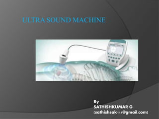
- 1. ULTRA SOUND MACHINE By SATHISHKUMAR G (sathishsak111@gmail.com)
- 2. HISTORY OF ULTRASOUND: In 1880 brothers JACQUES ET PIERRE curie noted that the electricity is created in the crystal of quartz under mechanical vibration. This phenomenon was described as the piezoelectric effect. Diagnosis medical application in use since late 1950’s like visualizing cerebral chamber.
- 4. Why ultrasound is used? ultrasound is not limited to diagnosis, but can also be used in screening for disease and to aid in treatment of diseases . another one is ultrasound is not expose any radiation.
- 5. PARTS OF ULTRASOUND MACHINE:
- 6. HOW DOES IT WORKS:
- 7. WHERE DO WE USE ULTRASOUND? Obstetrics liver gall bladder pancreas kidneys bladder prostate testicles uterus ovaries thyroid blood vessels brain ( in infants)
- 8. Modes of ultrasound: A- mode (amplitude) • The amplitude of reflected ultrasound is displayed. • It give one dimensional images only. • The A-mode is now used only in ophthalmology. M-mode (motion) • It reflects a motion of the heart structure over time. • Nowadays, 3D M-mode images is possible. • Accurate evaluation of rapid movements. B-mode (brightness) • An amplitude of reflected signal is converted into gray scale image . • 2D in echocardiography.
- 11. M- mode for heart scan
- 12. Doppler ultrasound: A Doppler ultrasound is a test that uses high- frequency sound waves to measure the amount of blood flow through your arteries and Veins, usually those that supply blood to your arms and lungs. IT MAY HELP DIAGNOSE MANY CONDITION, o Blood Clots o Venous Insufficiency o Congenital heart disease o Arterial Occlusion o peripheral artery disease o Carotid artery stenosis
- 14. 2d technology in imaging: It is the standard procedure used during Obstetric ultrasound. The 2D ultrasound imaging allows to view the baby in black & white technology. Traditional black & white is the most accurate Way to determine gender determination.
- 15. 2d images:
- 17. Clinical application of ultrasound: Abdominal ultrasound Gynaecology Obstetrics Echocardiography Small parts: thyroid, breast, scrotum musculoskeletal ultrasound
- 18. 1. Obstetric Ultrasound : Date the pregnancy Check the location of placenta Check for the number of fetes Check for physical abnormalities Check the sex of the baby Check for fetal movement, breathing, and heartbeat.
- 19. 2. Abdominal ultrasound: 1. To detect Gall stones 2. Determine the trauma in emergency 3. Fluid in the abdomen 4. Staging of neoplasis 5. Abdominal aneurysm 6. Bile duct disorders 7. Diffuse liver disease 8. Renal disorders
- 20. 3. Echocardiography Ultrasound: 1. Evaluate a heart murmur 2. Diagnose valve condition 3. Determine if fluid is collecting around the heart 4. Test blood flow through the heart. 5. Test heart function and diagnose heart and lung problem . 6. Look for blood clots within heart chambers.
- 21. 4. Musculoskeletal Ultrasound 1. To diagnose masses or fluid collections 2. To evaluate ligament sprains 3. Early changes of Rheumatoid Arthritis 4. Benign and Malignant soft tissue tumours. 5. Dislocation of the hip in infants 6. Neck muscle abnormalities in infants with torticollis.
- 22. Ultrasound scans involves no needless or injections. Less Expensive It uses no ionizing radiation. Give a clear picture of soft tissue It has been used to evaluate pregnancy It does not cause heath problems.
- 23. 1. Many cancers cannot be detected via an ultrasound. 2. An ultrasound requires a highly experienced and skilled operator to detect a malignant lump. 3. Ultrasound waves can heat the tissues slightly. 4. It can produce small pockets of gas in body fluids or tissues ( cavitations )