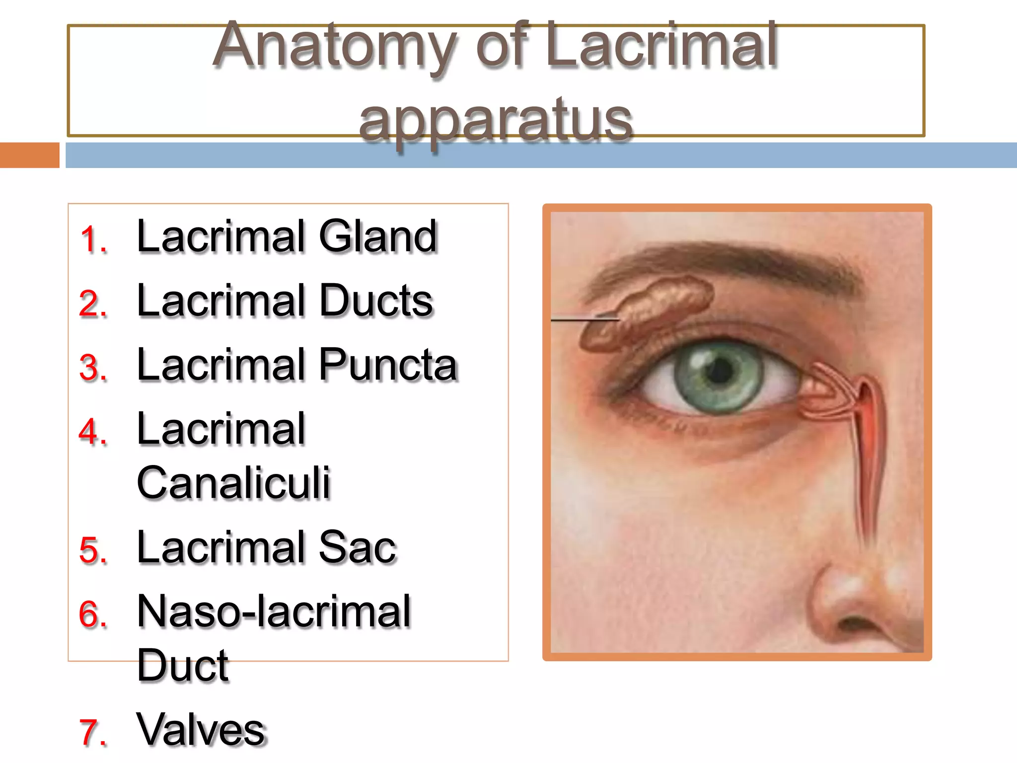
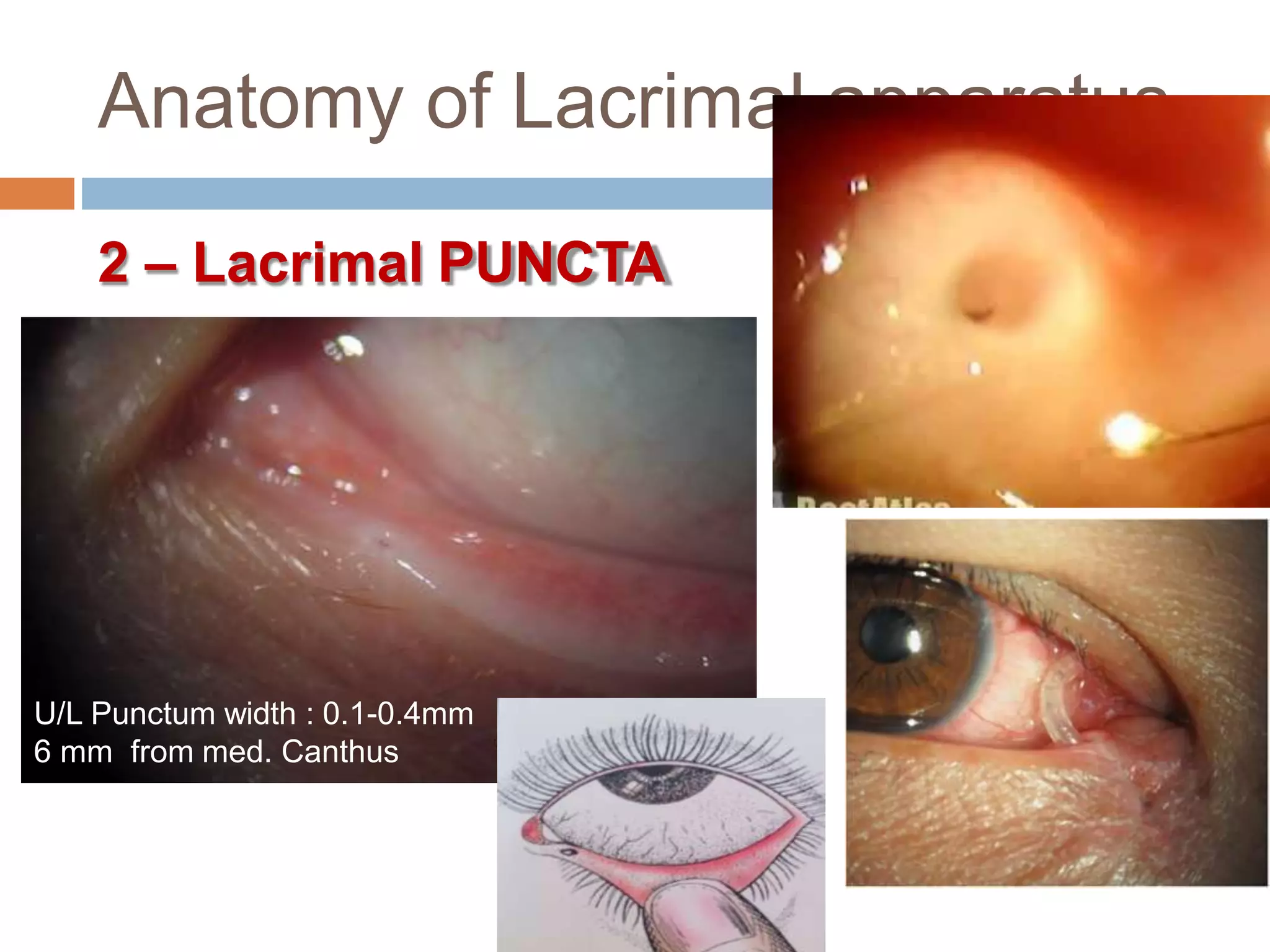
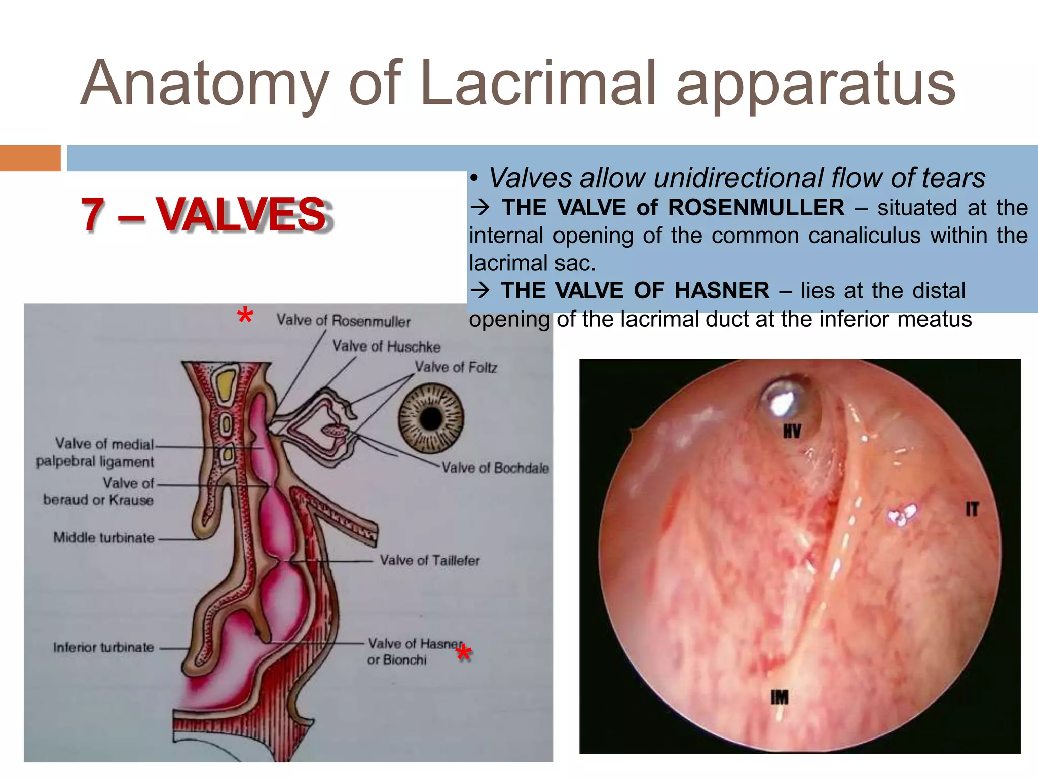
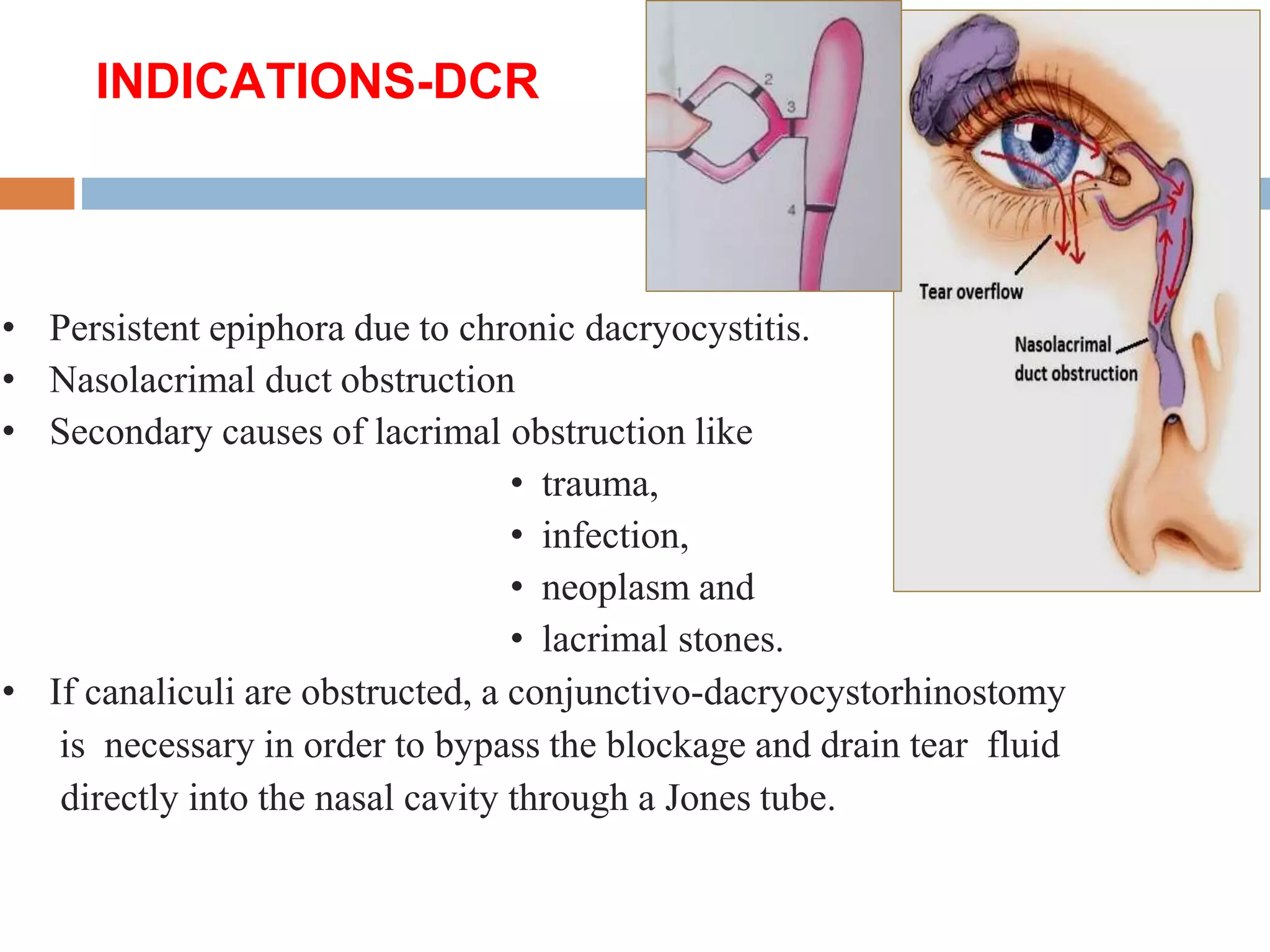
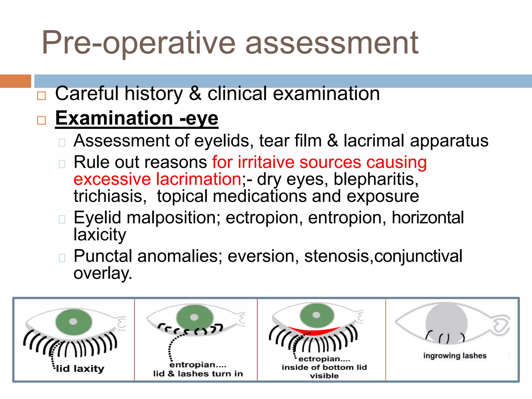
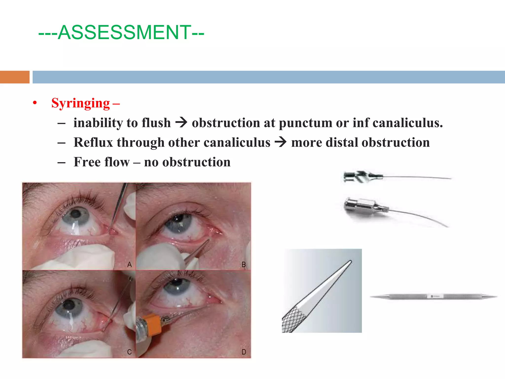
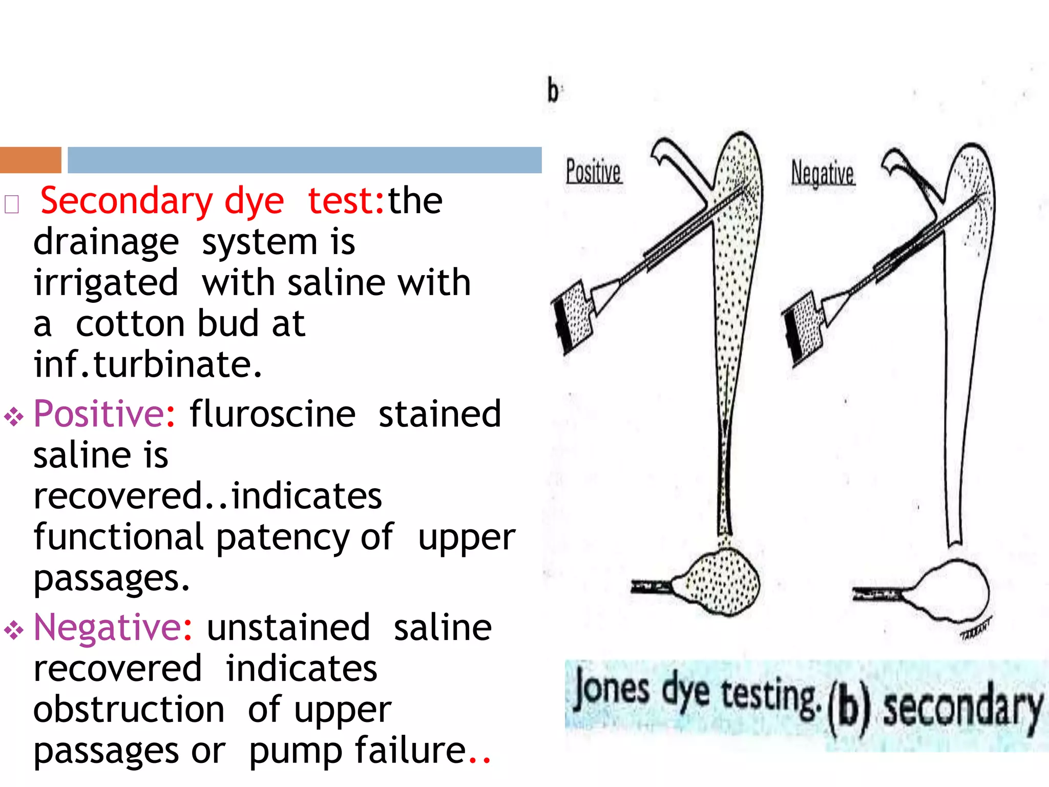
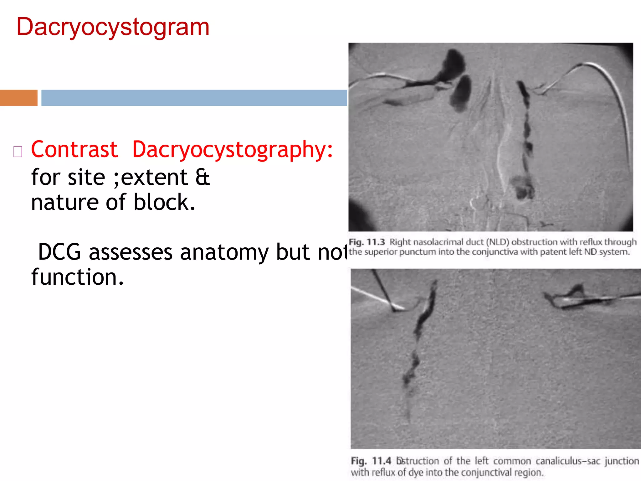
ENDOSCOPIC DCR
Anatomy of the
Lacrimal Apparatus
Anatomy of Lacrimal
apparatus
1. Lacrimal Gland
2. Lacrimal Ducts
3. Lacrimal Puncta
4. Lacrimal
Canaliculi
5. Lacrimal Sac
6. Naso-lacrimal
Duct
7. Valves
Anatomy of Lacrimal apparatus
1 – Lacrimal GLAND & Ducts
Anatomy of Lacrimal apparatus
2 – Lacrimal PUNCTA
U/L Punctum width : 0.1-0.4mm
6 mm from med. Canthus
Anatomy of Lacrimal apparatus
3 – Lacrimal CANALICULI Vertical part:2-2.5mm
Ampulla : V/H junction 90
Horizontal part : 7-10 mm
Common canaliculus : 1-3
mm
Lacrimal probing

Anatomy of Lacrimal apparatus
4 – The Lacrimal SAC
Lacrimal
sac
Anatomy of Lacrimal apparatus
4 – The Lacrimal SAC
Projection of Lacrimal system canal in the LNW of Rt. nasal
cavity.
L projection of the lacrimal system, MT middle turbinate, IT
inferior turbinate, ST superior turbinate
*
View through opening in the middle turbinate. FS frontal sinus, LC lamina
cribrosa, FP frontal process, UP uncinate process, HS hiatus semilunaris,
BE bulla ethmoidalis, MT middle turbinate, LD lacrimal duct, IT inferior
turbinate, SS sphenoid sinus
*
Sagittal view outlining the lacrimal sac (S) and duct (D).
Small arrows denote the common wall with the agger nasi cell.
Sagittal view with the lacrimal sac and duct marsupialized by completely
removing the medial bony and membranous wall.
(a) Lacrimal system,
Endoscopic view.
FP frontal process of maxilla,
ML maxillary line,
LB lacrimal bone.
*
(b) Dissection of the lacrimal system,
Endoscopic view.
FP frontal process of maxilla,
IT inferior turbinate, LD lacrimal duct,
LP lamina papyracea, S septum
UP uncinate process, MT middle turbinate,
Anatomy of Lacrimal apparatus
7 – VALVES
• Valves allow unidirectional flow of tears
 THE VALVE of ROSENMULLER – situated at the
internal opening of the common canaliculus within the
lacrimal sac.
 THE VALVE OF HASNER – lies at the distal
opening of the lacrimal duct at the inferior meatus
*
*
Epiphora
Epiphora --Obstrction is either anatomic (70%) or
functional (30%).
• This includes complete blockages anywhere from the
lacrimal punctum to the nasal cavity.
• Functional obstructions--result of either significant
narrowing within the lacrimal system that delays normal
lacrimal flow or a failure of the lacrimal pumping
mechanism.
Dacrocystorhinostomy (DCR) is performed to relieve
epiphora resulting from an obstruction of the nasolac-
rimal system.
The majority of nasolacrimal system obstruction is
unknown
History of DCR
• Caldwell – 1st intranasalDCR – 1893 – removed a portion
• of IT & followed NLD till the sac.
• Mosher – tried intranasal approach – 1921 – but later converted to
combined.
• Toti– First external DCR – 1940 – for relief of lacrimal obstruction.
• Advent of endoscope (1950s) – revived the intranasal approach – no scar
and excellent visualisation.
• Now, all DCRs performed with endoscope by ENT surgeon as 80% of
lacrimal pathway is nasal.
• Massaro – 1st reported use of laser (argon blue-green) to aid endonasal
DCR.
• Later Gonnering reported CO2 & KTP laser also.
INDICATIONS-DCR
• Persistent epiphora due to chronic dacryocystitis.
• Nasolacrimal duct obstruction
• Secondary causes of lacrimal obstruction like
• trauma,
• infection,
• neoplasm and
• lacrimal stones.
• If canaliculi are obstructed, a conjunctivo-dacryocystorhinostomy
is necessary in order to bypass the blockage and drain tear fluid
directly into the nasal cavity through a Jones tube.
 The key for a correct indication is to exclude a presaccal
stenosis, which is not suitable for an endoscopic procedure.
 The best method to assess the site of obstruction consists of
probing the lacrimal pathways: If it is possible to pass the
proximal canaliculi (superior and inferior) and to enter the
superior third of the lacrimal sac through the common
canaliculus, a presaccal obstruction can be excluded.
 External DCR is chosen over Endo-DCR
Trauma with medial canthal avulsion
Suspected lacrimal sac diverticuli
Lacrimal sac malignancy
Presacal obstruction
Pre-operative assessment
 Careful history & clinical examination
 Examination -eye
Assessment of eyelids, tear film & lacrimal apparatus
Rule out reasons for irritaive sources causing
excessive lacrimation;- dry eyes, blepharitis,
trichiasis, topical medications and exposure
Eyelid malposition; ectropion, entropion, horizontal
laxicity
Punctal anomalies; eversion, stenosis,conjunctival
overlay.
Epiphora
◆ Punctal stenosis. Obliteration or narrowing of the superior
or inferior punctum.
◆ Canalicular stenosis or obstruction. Superior or inferior
canalicular stenosis or obstruction may follow trauma or
viral infection.
◆ Nasolacrimal duct blockage. Usually from unknown cause.
---ASSESSMENT--
• Massaging or gentle pressure over sac –Regurgitation test
– If discharge from puncti  chronic dacryocystitis
– If bloody discharge  malignancy ?
• Swelling inferolateral to med canthus
 mucocoele
---ASSESSMENT--
• Syringing –
– inability to flush  obstruction at punctum or inf canaliculus.
– Reflux through other canaliculus  more distal obstruction
– Free flow – no obstruction
---ASSESSMENT--
• Probing with a ‘0’ bowman’s probe –
assist in confirming the level of obstruction.
Fluorescene dye test
Flourescein dye injected
into both conjuctval sacs &
observed for 2
minutes…normally no dye is
seen…
Prolonged retention
indicates obstruction to
lacrimal apparatus
Jones dye test
Primary test: a drop of 2%
fluoresceine is instilled into
conjunctiva..after 5 min.a
cotton bud is inserted under
inf.turbinate.
 Positive: Fluoresceine
recovered from nose
indicates patency of
drainage system.
negtive: no dye is
recovered ..indicates
partial obsruction or pump
failure
 Primary test differentiates
watering from partial
obstrctn from primary
hypersecretion of tears
Secondary dye test:the
drainage system is
irrigated with saline with
a cotton bud at
inf.turbinate.
 Positive: fluroscine stained
saline is
recovered..indicates
functional patency of upper
passages.
 Negative: unstained saline
recovered indicates
obstruction of upper
passages or pump failure..
Pre-operative assessment
 EXAMINATION –Nose
 Assessment of nasal cavity with nasal
speculum and endoscope.
 Identify pathologies like DNS/spur, AR, acute
infections, nasal polyps and malignancies etc.
 Treat any acute infection or severe allergic
rhinitis before surgery
Dacryocystogram
Contrast Dacryocystography:
for site ;extent &
nature of block.
DCG assesses anatomy but not
function.
Lacrimal Scintillography
Lacrimal
scintillography:
detects functional
efficiency of lacrimal
apparatus (Syringing
will be normal)-- Using
radionucleotide 99mTc
(detected using
gamma camera)
Operative Techniques
1. External DCR
2. Endoscopic DCR
3. Endonasal laser assisted DCR
Pre-operative considerations
 Surgery is performed under general anesthesia
(hypotensive anesthesia)/LA
 The nose is prepared with cottonoids soaked in
xylocaine & adrenaline .
Infiltrate 2% Xylocaine with adrenaline into the axilla
of the middle turbinate and frontal process of maxilla
 Avoid unnecessary manipulation of endoscope and
instruments during packing, avoid mucosal trauma
esp. MT
 A septoplasty is performed in case of an
obstructing septal deviation.
 The septal incision is ideally placed on the side
contralateral to the DCR:
This prevents inadvertent trauma to the septal
flap when the endoscope is inserted into the
nasal cavity.
It minimizes clouding of the endoscope with blood
from the septal incision.
Reduces the potential for the development of
postoperative synechiae between the septum and
LNW.
Instruments needed for
Endoscopic-DCR
 0 and 30 degree
endoscopes
 Light
source/Camera/monitor
 Suction
 No. 15 surgical blade
 Pointed diathermy
 Plester knife
 Rosen’s knife
 Sickle knife
 House (meatal) elevator
 Suction elevator, Freer’s
 Kerrison bone punch
 Hajek-Kofler punch
 Blakesely forceps
 Thru-cut forceps
 Ball probe
 Lacrimal probe
 Punctal dilator
 Lacrimal syringe
steps
• Incision- in the mucosa overlying ant lacrimal
crest (white vertical ridge immediately ant to
MT)
• Posteriorly based muco-perichondrial flap raised
• Ant lacrimal crest removed using a punch
• Post to ant lacrimal crest & just lat to UP lies thin
bone – resected
• Sac is exposed, has a dark red colour & firmer than nasal mucosa
• Sac divided vertically with either sickle knife or 45 degree beaver scalpel
• Aprobe placed within the sac, tenting it medially, facilitates incision
• Microscissors used sup & inf to create ant & post flaps
• These flaps of sac mucosa are then placed in continuity with nasal
mucosa
• Astent can be inserted if required.
• Adv - Lacrimal sac is widely exposed, common canaliculus can often be
seen
Inspection and
Identification of landmarks
Inspection and
Identification of landmarks
B
UP
Dimensions of the nasal mucosal flap
Dimensions of lacrimal sac/flap
The lacrimal sac extends approx.
10 mm above the axilla of MT.
Topical and local anesthesia
Perform a septoplasty if needed:
Limited access restricts surg.
Incision given by #15 blade
Elevation of flap
Removal of flakes of Lacrimal bone
Using Sickle and Rosen knifes
The lacrimal bone extends from the FP of maxilla anteriorly
to the attachment of the uncinate process posteriorly.
This retrolacrimal region of the lamina papyracea is
extremely thin, and inadvertent disturbance of the uncinate at
this point can lead to orbital penetration.
Remember that the lacrimal bone and sac lie anterior to the
orbit, and therefore the orbit is not at risk unless the surgeon
is inadvertently posterior to these landmarks.
Bone removal
using
Kerrison forceps
or
Hajek-Kofler punch
Kerrison forceps
Bone-punch
Hajek-Kofler punch
The Hajek-Koffler punch is faster at removing
bone than the DCR bur : Perform as much of the
removal of the hard bone of the frontal process of
the maxilla with the Hajek-Koffler punch
Superior bone removal
 When using the Hajek-Koffler punch,
release the jaws after each bite : this will
prevent inadvertent trauma to sac.
Use of DIAMOND BURR for bone removal
 Use diamond burr only when the punch
is unable to grip the bone adequately.
NLD
Fl
AN
LS
Very adequately exposed LS & NLD
Incising flap
using KERATOME
Tenting the sac
using lacrimal probe
Make an incision into the sac only when lacrimal
probe can be clearly seen through the sac wall.
When probing the lacrimal system, do so
delicately : avoid trauma and a false passage.
Sac completely opened
Flood of pus
f
Flap opened using 15 blade
Flap marsupialized and gelfoam placed
The common canaliculus opens
high up on the lateral wall of the
sac, and this area must be
exposed in DCR for best results.
Use of DCR tube
 Working as a team with an
oculoplastic surgeon : they have
requisite skills in probing and
examining the lacrimal system.
Causes of failure of DCR
 Inadequate osteotomy,
 Incomplete sac marsupialization,
 Cicatricial closure of the ostium
 Granuloma formation
Postoperative Care
•Saline nasal spray - within 3 to 4 hours of surgery.
clear blood clots
keep the nasal cavity moist
clear of secretions
•Avoid blowing of the nose
• Broad-spectrum antibiotics for 7 days
• Antibiotic eye drops for 3 weeks.
• If O’Donoghue tubes were placed, they are removed after 4
weeks
Patency of the nasolacrimal system checkedby placing a drop
of fluorescein in the conjunctiva and endoscopically monitoring
the flow of fluorescein from the conjunctiva to the nose.
• If granulations are present they should be removed.
Complications
• Epistaxis, occurring in 2% of patients
• obstruction and subsequent rhinosinusitis of the frontal or
maxillary
sinus,
• orbital penetration with damage to the extraocular muscles,
• orbital haematoma
• cerebrospinal fluid leak .
Advantages of Endoscopic
DCR
 It provides better aesthetic result with no external
scar.
 It allows a one-stage procedure to also correct
associated nasal pathology that may be causative.
 It avoids injury to the medial canthus /scar formation.
 It preserves the pumping mechanism of the orbicularis
oculi ms..
 Active infection of the lacrimal system is not a
contraindication to endoscopic surgery.
 It is superior to the external approach in revision
surgery.
 It is much less bloody than the external
Comparison of the 3 techniques of
DCR
T
THANK YOU

Endoscopic DCR

  • 1.
  • 2.
  • 3.
    Anatomy of Lacrimal apparatus 1.Lacrimal Gland 2. Lacrimal Ducts 3. Lacrimal Puncta 4. Lacrimal Canaliculi 5. Lacrimal Sac 6. Naso-lacrimal Duct 7. Valves
  • 4.
    Anatomy of Lacrimalapparatus 1 – Lacrimal GLAND & Ducts
  • 5.
    Anatomy of Lacrimalapparatus 2 – Lacrimal PUNCTA U/L Punctum width : 0.1-0.4mm 6 mm from med. Canthus
  • 6.
    Anatomy of Lacrimalapparatus 3 – Lacrimal CANALICULI Vertical part:2-2.5mm Ampulla : V/H junction 90 Horizontal part : 7-10 mm Common canaliculus : 1-3 mm
  • 7.
  • 8.
    Anatomy of Lacrimalapparatus 4 – The Lacrimal SAC Lacrimal sac
  • 9.
    Anatomy of Lacrimalapparatus 4 – The Lacrimal SAC
  • 10.
    Projection of Lacrimalsystem canal in the LNW of Rt. nasal cavity. L projection of the lacrimal system, MT middle turbinate, IT inferior turbinate, ST superior turbinate *
  • 11.
    View through openingin the middle turbinate. FS frontal sinus, LC lamina cribrosa, FP frontal process, UP uncinate process, HS hiatus semilunaris, BE bulla ethmoidalis, MT middle turbinate, LD lacrimal duct, IT inferior turbinate, SS sphenoid sinus *
  • 12.
    Sagittal view outliningthe lacrimal sac (S) and duct (D). Small arrows denote the common wall with the agger nasi cell.
  • 13.
    Sagittal view withthe lacrimal sac and duct marsupialized by completely removing the medial bony and membranous wall.
  • 15.
    (a) Lacrimal system, Endoscopicview. FP frontal process of maxilla, ML maxillary line, LB lacrimal bone. * (b) Dissection of the lacrimal system, Endoscopic view. FP frontal process of maxilla, IT inferior turbinate, LD lacrimal duct, LP lamina papyracea, S septum UP uncinate process, MT middle turbinate,
  • 16.
    Anatomy of Lacrimalapparatus 7 – VALVES • Valves allow unidirectional flow of tears  THE VALVE of ROSENMULLER – situated at the internal opening of the common canaliculus within the lacrimal sac.  THE VALVE OF HASNER – lies at the distal opening of the lacrimal duct at the inferior meatus * *
  • 18.
    Epiphora Epiphora --Obstrction iseither anatomic (70%) or functional (30%). • This includes complete blockages anywhere from the lacrimal punctum to the nasal cavity. • Functional obstructions--result of either significant narrowing within the lacrimal system that delays normal lacrimal flow or a failure of the lacrimal pumping mechanism. Dacrocystorhinostomy (DCR) is performed to relieve epiphora resulting from an obstruction of the nasolac- rimal system. The majority of nasolacrimal system obstruction is unknown
  • 19.
    History of DCR •Caldwell – 1st intranasalDCR – 1893 – removed a portion • of IT & followed NLD till the sac. • Mosher – tried intranasal approach – 1921 – but later converted to combined. • Toti– First external DCR – 1940 – for relief of lacrimal obstruction. • Advent of endoscope (1950s) – revived the intranasal approach – no scar and excellent visualisation. • Now, all DCRs performed with endoscope by ENT surgeon as 80% of lacrimal pathway is nasal. • Massaro – 1st reported use of laser (argon blue-green) to aid endonasal DCR. • Later Gonnering reported CO2 & KTP laser also.
  • 20.
    INDICATIONS-DCR • Persistent epiphoradue to chronic dacryocystitis. • Nasolacrimal duct obstruction • Secondary causes of lacrimal obstruction like • trauma, • infection, • neoplasm and • lacrimal stones. • If canaliculi are obstructed, a conjunctivo-dacryocystorhinostomy is necessary in order to bypass the blockage and drain tear fluid directly into the nasal cavity through a Jones tube.
  • 22.
     The keyfor a correct indication is to exclude a presaccal stenosis, which is not suitable for an endoscopic procedure.  The best method to assess the site of obstruction consists of probing the lacrimal pathways: If it is possible to pass the proximal canaliculi (superior and inferior) and to enter the superior third of the lacrimal sac through the common canaliculus, a presaccal obstruction can be excluded.  External DCR is chosen over Endo-DCR Trauma with medial canthal avulsion Suspected lacrimal sac diverticuli Lacrimal sac malignancy Presacal obstruction
  • 23.
    Pre-operative assessment  Carefulhistory & clinical examination  Examination -eye Assessment of eyelids, tear film & lacrimal apparatus Rule out reasons for irritaive sources causing excessive lacrimation;- dry eyes, blepharitis, trichiasis, topical medications and exposure Eyelid malposition; ectropion, entropion, horizontal laxicity Punctal anomalies; eversion, stenosis,conjunctival overlay.
  • 24.
    Epiphora ◆ Punctal stenosis.Obliteration or narrowing of the superior or inferior punctum. ◆ Canalicular stenosis or obstruction. Superior or inferior canalicular stenosis or obstruction may follow trauma or viral infection. ◆ Nasolacrimal duct blockage. Usually from unknown cause.
  • 25.
    ---ASSESSMENT-- • Massaging orgentle pressure over sac –Regurgitation test – If discharge from puncti  chronic dacryocystitis – If bloody discharge  malignancy ? • Swelling inferolateral to med canthus  mucocoele
  • 26.
    ---ASSESSMENT-- • Syringing – –inability to flush  obstruction at punctum or inf canaliculus. – Reflux through other canaliculus  more distal obstruction – Free flow – no obstruction
  • 27.
    ---ASSESSMENT-- • Probing witha ‘0’ bowman’s probe – assist in confirming the level of obstruction.
  • 28.
    Fluorescene dye test Flouresceindye injected into both conjuctval sacs & observed for 2 minutes…normally no dye is seen… Prolonged retention indicates obstruction to lacrimal apparatus
  • 29.
    Jones dye test Primarytest: a drop of 2% fluoresceine is instilled into conjunctiva..after 5 min.a cotton bud is inserted under inf.turbinate.  Positive: Fluoresceine recovered from nose indicates patency of drainage system. negtive: no dye is recovered ..indicates partial obsruction or pump failure  Primary test differentiates watering from partial obstrctn from primary hypersecretion of tears
  • 30.
    Secondary dye test:the drainagesystem is irrigated with saline with a cotton bud at inf.turbinate.  Positive: fluroscine stained saline is recovered..indicates functional patency of upper passages.  Negative: unstained saline recovered indicates obstruction of upper passages or pump failure..
  • 31.
    Pre-operative assessment  EXAMINATION–Nose  Assessment of nasal cavity with nasal speculum and endoscope.  Identify pathologies like DNS/spur, AR, acute infections, nasal polyps and malignancies etc.  Treat any acute infection or severe allergic rhinitis before surgery
  • 32.
    Dacryocystogram Contrast Dacryocystography: for site;extent & nature of block. DCG assesses anatomy but not function.
  • 33.
    Lacrimal Scintillography Lacrimal scintillography: detects functional efficiencyof lacrimal apparatus (Syringing will be normal)-- Using radionucleotide 99mTc (detected using gamma camera)
  • 34.
    Operative Techniques 1. ExternalDCR 2. Endoscopic DCR 3. Endonasal laser assisted DCR
  • 35.
    Pre-operative considerations  Surgeryis performed under general anesthesia (hypotensive anesthesia)/LA  The nose is prepared with cottonoids soaked in xylocaine & adrenaline . Infiltrate 2% Xylocaine with adrenaline into the axilla of the middle turbinate and frontal process of maxilla  Avoid unnecessary manipulation of endoscope and instruments during packing, avoid mucosal trauma esp. MT
  • 36.
     A septoplastyis performed in case of an obstructing septal deviation.  The septal incision is ideally placed on the side contralateral to the DCR: This prevents inadvertent trauma to the septal flap when the endoscope is inserted into the nasal cavity. It minimizes clouding of the endoscope with blood from the septal incision. Reduces the potential for the development of postoperative synechiae between the septum and LNW.
  • 37.
    Instruments needed for Endoscopic-DCR 0 and 30 degree endoscopes  Light source/Camera/monitor  Suction  No. 15 surgical blade  Pointed diathermy  Plester knife  Rosen’s knife  Sickle knife  House (meatal) elevator  Suction elevator, Freer’s  Kerrison bone punch  Hajek-Kofler punch  Blakesely forceps  Thru-cut forceps  Ball probe  Lacrimal probe  Punctal dilator  Lacrimal syringe
  • 38.
    steps • Incision- inthe mucosa overlying ant lacrimal crest (white vertical ridge immediately ant to MT) • Posteriorly based muco-perichondrial flap raised • Ant lacrimal crest removed using a punch • Post to ant lacrimal crest & just lat to UP lies thin bone – resected • Sac is exposed, has a dark red colour & firmer than nasal mucosa • Sac divided vertically with either sickle knife or 45 degree beaver scalpel • Aprobe placed within the sac, tenting it medially, facilitates incision • Microscissors used sup & inf to create ant & post flaps • These flaps of sac mucosa are then placed in continuity with nasal mucosa • Astent can be inserted if required. • Adv - Lacrimal sac is widely exposed, common canaliculus can often be seen
  • 39.
  • 40.
  • 42.
    Dimensions of thenasal mucosal flap
  • 43.
    Dimensions of lacrimalsac/flap The lacrimal sac extends approx. 10 mm above the axilla of MT.
  • 44.
    Topical and localanesthesia Perform a septoplasty if needed: Limited access restricts surg.
  • 45.
  • 46.
  • 48.
    Removal of flakesof Lacrimal bone Using Sickle and Rosen knifes
  • 49.
    The lacrimal boneextends from the FP of maxilla anteriorly to the attachment of the uncinate process posteriorly. This retrolacrimal region of the lamina papyracea is extremely thin, and inadvertent disturbance of the uncinate at this point can lead to orbital penetration. Remember that the lacrimal bone and sac lie anterior to the orbit, and therefore the orbit is not at risk unless the surgeon is inadvertently posterior to these landmarks.
  • 50.
  • 51.
    Kerrison forceps Bone-punch Hajek-Kofler punch TheHajek-Koffler punch is faster at removing bone than the DCR bur : Perform as much of the removal of the hard bone of the frontal process of the maxilla with the Hajek-Koffler punch
  • 52.
    Superior bone removal When using the Hajek-Koffler punch, release the jaws after each bite : this will prevent inadvertent trauma to sac.
  • 53.
    Use of DIAMONDBURR for bone removal  Use diamond burr only when the punch is unable to grip the bone adequately.
  • 54.
  • 58.
    Incising flap using KERATOME Tentingthe sac using lacrimal probe Make an incision into the sac only when lacrimal probe can be clearly seen through the sac wall.
  • 59.
    When probing thelacrimal system, do so delicately : avoid trauma and a false passage.
  • 60.
  • 62.
    f Flap opened using15 blade Flap marsupialized and gelfoam placed The common canaliculus opens high up on the lateral wall of the sac, and this area must be exposed in DCR for best results.
  • 65.
    Use of DCRtube  Working as a team with an oculoplastic surgeon : they have requisite skills in probing and examining the lacrimal system.
  • 66.
    Causes of failureof DCR  Inadequate osteotomy,  Incomplete sac marsupialization,  Cicatricial closure of the ostium  Granuloma formation
  • 67.
    Postoperative Care •Saline nasalspray - within 3 to 4 hours of surgery. clear blood clots keep the nasal cavity moist clear of secretions •Avoid blowing of the nose • Broad-spectrum antibiotics for 7 days • Antibiotic eye drops for 3 weeks. • If O’Donoghue tubes were placed, they are removed after 4 weeks Patency of the nasolacrimal system checkedby placing a drop of fluorescein in the conjunctiva and endoscopically monitoring the flow of fluorescein from the conjunctiva to the nose. • If granulations are present they should be removed.
  • 68.
    Complications • Epistaxis, occurringin 2% of patients • obstruction and subsequent rhinosinusitis of the frontal or maxillary sinus, • orbital penetration with damage to the extraocular muscles, • orbital haematoma • cerebrospinal fluid leak .
  • 69.
    Advantages of Endoscopic DCR It provides better aesthetic result with no external scar.  It allows a one-stage procedure to also correct associated nasal pathology that may be causative.  It avoids injury to the medial canthus /scar formation.  It preserves the pumping mechanism of the orbicularis oculi ms..  Active infection of the lacrimal system is not a contraindication to endoscopic surgery.  It is superior to the external approach in revision surgery.  It is much less bloody than the external
  • 71.
    Comparison of the3 techniques of DCR
  • 72.