Downloaded 28 times


![Revision
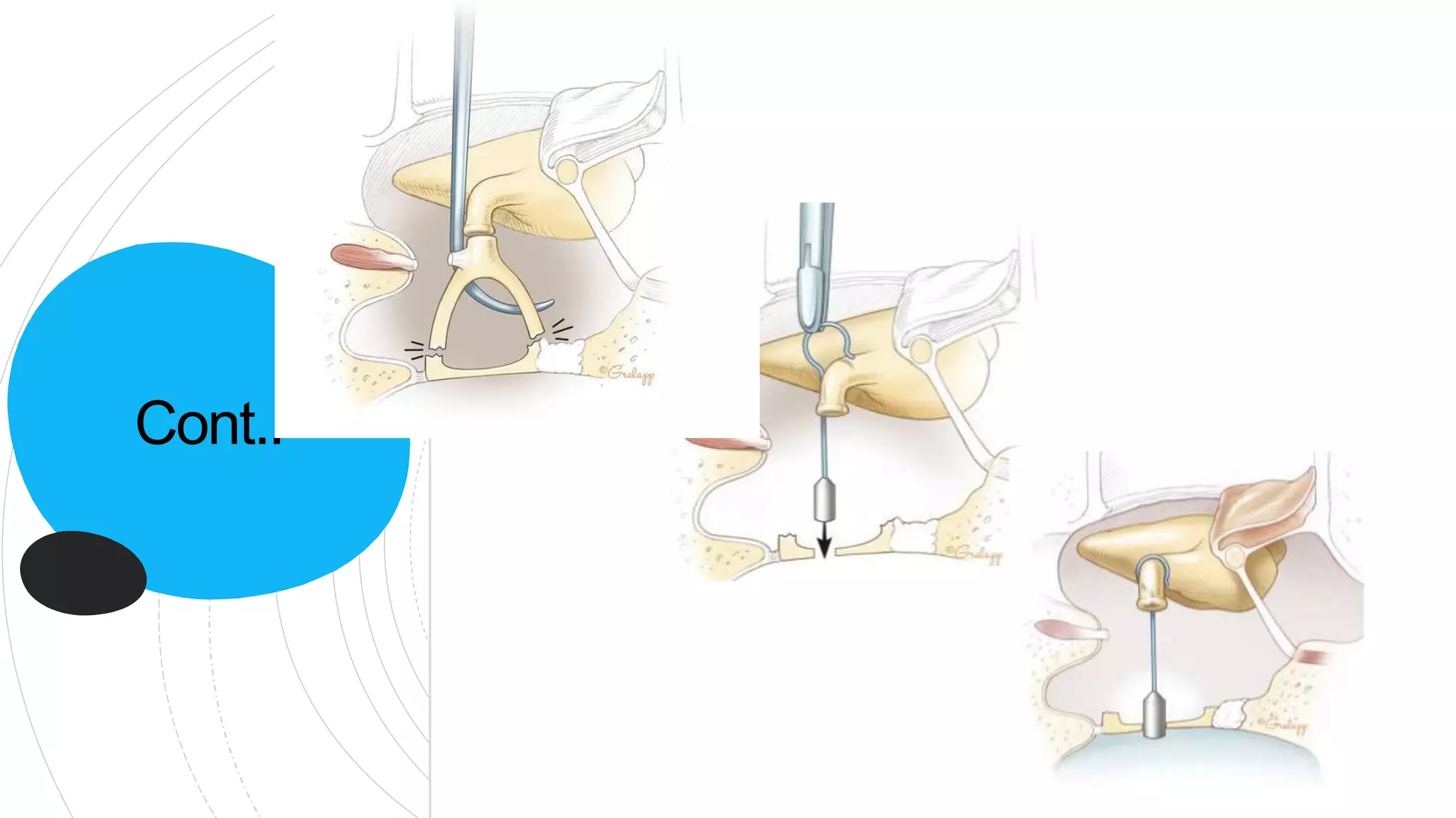
stapes
surgery
Prosthesis dislocation from oval window
Incus erosion and incus-prosthesis detachment
Short prosthesis
Postoperative fibrosis in the middle ear and re-ankyloses
Peri-lymphatic fistula
Insufficient fenestra and too tight prosthesis
Footplate re-sclerosis, incus subluxation, facial nerve
dehiscence and prosthesis friction, reparative granuloma
Yetiser S. Revision surgery for otosclerosis: An overview. World J Otorhinolaryngol 2015; 5(1): 21-29 [DOI:
10.5319/wjo.v5.i1.21]
Common causes](https://image.slidesharecdn.com/complicationsofstapessurgery-220405190623/75/Complications-of-Stapes-surgery-pptx-43-2048.jpg)


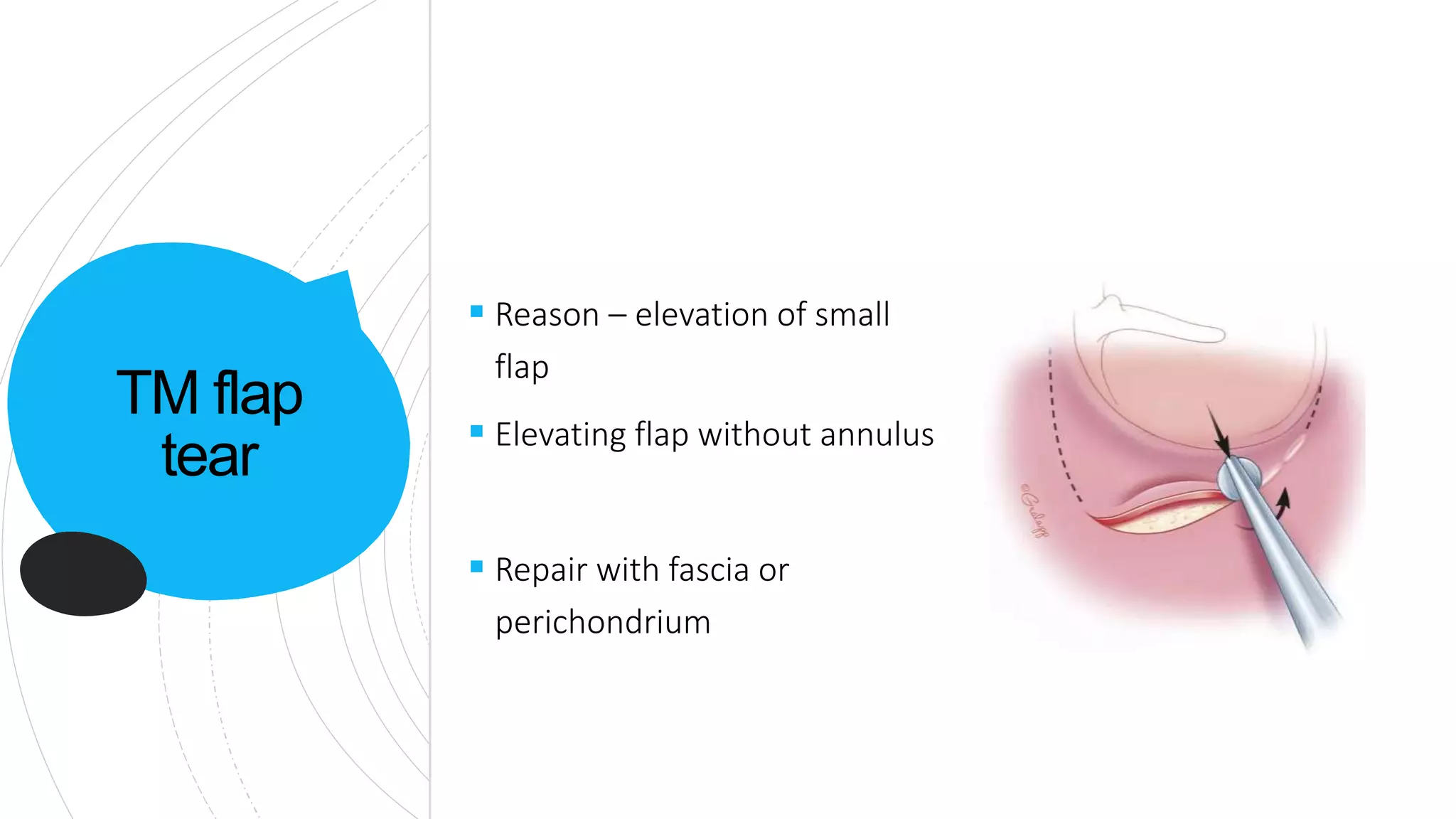
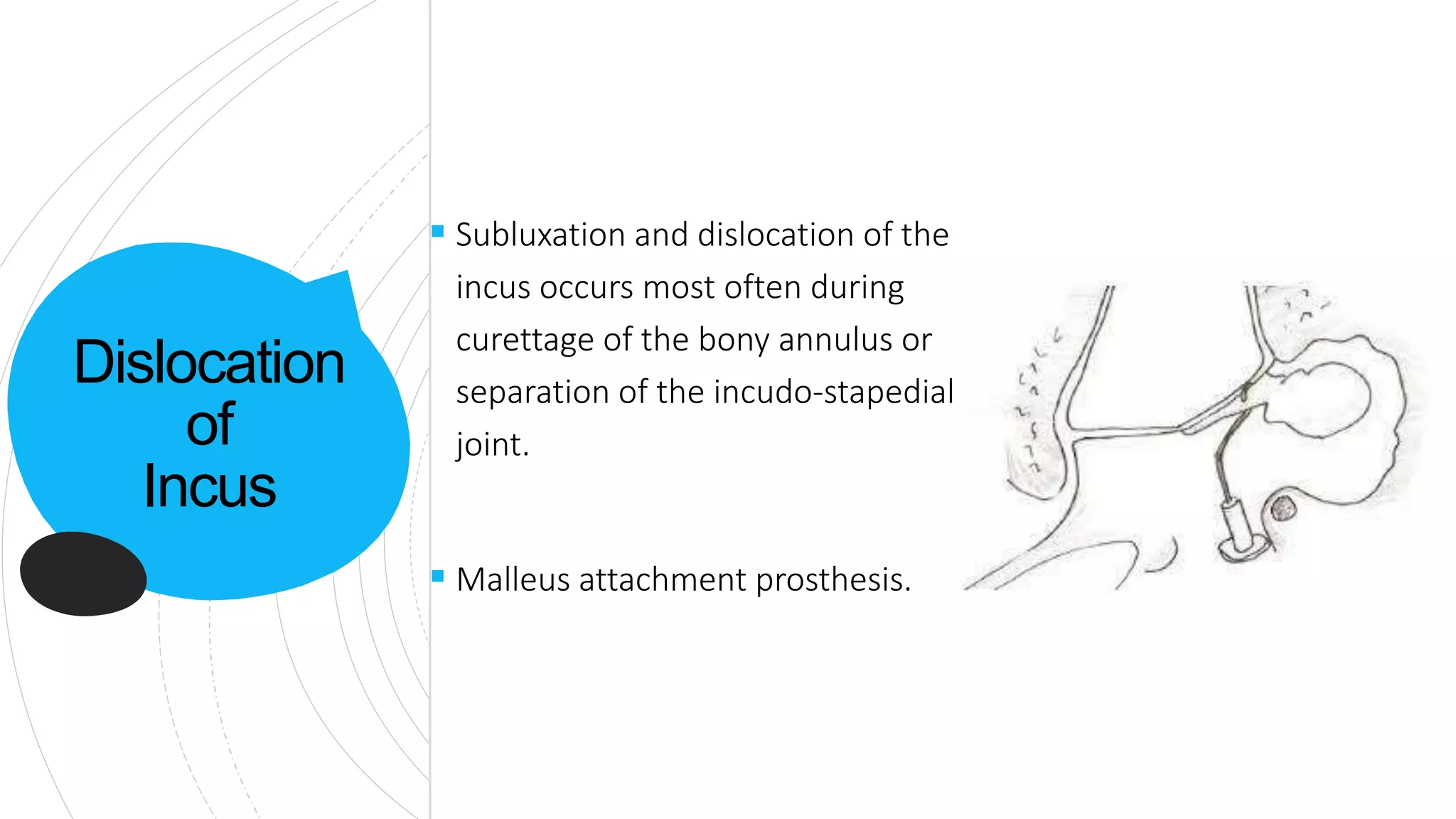
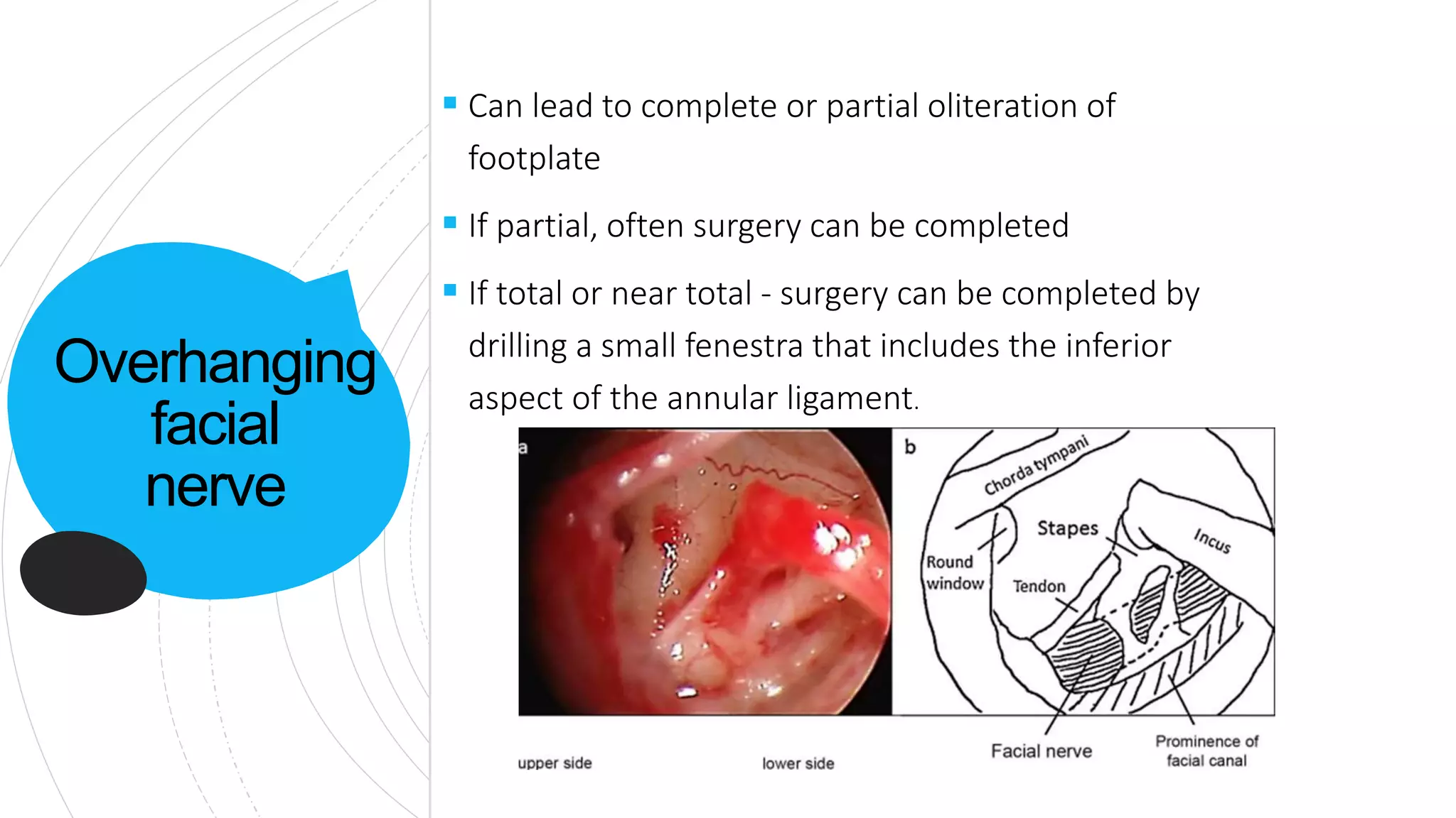
The document discusses complications that can occur with stapes surgery for otosclerosis. Some common intraoperative complications include tears in the tympanic membrane flap, dislocation of the incus, and overhanging of the facial nerve. Postoperative complications include otitis media, vertigo, facial palsy, sensorineural hearing loss, conductive hearing loss, and reparative granulomas. Rare complications include cerebrospinal fluid leaks. Revision stapes surgery carries higher risks and lower success rates than initial surgery due to postoperative fibrosis and other challenges.


