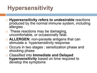
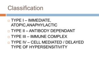
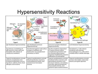
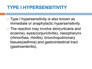
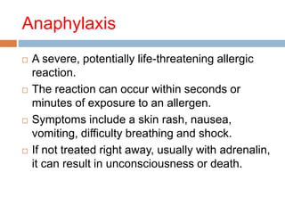
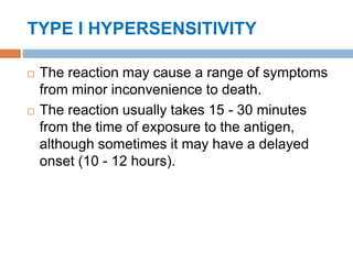
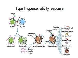
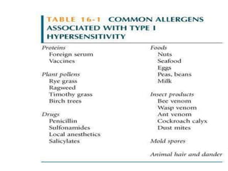
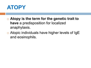
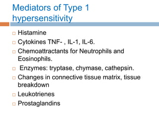
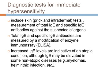
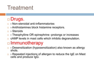
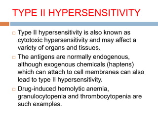
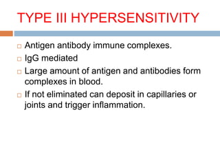
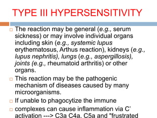
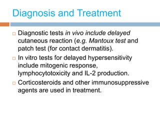
HYPERSENSITIVITY
DR.PRINCE C P
HOD & Associate Professor
Department of Microbiology
Mother Theresa Post Graduate & Research Institute of Health Sciences
(Government of Puducherry Institution)
Pondicherry
Hypersensitivity
 Hypersensitivity refers to undesirable reactions
produced by the normal immune system, including
allergies .
 These reactions may be damaging,
uncomfortable, or occasionally fatal.
 ALLERGEN: non-parasite antigens that can
stimulate a hypersensitivity response
 Occurs in two stages : sensitization phase and
shocking phase
 Classified into Immediate and Delayed
hypersensitivity based on time required to
develop the symptoms
Classification
 TYPE I – IMMEDIATE,
ATOPIC,ANAPHYLACTIC
 TYPE II – ANTIBODY DEPENDANT
 TYPE III – IMMUNE COMPLEX
 TYPE IV – CELL MEDIATED / DELAYED
TYPE OF HYPERSENSITIVITY
TYPE I HYPERSENSITIVITY
 Type I hypersensitivity is also known as
immediate or anaphylactic hypersensitivity.
 The reaction may involve skin(urticaria and
eczema), eyes(conjunctivitis), nasopharynx
(rhinorrhea, rhinitis), bronchopulmonary
tissues(asthma) and gastrointestinal tract
(gastroenteritis).
Anaphylaxis
 A severe, potentially life-threatening allergic
reaction.
 The reaction can occur within seconds or
minutes of exposure to an allergen.
 Symptoms include a skin rash, nausea,
vomiting, difficulty breathing and shock.
 If not treated right away, usually with adrenalin,
it can result in unconsciousness or death.
Angioedema of the face
TYPE I HYPERSENSITIVITY
 The reaction may cause a range of symptoms
from minor inconvenience to death.
 The reaction usually takes 15 - 30 minutes
from the time of exposure to the antigen,
although sometimes it may have a delayed
onset (10 - 12 hours).
TYPE I HYPERSENSITIVITY
 Mediated by IgE antibody to specific antigens
 The primary cellular component in this
hypersensitivity is the mast cell or basophil.
 The reaction is amplified and/or modified by
platelets, neutrophils and eosinophils.
 Mast cells stimulated and release histamine
ATOPY
 Atopy is the term for the genetic trait to
have a predisposition for localized
anaphylaxis.
 Atopic individuals have higher levels of IgE
and eosinophils.
Mediators of Type 1
hypersensitivity
 Histamine
 Cytokines TNF- , IL-1, IL-6.
 Chemoattractants for Neutrophils and
Eosinophils.
 Enzymes: tryptase, chymase, cathepsin.
 Changes in connective tissue matrix, tissue
breakdown
 Leukotrienes
 Prostaglandins
Diagnostic tests for immediate
hypersensitivity
 include skin (prick and intradermal) tests ,
measurement of total IgE and specific IgE
antibodies against the suspected allergens.
 Total IgE and specific IgE antibodies are
measured by a modification of enzyme
immunoassay (ELISA).
 Increased IgE levels are indicative of an atopic
condition, although IgE may be elevated in
some non-atopic diseases (e.g.,myelomas,
helminthic infection, etc.).
Treatment
 Drugs.
 › Non-steroidal anti-inflammatories
 › Antihistamines block histamine receptors.
 › Steroids
 › Theophylline OR epinephrine -prolongs or increases
 cAMP levels in mast cells which inhibits degranulation.
 Immunotherapy
 › Desensitization (hyposensitization) also known as allergy
shots.
 › Repeated injections of allergen to reduce the IgE on Mast
cells and produce IgG.
TYPE II HYPERSENSITIVITY
 Type II hypersensitivity is also known as
cytotoxic hypersensitivity and may affect a
variety of organs and tissues.
 The antigens are normally endogenous,
although exogenous chemicals (haptens)
which can attach to cell membranes can also
lead to type II hypersensitivity.
 Drug-induced hemolytic anemia,
granulocytopenia and thrombocytopenia are
such examples.
TYPE III HYPERSENSITIVITY
 Antigen antibody immune complexes.
 IgG mediated
 Large amount of antigen and antibodies form
complexes in blood.
 If not eliminated can deposit in capillaries or
joints and trigger inflammation.
TYPE III HYPERSENSITIVITY
 The reaction may be general (e.g., serum
sickness) or may involve individual organs
including skin (e.g., systemic lupus
erythematosus, Arthus reaction), kidneys (e.g.,
lupus nephritis), lungs (e.g., aspergillosis),
joints (e.g., rheumatoid arthritis) or other
organs.
 This reaction may be the pathogenic
mechanism of diseases caused by many
microorganisms.
 If unable to phagocytize the immune
 complexes can cause inflammation via C’
activation ---> C3a C4a, C5a and "frustrated
 Reaction involves sensitized T cells and
release of its lymphokines as mediators and
amplifiers
 Mediated by cells rather than antibodies
 Clinical states: Contact dermatitis,
Transplant rejection, Granuloma
 T cells release cytokines to activate
macrophages causing inflammation and tissue
damage.
 Continued macrophage activation can cause
chronic inflammation resulting in tissue
lesions,scarring, and granuloma formation.
 Response starts after 48 -72 hrs
Diagnosis and Treatment
 Diagnostic tests in vivo include delayed
cutaneous reaction (e.g. Mantoux test and
patch test (for contact dermatitis).
 In vitro tests for delayed hypersensitivity
include mitogenic response,
lymphocytotoxicity and IL-2 production.
 Corticosteroids and other immunosuppressive
agents are used in treatment.
Tuberculin test
 Tuberculin test is a delayed hypersensitivity (Type
IV) to intradermal injection of purified protein
derivative (PPD) of Tuberculin, to test for cell
mediated immunity against Mycobacteria.
 The tuberculin test is based on the fact that
infection with M. tuberculosis produces sensitivity
to certain antigenic components of the organism
that are contained in culture extracts called
"tuberculins."
 Hypersensitivity to tuberculin, which is
demonstrated by the development of a positive
reaction to the tuberculin skin test, develops 2 to
Tuberculin test
 The Mantoux test or Mendel–Mantoux
test (also known as the Mantoux screening
test, tuberculin sensitivity test, Pirquet test,
or PPD test for purified protein derivative) is a
tool for screening for tuberculosis (TB) and
for tuberculosis diagnosis. It is one of the
major tuberculin skin tests used around the
world, largely replacing multiple-puncture tests
such as the Tine test and The Heaf test
Tuberculin test
Thank you

Hypersensitivity/ Allergy ppt by DR.C.P.PRINCE

  • 1.
    HYPERSENSITIVITY DR.PRINCE C P HOD& Associate Professor Department of Microbiology Mother Theresa Post Graduate & Research Institute of Health Sciences (Government of Puducherry Institution) Pondicherry
  • 2.
    Hypersensitivity  Hypersensitivity refersto undesirable reactions produced by the normal immune system, including allergies .  These reactions may be damaging, uncomfortable, or occasionally fatal.  ALLERGEN: non-parasite antigens that can stimulate a hypersensitivity response  Occurs in two stages : sensitization phase and shocking phase  Classified into Immediate and Delayed hypersensitivity based on time required to develop the symptoms
  • 3.
    Classification  TYPE I– IMMEDIATE, ATOPIC,ANAPHYLACTIC  TYPE II – ANTIBODY DEPENDANT  TYPE III – IMMUNE COMPLEX  TYPE IV – CELL MEDIATED / DELAYED TYPE OF HYPERSENSITIVITY
  • 5.
    TYPE I HYPERSENSITIVITY Type I hypersensitivity is also known as immediate or anaphylactic hypersensitivity.  The reaction may involve skin(urticaria and eczema), eyes(conjunctivitis), nasopharynx (rhinorrhea, rhinitis), bronchopulmonary tissues(asthma) and gastrointestinal tract (gastroenteritis).
  • 6.
    Anaphylaxis  A severe,potentially life-threatening allergic reaction.  The reaction can occur within seconds or minutes of exposure to an allergen.  Symptoms include a skin rash, nausea, vomiting, difficulty breathing and shock.  If not treated right away, usually with adrenalin, it can result in unconsciousness or death.
  • 8.
  • 9.
    TYPE I HYPERSENSITIVITY The reaction may cause a range of symptoms from minor inconvenience to death.  The reaction usually takes 15 - 30 minutes from the time of exposure to the antigen, although sometimes it may have a delayed onset (10 - 12 hours).
  • 10.
    TYPE I HYPERSENSITIVITY Mediated by IgE antibody to specific antigens  The primary cellular component in this hypersensitivity is the mast cell or basophil.  The reaction is amplified and/or modified by platelets, neutrophils and eosinophils.  Mast cells stimulated and release histamine
  • 15.
    ATOPY  Atopy isthe term for the genetic trait to have a predisposition for localized anaphylaxis.  Atopic individuals have higher levels of IgE and eosinophils.
  • 16.
    Mediators of Type1 hypersensitivity  Histamine  Cytokines TNF- , IL-1, IL-6.  Chemoattractants for Neutrophils and Eosinophils.  Enzymes: tryptase, chymase, cathepsin.  Changes in connective tissue matrix, tissue breakdown  Leukotrienes  Prostaglandins
  • 17.
    Diagnostic tests forimmediate hypersensitivity  include skin (prick and intradermal) tests , measurement of total IgE and specific IgE antibodies against the suspected allergens.  Total IgE and specific IgE antibodies are measured by a modification of enzyme immunoassay (ELISA).  Increased IgE levels are indicative of an atopic condition, although IgE may be elevated in some non-atopic diseases (e.g.,myelomas, helminthic infection, etc.).
  • 18.
    Treatment  Drugs.  ›Non-steroidal anti-inflammatories  › Antihistamines block histamine receptors.  › Steroids  › Theophylline OR epinephrine -prolongs or increases  cAMP levels in mast cells which inhibits degranulation.  Immunotherapy  › Desensitization (hyposensitization) also known as allergy shots.  › Repeated injections of allergen to reduce the IgE on Mast cells and produce IgG.
  • 19.
    TYPE II HYPERSENSITIVITY Type II hypersensitivity is also known as cytotoxic hypersensitivity and may affect a variety of organs and tissues.  The antigens are normally endogenous, although exogenous chemicals (haptens) which can attach to cell membranes can also lead to type II hypersensitivity.  Drug-induced hemolytic anemia, granulocytopenia and thrombocytopenia are such examples.
  • 20.
    TYPE III HYPERSENSITIVITY Antigen antibody immune complexes.  IgG mediated  Large amount of antigen and antibodies form complexes in blood.  If not eliminated can deposit in capillaries or joints and trigger inflammation.
  • 21.
    TYPE III HYPERSENSITIVITY The reaction may be general (e.g., serum sickness) or may involve individual organs including skin (e.g., systemic lupus erythematosus, Arthus reaction), kidneys (e.g., lupus nephritis), lungs (e.g., aspergillosis), joints (e.g., rheumatoid arthritis) or other organs.  This reaction may be the pathogenic mechanism of diseases caused by many microorganisms.  If unable to phagocytize the immune  complexes can cause inflammation via C’ activation ---> C3a C4a, C5a and "frustrated
  • 22.
     Reaction involvessensitized T cells and release of its lymphokines as mediators and amplifiers  Mediated by cells rather than antibodies  Clinical states: Contact dermatitis, Transplant rejection, Granuloma
  • 23.
     T cellsrelease cytokines to activate macrophages causing inflammation and tissue damage.  Continued macrophage activation can cause chronic inflammation resulting in tissue lesions,scarring, and granuloma formation.  Response starts after 48 -72 hrs
  • 26.
    Diagnosis and Treatment Diagnostic tests in vivo include delayed cutaneous reaction (e.g. Mantoux test and patch test (for contact dermatitis).  In vitro tests for delayed hypersensitivity include mitogenic response, lymphocytotoxicity and IL-2 production.  Corticosteroids and other immunosuppressive agents are used in treatment.
  • 29.
    Tuberculin test  Tuberculintest is a delayed hypersensitivity (Type IV) to intradermal injection of purified protein derivative (PPD) of Tuberculin, to test for cell mediated immunity against Mycobacteria.  The tuberculin test is based on the fact that infection with M. tuberculosis produces sensitivity to certain antigenic components of the organism that are contained in culture extracts called "tuberculins."  Hypersensitivity to tuberculin, which is demonstrated by the development of a positive reaction to the tuberculin skin test, develops 2 to
  • 30.
    Tuberculin test  TheMantoux test or Mendel–Mantoux test (also known as the Mantoux screening test, tuberculin sensitivity test, Pirquet test, or PPD test for purified protein derivative) is a tool for screening for tuberculosis (TB) and for tuberculosis diagnosis. It is one of the major tuberculin skin tests used around the world, largely replacing multiple-puncture tests such as the Tine test and The Heaf test
  • 31.
  • 32.