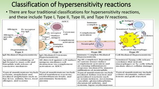
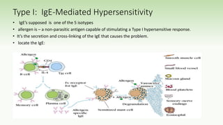
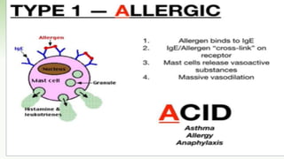
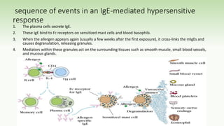
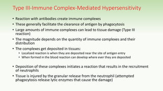
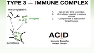
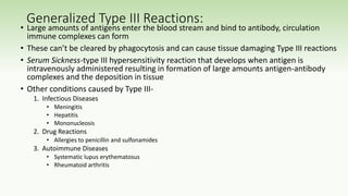
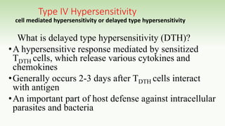
SEMINAR PRESENTATION ON:
HYPERSENSITIVITY REACTIONS
HARAMAYA UNVERSITY
COLLEGE OF HEALTH AND MEDICAL SCIENCE
SCHOOL OF NURSING AND MIDWIFERY
POST GRADUATE STUDY PROGRAMIES
PREPARED BY: GROUP One MEMBERS
1. Henock Shiferaw
2. Kadir Abdu
3. Jemal Yusuf
MODERATOR : Dr Adissu Alemu (MD, Pathologist)
1
Presentation outline
• Objectives
• Introduction
• Mechanisms and examples of hypersensitivity reaction
• Classification of hypersensitivity reaction
• Some of the complication of hypersensitivity reaction
• Summary
• Reference
OBJECTIVES
• At the end of the topic the participant are expected to learn;
• Define hypersensitivity reaction
• Mechanisms and examples of hypersensitivity reaction
• Classification of hypersensitivity reaction
• Some of the complication of hypersensitivity reaction
INTRODUCTION
• The immune system plays a crucial role in maintaining
health and protecting the human body against microbial
invasions.
• However, this same system can lead to exaggerated immune
and inflammatory responses that result in adverse outcomes
known as hypersensitivity reactions.
• Hypersensitivity reactions are exaggerated or
inappropriate immune responses to benign antigens. It is the
immune response, not the antigens that are harmful to the
host.
• Hypersensitivity reactions: An immunologic reaction which produces
tissue damage on re exposure to antigens
Hypersensitivity reactions
•In general, hypersensitivity reactions occur in response
to external stimuli (antigens), whereas autoimmune
reactions occur in response to internal stimuli (antigens).
•Hypersensitivity reactions is pathologic features of one or
more of many autoimmune diseases
Classification of hypersensitivity reactions
• There are four traditional classifications for hypersensitivity reactions,
and these include Type I, Type II, Type III, and Type IV reactions.
• Type I hypersensitivity is also known as an immediate reaction and involves
immunoglobulin E (IgE) mediated release of antibodies against the soluble
antigen. This results in mast cell degranulation and release of histamine and
other inflammatory mediators.
• Type II hypersensitivity is also known as cytotoxic reactions and engages IgG
and IgM antibodies, leading to the complement system activation and cell
damage or lysis.
• Type III hypersensitivity (immune complex reactions) and involves IgG, IgM,
and sometimes IgA antibodies. The build-up immune complexes results
in complement system activation, which leads to polymorphonuclear
leukocytes (PMNs) chemotaxis and eventually causing tissue damage.
• Type IV hypersensitivity is also known as delayed-type and involves of T-cell-
mediated reactions. T-cells or macrophages are activated as a result of
cytokine release, leading to tissue damage.
Type I: IgE-Mediated Hypersensitivity
• IgE’s supposed is one of the 5 isotypes
• allergen is – a non-parasitic antigen capable of stimulating a Type I hypersensitive response.
• It’s the secretion and cross-linking of the IgE that causes the problem.
• locate the IgE:
sequence of events in an IgE-mediated hypersensitive
response
1. The plasma cells secrete IgE.
2. These IgE bind to Fc receptors on sensitized mast cells and blood basophils.
3. When the allergen appears again (usually a few weeks after the first exposure), it cross-links the mIgEs and
causes degranulation, releasing granules.
4. Mediators within these granules act on the surrounding tissues such as smooth muscle, small blood vessels,
and mucous glands.
•The chemically active effectors within the
granules released via degranulation are
called mediators. This group includes:
• Histamines
• Leukotrienes
• Prostaglandins
• Cytokines
• Vasodilation and increased vascular permeability usher in plasma and
inflammatory cells (such as esinophils, neutrophils) to attack the pathogen
However, when these effects go overboard, the result is
problematic:
• Anaphylactic shock – extreme smooth muscle contraction compromises
control of the bladder and GI tract and causes bronchiole constriction
• Allergic rhinitis – excess mucous is released. More commonly known as
hay fever, this ailment affects 10% of the US population
• Food allergies – a variety of symptoms
• Asthma – bronchoconstriction and excess mucous
•In case of asthmatic patients
• They can suffer from a late-phase reaction that develops 4-6 hours
after the initial reaction and persists for 1-2 days.
• The late-phase reaction is caused by the heavy infiltration of
inflammatory cells and the release of cytokines from mast cells, which
increases the adhesion of these inflammatory cells to epithelial
linings of smooth muscle.
• Epithelial damage, bronchoconstriction, and inflamed bronchiole
tubes results.
Type II-Antibody-Mediated Cytotoxic
Hypersensitivity
•Involves the antibody mediated destruction of cells
•Can mediated cell destruction by activating the
complement system to create pores in the
membrane of the foreign cell
•Can also mediated by Antibody-Dependent Cell-
Mediated Cytotoxicity (ADCC) where the Fc receptors
bind to Fc receptor of antibody on the target cell and
promote killing
Transfusion reactions:
• Antibodies of the A,B, and O
antigens are usually of the IgM
class (these antigens are call
isohemagglutinins)
• For example an A individual
produce isohemagglutinins to B-
like epitopes but not to A epitopes
because they are self
• Person who are transfused with
the wrong blood type will produce
anti-hemmagglutinins causing
complement mediated lysis
• Antibodies are usually of the IgG
class
• Transfusion reactions can be delayed or
immediate but have different Ig
isohemagglutinins
• Immediate reactions has a complement-
mediated lysis triggered by IgM
isohemagglutinins
• Delayed reactions induce clonal selection and
the productions of IgG which is less effective
in activating the complement
• This leads to incomplete complement-
mediated lysis
• Cross-matching can detect antibodies in the
sera to prevent this
Hemolytic Disease of the Newborn
• This is where maternal IgG antibodies specific for fetal blood group antigens cross the
placenta and destroy fetal RBC’s
• Erythroblastosis fetalis-severe hemolytic disease of newborns
• Most commonly develops when an Rh+ fetus expresses an Rh antigen on it’s blood that and Rh-
mother doesn’t recognize
Erythroblastosis fetalis
• During the 1st pregnancy small amounts of fetal blood pass through the
placenta but not enough to induce a responses
• During delivery larger amounts of fetal blood cross the placenta causing an
activation of B-cells that are Rh specific thus leading to memory B-cells (anti-
Rh antibodies)
• The IgM antibody clears the Rh+ cells from the mother
• In subsequent pregnancies with an Rh+ fetus, the Rh+ RBC cross the placenta
activating the memory B-cells
• These in turn cross the placenta and damage the fetal RBC because they are
seen as “foreign”
Drug-Induced Hemolytic Anemia
• This is where certain antibiotics can absorb nonspecifically to the
proteins on RBC membranes
• Examples: penicillin, streptomycin
• Sometimes antibodies form inducing complement-mediated lysis and
thus progressive anemia
• When drug is withdrawn the hemolytic anemia disappears
Type III-Immune Complex-Mediated Hypersensitivity
• Reaction with antibodies create immune complexes
• These generally facilitate the clearance of antigen by phagocytosis
• Large amounts of immune complexes can lead to tissue damage (Type III
reaction)
• The magnitude depends on the quantity of immune complexes and their
distribution
• The complexes get deposited in tissues:
• Localized reaction is when they are deposited near the site of antigen entry
• When formed in the blood reaction can develop where ever they are deposited
• Deposition of these complexes initiates a reaction that results in the recruitment
of neutrophils
• Tissue is injured by the granular release from the neutrophil (attempted
phagocytosis release lytic enzymes that cause the damage)
Localized Type III Reactions:
1. Injection of an Antigen:
• Can lead to an acute Arthus
reaction within 4-8 hours
• Localized tissue and vascular
damage result from accumulation
of fluid (edema) and RBC
(erythema)
• Severity can vary from mild
swelling to redness to tissue
necrosis
2. Insect bite:
• May first have a rapid type I
reaction
• Some 4-8 hours later a typical
Arthus reaction develops
Generalized Type III Reactions:
• Large amounts of antigens enter the blood stream and bind to antibody, circulation
immune complexes can form
• These can’t be cleared by phagocytosis and can cause tissue damaging Type III reactions
• Serum Sickness-type III hypersensitivity reaction that develops when antigen is
intravenously administered resulting in formation of large amounts antigen-antibody
complexes and the deposition in tissue
• Other conditions caused by Type III-
1. Infectious Diseases
• Meningitis
• Hepatitis
• Mononucleosis
2. Drug Reactions
• Allergies to penicillin and sulfonamides
3. Autoimmune Diseases
• Systematic lupus erythematosus
• Rheumatoid arthritis
Type IV Hypersensitivity
cell mediated hypersensitivity or delayed type hypersensitivity
What is delayed type hypersensitivity (DTH)?
•A hypersensitive response mediated by sensitized
TDTH cells, which release various cytokines and
chemokines
•Generally occurs 2-3 days after TDTH cells interact
with antigen
•An important part of host defense against intracellular
parasites and bacteria
•Delayed or type IV hypersensitivity was initially
described by its period course in which the
responses took 12-24 hours of time to progress
and persevered for 2-3 days.
•Cell-mediated responses are introduced by T-
lymphocytes and intermediated by effector T-cells
and macrophages.
•including tuberculosis exhibits delayed type
hypersensitivity
Phases of the DTH Response
Sensitization phase: occurs 1-2 weeks
after primary contact with Ag
What happens during this phase?
• TH cells are activated and clonally
expanded by Ag presented
together with class II MHC on an
appropriate APC, such as
macrophages or Langerhan cell
(dendritic epidermal cell)
• Generally CD4+ cells of the TH1
subtype are activated during
sensitization and designated as
TDTH cells
Phases of the DTH Response
Effector phase: occurs upon
subsequent exposure to the Ag
• TDTH cells secrete a variety of
cytokines and chemokines, which
recruit and activate macrophages
• Macrophage activation promotes
phagocytic activity and increased
concentration of lytic enzymes for
more effective killing
• Activated macrophages are also more
effective in presenting Ag and
function as the primary effector cell
What happens if the DTH response is
prolonged?
A granuloma develops…
• Continuous activation of macrophages
induces the macrophages to adhere
closely to one another, assuming an
epithelioid shape and sometimes fusing
together to form giant, multinucleated
cells.
Advantages of DTH Response
Protective Role of DTH Response
• A variety of intracellular pathogens and contact antigens can
induce a DTH response.
• Cells harboring intracellular pathogens are rapidly destroyed
by lytic enzymes released by activated macrophages
Detrimental Effects of DTH Response
• The initial response of the DTH is nonspecific and often results in significant
damage to healthy tissue
• In some cases, a DTH response can cause such extensive tissue damage that
the response itself is pathogenic
• Example: Mycobacterium tuberculosis – an accumulation of activated
macrophages whose lysosomal enzymes destroy healthy lung tissue
• In this case, tissue damage far outweighs any beneficial effects.
• This reaction contains the antigens attached to the surface of lymphocytes. The pre-
sensitized lymphocytes can induce cytokines, which can damage cells .
• Many long-lasting diseases, including tuberculosis exhibits delayed type hypersensitivity
Summay
• An allergy is an adaptive immune response, sometimes life-threatening, to
an allergen.
• Type I hypersensitivity: requires sensitization of mast cells with IgE, involving
an initial IgE antibody response and IgE attachment to mast cells.
• On second exposure to an allergen, cross-linking of IgE molecules on mast cells
triggers degranulation and release of preformed and newly formed chemical
mediators of inflammation.
• Type I hypersensitivity may be localized and relatively minor (hives and hay
fever) or system-wide and dangerous (systemic anaphylaxis).
Summary cont’d…
• Type II hypersensitivities result from antibodies binding to antigens
on cells and initiating cytotoxic responses.
• Examples include hemolytic transfusion reaction and hemolytic
disease of the newborn.
•Type III hypersensitivities : result from formation and
accumulation of immune complexes in tissues, stimulating
damaging inflammatory responses.
• Type IV hypersensitivities are not mediated by antibodies, but by
helper T-cell activation of macrophages, eosinophils, and cytotoxic T
cells.
Reference
• Mac Sween RNM, Whaley K. Muir’s Textbook of pathology. London,
Edward Arlond 13th edition 1992
• Dey NC, Dey TK. A Textbook of Pathology Calcatta, Messers Allied
agency 10th edition 1994.
• Kumar, Vinay; Abbas, Abul K.; Aster, Jon C., eds. (2014).
"Hypersensitivity: Immunologicaly Mediated Tissue Injury".
• Robbins & Cotran Pathologic Basis of Disease (9th ed.). Elsevier
Health Sciences. pp. 200–11. ISBN 978-0-323-29635-9
Thank you !!!

Hypersensitivity reactions BY GROUP 1.pptx

  • 1.
    SEMINAR PRESENTATION ON: HYPERSENSITIVITYREACTIONS HARAMAYA UNVERSITY COLLEGE OF HEALTH AND MEDICAL SCIENCE SCHOOL OF NURSING AND MIDWIFERY POST GRADUATE STUDY PROGRAMIES PREPARED BY: GROUP One MEMBERS 1. Henock Shiferaw 2. Kadir Abdu 3. Jemal Yusuf MODERATOR : Dr Adissu Alemu (MD, Pathologist) 1
  • 2.
    Presentation outline • Objectives •Introduction • Mechanisms and examples of hypersensitivity reaction • Classification of hypersensitivity reaction • Some of the complication of hypersensitivity reaction • Summary • Reference
  • 3.
    OBJECTIVES • At theend of the topic the participant are expected to learn; • Define hypersensitivity reaction • Mechanisms and examples of hypersensitivity reaction • Classification of hypersensitivity reaction • Some of the complication of hypersensitivity reaction
  • 4.
    INTRODUCTION • The immunesystem plays a crucial role in maintaining health and protecting the human body against microbial invasions. • However, this same system can lead to exaggerated immune and inflammatory responses that result in adverse outcomes known as hypersensitivity reactions. • Hypersensitivity reactions are exaggerated or inappropriate immune responses to benign antigens. It is the immune response, not the antigens that are harmful to the host. • Hypersensitivity reactions: An immunologic reaction which produces tissue damage on re exposure to antigens
  • 5.
    Hypersensitivity reactions •In general,hypersensitivity reactions occur in response to external stimuli (antigens), whereas autoimmune reactions occur in response to internal stimuli (antigens). •Hypersensitivity reactions is pathologic features of one or more of many autoimmune diseases
  • 6.
    Classification of hypersensitivityreactions • There are four traditional classifications for hypersensitivity reactions, and these include Type I, Type II, Type III, and Type IV reactions.
  • 7.
    • Type Ihypersensitivity is also known as an immediate reaction and involves immunoglobulin E (IgE) mediated release of antibodies against the soluble antigen. This results in mast cell degranulation and release of histamine and other inflammatory mediators. • Type II hypersensitivity is also known as cytotoxic reactions and engages IgG and IgM antibodies, leading to the complement system activation and cell damage or lysis. • Type III hypersensitivity (immune complex reactions) and involves IgG, IgM, and sometimes IgA antibodies. The build-up immune complexes results in complement system activation, which leads to polymorphonuclear leukocytes (PMNs) chemotaxis and eventually causing tissue damage. • Type IV hypersensitivity is also known as delayed-type and involves of T-cell- mediated reactions. T-cells or macrophages are activated as a result of cytokine release, leading to tissue damage.
  • 8.
    Type I: IgE-MediatedHypersensitivity • IgE’s supposed is one of the 5 isotypes • allergen is – a non-parasitic antigen capable of stimulating a Type I hypersensitive response. • It’s the secretion and cross-linking of the IgE that causes the problem. • locate the IgE:
  • 10.
    sequence of eventsin an IgE-mediated hypersensitive response 1. The plasma cells secrete IgE. 2. These IgE bind to Fc receptors on sensitized mast cells and blood basophils. 3. When the allergen appears again (usually a few weeks after the first exposure), it cross-links the mIgEs and causes degranulation, releasing granules. 4. Mediators within these granules act on the surrounding tissues such as smooth muscle, small blood vessels, and mucous glands.
  • 11.
    •The chemically activeeffectors within the granules released via degranulation are called mediators. This group includes: • Histamines • Leukotrienes • Prostaglandins • Cytokines
  • 13.
    • Vasodilation andincreased vascular permeability usher in plasma and inflammatory cells (such as esinophils, neutrophils) to attack the pathogen However, when these effects go overboard, the result is problematic: • Anaphylactic shock – extreme smooth muscle contraction compromises control of the bladder and GI tract and causes bronchiole constriction • Allergic rhinitis – excess mucous is released. More commonly known as hay fever, this ailment affects 10% of the US population • Food allergies – a variety of symptoms • Asthma – bronchoconstriction and excess mucous
  • 14.
    •In case ofasthmatic patients • They can suffer from a late-phase reaction that develops 4-6 hours after the initial reaction and persists for 1-2 days. • The late-phase reaction is caused by the heavy infiltration of inflammatory cells and the release of cytokines from mast cells, which increases the adhesion of these inflammatory cells to epithelial linings of smooth muscle. • Epithelial damage, bronchoconstriction, and inflamed bronchiole tubes results.
  • 16.
    Type II-Antibody-Mediated Cytotoxic Hypersensitivity •Involvesthe antibody mediated destruction of cells •Can mediated cell destruction by activating the complement system to create pores in the membrane of the foreign cell •Can also mediated by Antibody-Dependent Cell- Mediated Cytotoxicity (ADCC) where the Fc receptors bind to Fc receptor of antibody on the target cell and promote killing
  • 18.
    Transfusion reactions: • Antibodiesof the A,B, and O antigens are usually of the IgM class (these antigens are call isohemagglutinins) • For example an A individual produce isohemagglutinins to B- like epitopes but not to A epitopes because they are self • Person who are transfused with the wrong blood type will produce anti-hemmagglutinins causing complement mediated lysis • Antibodies are usually of the IgG class • Transfusion reactions can be delayed or immediate but have different Ig isohemagglutinins • Immediate reactions has a complement- mediated lysis triggered by IgM isohemagglutinins • Delayed reactions induce clonal selection and the productions of IgG which is less effective in activating the complement • This leads to incomplete complement- mediated lysis • Cross-matching can detect antibodies in the sera to prevent this
  • 19.
    Hemolytic Disease ofthe Newborn • This is where maternal IgG antibodies specific for fetal blood group antigens cross the placenta and destroy fetal RBC’s • Erythroblastosis fetalis-severe hemolytic disease of newborns • Most commonly develops when an Rh+ fetus expresses an Rh antigen on it’s blood that and Rh- mother doesn’t recognize
  • 20.
    Erythroblastosis fetalis • Duringthe 1st pregnancy small amounts of fetal blood pass through the placenta but not enough to induce a responses • During delivery larger amounts of fetal blood cross the placenta causing an activation of B-cells that are Rh specific thus leading to memory B-cells (anti- Rh antibodies) • The IgM antibody clears the Rh+ cells from the mother • In subsequent pregnancies with an Rh+ fetus, the Rh+ RBC cross the placenta activating the memory B-cells • These in turn cross the placenta and damage the fetal RBC because they are seen as “foreign”
  • 21.
    Drug-Induced Hemolytic Anemia •This is where certain antibiotics can absorb nonspecifically to the proteins on RBC membranes • Examples: penicillin, streptomycin • Sometimes antibodies form inducing complement-mediated lysis and thus progressive anemia • When drug is withdrawn the hemolytic anemia disappears
  • 22.
    Type III-Immune Complex-MediatedHypersensitivity • Reaction with antibodies create immune complexes • These generally facilitate the clearance of antigen by phagocytosis • Large amounts of immune complexes can lead to tissue damage (Type III reaction) • The magnitude depends on the quantity of immune complexes and their distribution • The complexes get deposited in tissues: • Localized reaction is when they are deposited near the site of antigen entry • When formed in the blood reaction can develop where ever they are deposited • Deposition of these complexes initiates a reaction that results in the recruitment of neutrophils • Tissue is injured by the granular release from the neutrophil (attempted phagocytosis release lytic enzymes that cause the damage)
  • 24.
    Localized Type IIIReactions: 1. Injection of an Antigen: • Can lead to an acute Arthus reaction within 4-8 hours • Localized tissue and vascular damage result from accumulation of fluid (edema) and RBC (erythema) • Severity can vary from mild swelling to redness to tissue necrosis 2. Insect bite: • May first have a rapid type I reaction • Some 4-8 hours later a typical Arthus reaction develops
  • 25.
    Generalized Type IIIReactions: • Large amounts of antigens enter the blood stream and bind to antibody, circulation immune complexes can form • These can’t be cleared by phagocytosis and can cause tissue damaging Type III reactions • Serum Sickness-type III hypersensitivity reaction that develops when antigen is intravenously administered resulting in formation of large amounts antigen-antibody complexes and the deposition in tissue • Other conditions caused by Type III- 1. Infectious Diseases • Meningitis • Hepatitis • Mononucleosis 2. Drug Reactions • Allergies to penicillin and sulfonamides 3. Autoimmune Diseases • Systematic lupus erythematosus • Rheumatoid arthritis
  • 26.
    Type IV Hypersensitivity cellmediated hypersensitivity or delayed type hypersensitivity What is delayed type hypersensitivity (DTH)? •A hypersensitive response mediated by sensitized TDTH cells, which release various cytokines and chemokines •Generally occurs 2-3 days after TDTH cells interact with antigen •An important part of host defense against intracellular parasites and bacteria
  • 28.
    •Delayed or typeIV hypersensitivity was initially described by its period course in which the responses took 12-24 hours of time to progress and persevered for 2-3 days. •Cell-mediated responses are introduced by T- lymphocytes and intermediated by effector T-cells and macrophages. •including tuberculosis exhibits delayed type hypersensitivity
  • 29.
    Phases of theDTH Response Sensitization phase: occurs 1-2 weeks after primary contact with Ag What happens during this phase? • TH cells are activated and clonally expanded by Ag presented together with class II MHC on an appropriate APC, such as macrophages or Langerhan cell (dendritic epidermal cell) • Generally CD4+ cells of the TH1 subtype are activated during sensitization and designated as TDTH cells
  • 30.
    Phases of theDTH Response Effector phase: occurs upon subsequent exposure to the Ag • TDTH cells secrete a variety of cytokines and chemokines, which recruit and activate macrophages • Macrophage activation promotes phagocytic activity and increased concentration of lytic enzymes for more effective killing • Activated macrophages are also more effective in presenting Ag and function as the primary effector cell
  • 31.
    What happens ifthe DTH response is prolonged? A granuloma develops… • Continuous activation of macrophages induces the macrophages to adhere closely to one another, assuming an epithelioid shape and sometimes fusing together to form giant, multinucleated cells.
  • 32.
    Advantages of DTHResponse Protective Role of DTH Response • A variety of intracellular pathogens and contact antigens can induce a DTH response. • Cells harboring intracellular pathogens are rapidly destroyed by lytic enzymes released by activated macrophages
  • 33.
    Detrimental Effects ofDTH Response • The initial response of the DTH is nonspecific and often results in significant damage to healthy tissue • In some cases, a DTH response can cause such extensive tissue damage that the response itself is pathogenic • Example: Mycobacterium tuberculosis – an accumulation of activated macrophages whose lysosomal enzymes destroy healthy lung tissue • In this case, tissue damage far outweighs any beneficial effects. • This reaction contains the antigens attached to the surface of lymphocytes. The pre- sensitized lymphocytes can induce cytokines, which can damage cells . • Many long-lasting diseases, including tuberculosis exhibits delayed type hypersensitivity
  • 34.
    Summay • An allergyis an adaptive immune response, sometimes life-threatening, to an allergen. • Type I hypersensitivity: requires sensitization of mast cells with IgE, involving an initial IgE antibody response and IgE attachment to mast cells. • On second exposure to an allergen, cross-linking of IgE molecules on mast cells triggers degranulation and release of preformed and newly formed chemical mediators of inflammation. • Type I hypersensitivity may be localized and relatively minor (hives and hay fever) or system-wide and dangerous (systemic anaphylaxis).
  • 35.
    Summary cont’d… • TypeII hypersensitivities result from antibodies binding to antigens on cells and initiating cytotoxic responses. • Examples include hemolytic transfusion reaction and hemolytic disease of the newborn. •Type III hypersensitivities : result from formation and accumulation of immune complexes in tissues, stimulating damaging inflammatory responses. • Type IV hypersensitivities are not mediated by antibodies, but by helper T-cell activation of macrophages, eosinophils, and cytotoxic T cells.
  • 36.
    Reference • Mac SweenRNM, Whaley K. Muir’s Textbook of pathology. London, Edward Arlond 13th edition 1992 • Dey NC, Dey TK. A Textbook of Pathology Calcatta, Messers Allied agency 10th edition 1994. • Kumar, Vinay; Abbas, Abul K.; Aster, Jon C., eds. (2014). "Hypersensitivity: Immunologicaly Mediated Tissue Injury". • Robbins & Cotran Pathologic Basis of Disease (9th ed.). Elsevier Health Sciences. pp. 200–11. ISBN 978-0-323-29635-9
  • 37.

Editor's Notes

  • #8 There are four types of reactions: Type I-IgE mediated Type II-Antibody-Mediated Type III-Immune Complex-Mediated Type IV-Delayed-Type Hypersensitivity (DTH)
  • #21 This type of reaction can be prevented by administering antibodies against the Rh antigen within 25-48 hours after the 1st delivery Rhogam-is the antibody that is injected it will bind to the fetal RBC that enter the mother’s circulation and facilitate the clearance of them before B-cell activation In subsequent pregnancies the mother is unlikely to produce IgG anti-Rh antibodies If the mother doesn’t receive this injection there are other ways to treat this, depending on the severity
  • #33 Intracellular bacteria Intracellular viruses Contact Antigens Mycobacterium tuberculosis Herpes simplex virus Hair dyes Mycobacterium leprae Measles virus Poison ivy