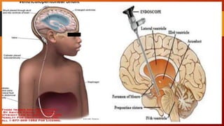
HYDROCEPHALUS
IBRAHIM
ADAMU
UG15/MDMD/
1014
GOMBE STATE UNIVERSITY
COLLEGE OF MEDICAL SCIENCES
DEPARTMENT OF PAEDIATRICS
Moderator: Dr. Ahmed Bappa Bakura
MEDICAL STUDENTS’
PRESENTATION
OUTLINE
• Introduction
• Epidemiology
• Relevant anatomy & CSF Physiology
• Pathophysiology
• Classifications
• Aetiology
• Clinical features
• Special Types
• Investigations
• Treatment
• Complications
• Prognosis
• Conclusion
INTRODUCTION
• Derived from Greek-
Hydro- water
Kefale- head
• Literally means “water in the head”
• Hydrocephalus refers to the excessive accumulation of CSF within the
ventricular system leading to enlargement of the ventricles (with or without
increase CSF pressure)
• Hydrocephalus result from disturbance of CSF production, flow, or absorption
leading to an abnormal increase in volume of CSF within the CNS
INTRODUCTION
• It is a common complication of Tuberculous meningitis; upon presentation may
indicate a higher risk of symptomatic cerebral infarcts
• As a clinical entity, hydrocephalus dates back to antiquity.
• In 5th century, Hippocrates describes it as water in the head.
• Galen described relationship of choroid plexus
• 17th century AD, Willis- CSF physiology
NATURAL HISTORY
• Hydrocephalus is a fatal disease condition
• Fifty-five percent progresses to brain herniation, coma and eventually death
• However in up to 45%, there is arrest of ventricular enlargement; compensated
hydrocephalus
• Children with compensated hydrocephalus do not require CSF diversion,
however many factors; fever, infection, trauma can offset this balance causing
decompensation
• Decompensated hydrocephalus would require CSF diversion in about 70% of
cases
EPIDEMIOLOGY
• The global estimated annual incidence of pediatric hydrocephalus is 400,000
new cases each year
• Prevalence higher in Africa and Latin America 145 and 136 per 100,000 births
respectively and lowest in U.S about 68 per 100,000 births
• The incidence is about 0.9-1.8/1000 birth in Nigeria
• Slight male preponderance
RELEVANT ANATOMY
CSF PHYSIOLOGY
• Formation of CSF is by:
• 80% by choroid plexus
• 20% by ventricular ependymal cells and brain tissue
• Formation is energy dependent process requiring carbonic anhydrase, with total
daily output of 450mls, 0.3ml/min
• The normal total volume of CSF in the CNS is about 150ml in adult and 25mls in
neonates, 50mls in infants
• After formation, CSF passes via foramen of Monro to the 3rd ventricle then via
aqueduct of Sylvius to the 4th ventricle then via foramena of luscka & Magendi
into the cortical and spinal cord subarachnoid space
CSF PHYSIOLOGY
• CSF in subarachnoid space is absorbed by arachnoid villi to dural venous sinuses
• In contrast, absorption is energy independent, it is driven by pressure gradient
from subarachnoid space across the arachnoid villi into venous sinuses
• The normal ICP is 100 – 180 mmH20 (8-15mmHg)
• When ICP is <7mmHg, CSF is produced at a rate of 0.3ml/min but no absorption
occur.
• Other proposed pathway for CSF absorption includes the nerve root sleeve of
cranial nerves, paranasal sinuses and nasal mucosal
PATHOPHYSIOLOGY
• Obstruction of ventricular passage way, CSF over production or impaired
absorption causes ventricular dilatation and increased pressure
• Trans-ependymal CSF leakage into periventricular tissue results to subsequent
scarring, elevated ICP, brain herniaton, coma and death
Obstruction of CSF flow within the ventricular system
Obstructive (non communicating type)
PATHOPHYSIOLOGY
Dysfunction in absorption or CSF over secretion
Non obstructive (communicating type)
CLASSIFICATION
• Idiopathic or Secondary
• Acute or Chronic
• Congenital or Acquired
• Communicating or Non-communicating
AETIOLOGY
• OBSTRUCTIVE
a. Obstruction of aqueduct of sylvius
 Congenital atresia: - may be sex linked recessive or may be
associated with spina bifida
 Obstruction from outside by: brain tumors
 Obstruction from inside:- Post hemorrhagic (specially in premature),
Post infection (T.B., pneumocci, mumps)
b. Congenital atresia of:
 Foramen of Monro
 Foramina of Luscka & Magendi: with cystic dilatation of 4th
ventricle usually with agenesis of cerebellar vermis (Dandy Walker
malformation)
AETIOLOGY
c. Arnold Chiari malformation: Congenital downward displacement of
cerebellum, pons & medulla consequently elongation of the 4th ventricle
d. Congenital infection especially toxoplasmosis
NON-OBSTRUCTIVE
a. Defective CSF absorption
 Subarachnoid space adhesions; due to Post hemorrhagic (IVH) or
Post meningitis
 Leukemic infiltration
 Dural sinus thrombosis
AETIOLOGY
b. Excessive CSF secretion
 Choroid plexus papilloma
 Choroid plexus congestion as in meningitis
 Ependymoma
CLINICAL FEATURES
INFANT
• Large head with progressive increase in size (increasing
head circumference on serial measurement).
• Fontanels are widely opened & bulging.
• Sutures are widely separated.
• Dilated scalp veins.
• Eyes deviated downwards ~ sunset appearance
• Skull percussion ~ cracked pot sound (Macewen sign).
• Craniotabes
• Back of the skull:- Promeninet occiput esp. in Dandy
Walker.
• Mild vomiting may be present with irritability and poor feeding
• Delayed motor milestones and mental retardation in severe cases
OLDER CHILDREN
• Marked neurologic manifestations as the sutures are not easily separated with
subsequent marked increase intracranial tension
• Bursting headache (severe in the morning) and blurring of vision
• Projectile vomiting (unrelated to meals)
• Deterioration in school performance
• Bradycardia, irregular respiration & hypertension (Cushing triad)
CLINICAL FEATURES
SPECIAL TYPES
• Hydrocephalus ex-vacuo
This condition occurs when there is brain injury with subsequent shrinkage of the
brain substance with increase in CSF volume although the pressure may be normal
• Normal pressure hydrocephalus
The term Normal Pressure Hydrocephalus NPH describes a condition that rarely
occurs in younger patients with an enlarged ventricles and normal CSF pressure at
lumbar puncture
NB: Hydranencephaly should not be confused with hydrocephalus which is a rare
condition in which the brains cerebral hemispheres are absent and replaced by
sacs filled with CSF
DIAGNOSIS
Clinical picture
• History
• General examination
Investigation
INVESTIGATION
1. Cranial X-ray
• Before closure of sutures -
Wide fontanels, wide
separation of sutures.
Craniofacial disproportion
with large cranium.
• After closure of sutures
increase intra cranial
tension (beaten silver
appearance, wide sella)
Beaten Silver Appearance
Ultrasound
INVESTIGATION
• 5. Simultaneous lumbar & ventricular manometry:
-Normally, both are equal
-Ventricular pressure> spinal pressure in obstructive hydrocephalus
• 6. Lumbar Puncture and CSF examination: xanthochromia and increased
pressure
DIFFERENTIALS
• Constitutional
• Hydrancephaly
• Space occupying lesion e.g. tumor
• Subdural heamatoma
TREATMENT
The goal of treatment is to achieve optimum neurologic function and prevent or
reverse the neurologic damage caused by distortion of the brain from ventricular
dilatation
 Medical
 Surgical
MEDICAL TREATMENT
• Carbonic anhydrase inhibitors: acetazolamide
• Loop diuretics: Frosemide
• Osmotic diuretics: Mannitol
Draw backs: - Transient effect and electrolytes & pH disturbances.
TREATMENT
SURGICAL TREATMENT
• Surgical options depends on the type of the hydrocephalus
1. Shunting
Used for communicating hydrocephalus and some non
communicating hydrocephalus
TREATMENT
Sites
• Peritoneal cavity
• Atrium
• Pleural cavity
• Others- bladder, ureter
Complications
• Mechanical failure due to either Peritoneal end obstruction, Shunt
fracture or Shunt migration
• Shunt infection 5-15% of Shunts get infected by organism like
S.epidermidis, S. aureus, enterococcus, streptococcus
• Shunt overdrainage
• Hemorrhage
TREATMENT
TREATMENT
2. Endoscopic third ventriculostomy (ETV) - Option for non- communicating
hydrocephalus.
Others
i. Excision of cyst and tumour
ii. Choroid plexectomy or endoscopic diathermy coagulation of choroid plexus
PROGNOSIS
• Natural history is poor with 50% dying before 3 years
• Only 20 – 23% reach adult life and only 38% of these have normal intelligence
• About 50-55% of shunted hydrocephalus have IQ > 80
• Shunt complication further worsen the prognosis
CONCLUSION
• Hydrocephalus is a fatal disease condition
• It refers to the excessive accumulation of CSF within the ventricular system
leading to enlargement of the ventricles
• Early detection and treatment is key to preventing long term sequalae
• Prognosis is poor with 50% dying before 3 years
REFERENCES
• Nelson Textbook of Paediatrics 19th Edition
• Robert M. Kliegman, Bonita F. Stanton, Joseph W. St. Geme III, Nina F. Schor, &
Richard E. Behrman Pediatric Textbook
• https://www.thisdaylive.com/index.php/2017/04/09/hydrocephalus/
• Lecture note on Hydrocephalus by Dr Sani Adamu, Department of Surgery,
FTHG
THANK YOU FOR
LISTENING

Hydrocephalus.pptx

  • 1.
    HYDROCEPHALUS IBRAHIM ADAMU UG15/MDMD/ 1014 GOMBE STATE UNIVERSITY COLLEGEOF MEDICAL SCIENCES DEPARTMENT OF PAEDIATRICS Moderator: Dr. Ahmed Bappa Bakura MEDICAL STUDENTS’ PRESENTATION
  • 2.
    OUTLINE • Introduction • Epidemiology •Relevant anatomy & CSF Physiology • Pathophysiology • Classifications • Aetiology • Clinical features • Special Types • Investigations • Treatment • Complications • Prognosis • Conclusion
  • 3.
    INTRODUCTION • Derived fromGreek- Hydro- water Kefale- head • Literally means “water in the head” • Hydrocephalus refers to the excessive accumulation of CSF within the ventricular system leading to enlargement of the ventricles (with or without increase CSF pressure) • Hydrocephalus result from disturbance of CSF production, flow, or absorption leading to an abnormal increase in volume of CSF within the CNS
  • 4.
    INTRODUCTION • It isa common complication of Tuberculous meningitis; upon presentation may indicate a higher risk of symptomatic cerebral infarcts • As a clinical entity, hydrocephalus dates back to antiquity. • In 5th century, Hippocrates describes it as water in the head. • Galen described relationship of choroid plexus • 17th century AD, Willis- CSF physiology
  • 5.
    NATURAL HISTORY • Hydrocephalusis a fatal disease condition • Fifty-five percent progresses to brain herniation, coma and eventually death • However in up to 45%, there is arrest of ventricular enlargement; compensated hydrocephalus • Children with compensated hydrocephalus do not require CSF diversion, however many factors; fever, infection, trauma can offset this balance causing decompensation • Decompensated hydrocephalus would require CSF diversion in about 70% of cases
  • 6.
    EPIDEMIOLOGY • The globalestimated annual incidence of pediatric hydrocephalus is 400,000 new cases each year • Prevalence higher in Africa and Latin America 145 and 136 per 100,000 births respectively and lowest in U.S about 68 per 100,000 births • The incidence is about 0.9-1.8/1000 birth in Nigeria • Slight male preponderance
  • 7.
  • 8.
    CSF PHYSIOLOGY • Formationof CSF is by: • 80% by choroid plexus • 20% by ventricular ependymal cells and brain tissue • Formation is energy dependent process requiring carbonic anhydrase, with total daily output of 450mls, 0.3ml/min • The normal total volume of CSF in the CNS is about 150ml in adult and 25mls in neonates, 50mls in infants • After formation, CSF passes via foramen of Monro to the 3rd ventricle then via aqueduct of Sylvius to the 4th ventricle then via foramena of luscka & Magendi into the cortical and spinal cord subarachnoid space
  • 9.
    CSF PHYSIOLOGY • CSFin subarachnoid space is absorbed by arachnoid villi to dural venous sinuses • In contrast, absorption is energy independent, it is driven by pressure gradient from subarachnoid space across the arachnoid villi into venous sinuses • The normal ICP is 100 – 180 mmH20 (8-15mmHg) • When ICP is <7mmHg, CSF is produced at a rate of 0.3ml/min but no absorption occur. • Other proposed pathway for CSF absorption includes the nerve root sleeve of cranial nerves, paranasal sinuses and nasal mucosal
  • 10.
    PATHOPHYSIOLOGY • Obstruction ofventricular passage way, CSF over production or impaired absorption causes ventricular dilatation and increased pressure • Trans-ependymal CSF leakage into periventricular tissue results to subsequent scarring, elevated ICP, brain herniaton, coma and death Obstruction of CSF flow within the ventricular system Obstructive (non communicating type)
  • 11.
    PATHOPHYSIOLOGY Dysfunction in absorptionor CSF over secretion Non obstructive (communicating type)
  • 12.
    CLASSIFICATION • Idiopathic orSecondary • Acute or Chronic • Congenital or Acquired • Communicating or Non-communicating
  • 13.
    AETIOLOGY • OBSTRUCTIVE a. Obstructionof aqueduct of sylvius  Congenital atresia: - may be sex linked recessive or may be associated with spina bifida  Obstruction from outside by: brain tumors  Obstruction from inside:- Post hemorrhagic (specially in premature), Post infection (T.B., pneumocci, mumps) b. Congenital atresia of:  Foramen of Monro  Foramina of Luscka & Magendi: with cystic dilatation of 4th ventricle usually with agenesis of cerebellar vermis (Dandy Walker malformation)
  • 14.
    AETIOLOGY c. Arnold Chiarimalformation: Congenital downward displacement of cerebellum, pons & medulla consequently elongation of the 4th ventricle d. Congenital infection especially toxoplasmosis NON-OBSTRUCTIVE a. Defective CSF absorption  Subarachnoid space adhesions; due to Post hemorrhagic (IVH) or Post meningitis  Leukemic infiltration  Dural sinus thrombosis
  • 15.
    AETIOLOGY b. Excessive CSFsecretion  Choroid plexus papilloma  Choroid plexus congestion as in meningitis  Ependymoma
  • 16.
    CLINICAL FEATURES INFANT • Largehead with progressive increase in size (increasing head circumference on serial measurement). • Fontanels are widely opened & bulging. • Sutures are widely separated. • Dilated scalp veins. • Eyes deviated downwards ~ sunset appearance • Skull percussion ~ cracked pot sound (Macewen sign). • Craniotabes • Back of the skull:- Promeninet occiput esp. in Dandy Walker.
  • 17.
    • Mild vomitingmay be present with irritability and poor feeding • Delayed motor milestones and mental retardation in severe cases OLDER CHILDREN • Marked neurologic manifestations as the sutures are not easily separated with subsequent marked increase intracranial tension • Bursting headache (severe in the morning) and blurring of vision • Projectile vomiting (unrelated to meals) • Deterioration in school performance • Bradycardia, irregular respiration & hypertension (Cushing triad) CLINICAL FEATURES
  • 18.
    SPECIAL TYPES • Hydrocephalusex-vacuo This condition occurs when there is brain injury with subsequent shrinkage of the brain substance with increase in CSF volume although the pressure may be normal • Normal pressure hydrocephalus The term Normal Pressure Hydrocephalus NPH describes a condition that rarely occurs in younger patients with an enlarged ventricles and normal CSF pressure at lumbar puncture NB: Hydranencephaly should not be confused with hydrocephalus which is a rare condition in which the brains cerebral hemispheres are absent and replaced by sacs filled with CSF
  • 19.
    DIAGNOSIS Clinical picture • History •General examination Investigation
  • 20.
    INVESTIGATION 1. Cranial X-ray •Before closure of sutures - Wide fontanels, wide separation of sutures. Craniofacial disproportion with large cranium. • After closure of sutures increase intra cranial tension (beaten silver appearance, wide sella)
  • 21.
  • 22.
  • 23.
    INVESTIGATION • 5. Simultaneouslumbar & ventricular manometry: -Normally, both are equal -Ventricular pressure> spinal pressure in obstructive hydrocephalus • 6. Lumbar Puncture and CSF examination: xanthochromia and increased pressure
  • 24.
    DIFFERENTIALS • Constitutional • Hydrancephaly •Space occupying lesion e.g. tumor • Subdural heamatoma
  • 25.
    TREATMENT The goal oftreatment is to achieve optimum neurologic function and prevent or reverse the neurologic damage caused by distortion of the brain from ventricular dilatation  Medical  Surgical MEDICAL TREATMENT • Carbonic anhydrase inhibitors: acetazolamide • Loop diuretics: Frosemide • Osmotic diuretics: Mannitol Draw backs: - Transient effect and electrolytes & pH disturbances.
  • 26.
    TREATMENT SURGICAL TREATMENT • Surgicaloptions depends on the type of the hydrocephalus 1. Shunting Used for communicating hydrocephalus and some non communicating hydrocephalus
  • 28.
    TREATMENT Sites • Peritoneal cavity •Atrium • Pleural cavity • Others- bladder, ureter Complications • Mechanical failure due to either Peritoneal end obstruction, Shunt fracture or Shunt migration • Shunt infection 5-15% of Shunts get infected by organism like S.epidermidis, S. aureus, enterococcus, streptococcus • Shunt overdrainage • Hemorrhage
  • 29.
  • 30.
    TREATMENT 2. Endoscopic thirdventriculostomy (ETV) - Option for non- communicating hydrocephalus. Others i. Excision of cyst and tumour ii. Choroid plexectomy or endoscopic diathermy coagulation of choroid plexus
  • 31.
    PROGNOSIS • Natural historyis poor with 50% dying before 3 years • Only 20 – 23% reach adult life and only 38% of these have normal intelligence • About 50-55% of shunted hydrocephalus have IQ > 80 • Shunt complication further worsen the prognosis
  • 32.
    CONCLUSION • Hydrocephalus isa fatal disease condition • It refers to the excessive accumulation of CSF within the ventricular system leading to enlargement of the ventricles • Early detection and treatment is key to preventing long term sequalae • Prognosis is poor with 50% dying before 3 years
  • 33.
    REFERENCES • Nelson Textbookof Paediatrics 19th Edition • Robert M. Kliegman, Bonita F. Stanton, Joseph W. St. Geme III, Nina F. Schor, & Richard E. Behrman Pediatric Textbook • https://www.thisdaylive.com/index.php/2017/04/09/hydrocephalus/ • Lecture note on Hydrocephalus by Dr Sani Adamu, Department of Surgery, FTHG
  • 34.