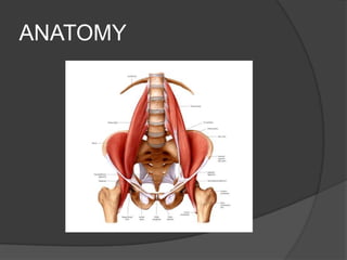
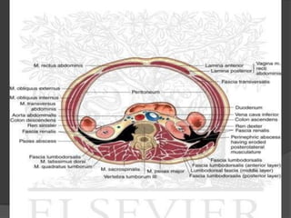
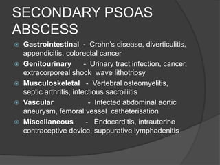
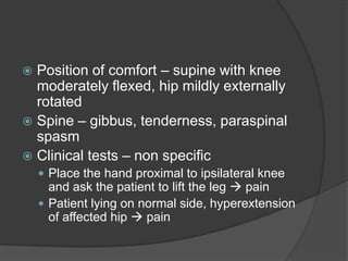
ANATOMY
 Extraperitoneal space
 Psoas major and iliacus
 Psoas major – fusiform muscle from
  lower border of T12 to upper border or
  L5
 Passes along pelvic brim and beneath
  inguinal ligament into thigh
 Attached to lesser trochanter of femur
 L2 , L3, L4
 Lies in close proximity to organs such as
  the sigmoid
  colon, appendix, jejunum, ureters, abdomin
  al aorta, kidneys, pancreas, spine, and iliac
  lymph nodes
 Psoas fascia ( part of iliac fascia) invests
  the surface of the muscle
 Attached to vertebral bodies, fibrous
  arches and transverse processes and to
  iliopubic eminence
 Retains the pus of psoas abscess
Psoas abscess
 Iliopsoas abscess is a collection of pus
  in the iliopsoas compartment.
 First described by Mynter in 1881 who
  referred it as ‘psoitis’
 Classification
     Primary and secondary
Aetiology
   Primary psoas abscess :
     Hematogenous spread from an occult
      source of infection
     Occurs in patients with
      immunocompromised state
     Diabetes mellitus, AIDS, IV drug
      abusers, renal failure
   Secondary abscess :
     Due to infection from an adjacent organ
     Can be pyogenic or tuberculous
     Tuberculosis of the spine
     Crohn’s disease
SECONDARY PSOAS
ABSCESS
   Gastrointestinal - Crohn’s disease, diverticulitis,
    appendicitis, colorectal cancer
   Genitourinary         - Urinary tract infection, cancer,
    extracorporeal shock wave lithotripsy
   Musculoskeletal - Vertebral osteomyelitis,
    septic arthritis, infectious sacroiliitis
   Vascular                - Infected abdominal aortic
    aneurysm, femoral vessel catheterisation
   Miscellaneous          - Endocarditis, intrauterine
    contraceptive device, suppurative lymphadenitis
BACTERIOLOGY
   Related to cause

   Mycobacterium tuberculosis

   Primary (hematogenous) – usually Staphylococcus aureus

   Renal source – usually monomicrobial ; E. coli, Proteus
    mirabilis

   Gastrointestinal – polymicrobial; E. coli, Enterobacter spp.,
    enterococci and anaerobes such as bactroides;

   Salmonella, Mycobacterium kansasii and Mycobacterium
    xenopi are other rare causative organisms
 Common in males than females
 Right > left ; bilateral (3%)
 Mortality
     Secondary > primary
     Untreated – 100%
CLINICAL FEATURES
   Classical triad ( 30% )
     Fever
     Back pain
     Limp
    Variable and non-specific features
     Abdominal or flank pain
     Malaise
     Weight loss
     Nausea
     Referred pain to the groin or knee
     Painless swelling in the inguinal region
     Duration – usually longer than one week
 Position of comfort – supine with knee
  moderately flexed, hip mildly externally
  rotated
 Spine – gibbus, tenderness, paraspinal
  spasm
 Clinical tests – non specific
     Place the hand proximal to ipsilateral knee
      and ask the patient to lift the leg  pain
     Patient lying on normal side, hyperextension
      of affected hip  pain
DIFFERENTIAL
DIAGNOSIS

 Femoral hernia
 Enlarged inguinal nodes
 Tumors arising from the pelvis or lumbar
  area
 Iliac artery aneurysm
Complications


 Intra peritoneal rupture
 Hydroureteronephrosis
 Deep venous thrombosis
 Septicemia
Investigations
 CBC - Raised white cell count, Anaemia
 ↑ ESR
 ↑CRP
 Blood culture, urine culture
 Radiography of abdomen, kidney, spine
 X ray abdomen erect – bulge in psoas
  shadow
 USG – operator dependant; gas
  shadows obscure retoperitoneum
Investigations
   CT abdomen – ‘gold standard’; low density
    mass in retroperitoneum; info on location
    and relation with adjacent organs
   MRI
   IVP
   Mantoux
   Screening for diabetes, HIV, kidney
    disorders
   Pus culture and sensitivity
   Pus for AFB and Gram staining
Management
 Appropriate antibiotics and adequate
  drainage
 Antibiotics :
     Culture specific
     Primary : empirical anti-staphylococcal
   Anti tuberculous drugs
   Drainage :
     Image guided ( CT ) percutaneous drainage
     Open extraperitoneal drainage:
      ○ Through lateral loin incision
      ○ Psoas region reached extraperitoneally
      ○ Pus drained – drainage tube kept
 Relatively uncommon condition
 Vague clinical features
 Insiduous onset and occult nature –
  diagnostic delays
 High mortality and morbidity
 High index of suspicion required
Psoas abscess

Psoas abscess

  • 2.
  • 3.
     Extraperitoneal space Psoas major and iliacus  Psoas major – fusiform muscle from lower border of T12 to upper border or L5  Passes along pelvic brim and beneath inguinal ligament into thigh  Attached to lesser trochanter of femur  L2 , L3, L4
  • 5.
     Lies inclose proximity to organs such as the sigmoid colon, appendix, jejunum, ureters, abdomin al aorta, kidneys, pancreas, spine, and iliac lymph nodes  Psoas fascia ( part of iliac fascia) invests the surface of the muscle  Attached to vertebral bodies, fibrous arches and transverse processes and to iliopubic eminence  Retains the pus of psoas abscess
  • 6.
    Psoas abscess  Iliopsoasabscess is a collection of pus in the iliopsoas compartment.  First described by Mynter in 1881 who referred it as ‘psoitis’  Classification  Primary and secondary
  • 8.
    Aetiology  Primary psoas abscess :  Hematogenous spread from an occult source of infection  Occurs in patients with immunocompromised state  Diabetes mellitus, AIDS, IV drug abusers, renal failure
  • 9.
    Secondary abscess :  Due to infection from an adjacent organ  Can be pyogenic or tuberculous  Tuberculosis of the spine  Crohn’s disease
  • 10.
    SECONDARY PSOAS ABSCESS  Gastrointestinal - Crohn’s disease, diverticulitis, appendicitis, colorectal cancer  Genitourinary - Urinary tract infection, cancer, extracorporeal shock wave lithotripsy  Musculoskeletal - Vertebral osteomyelitis, septic arthritis, infectious sacroiliitis  Vascular - Infected abdominal aortic aneurysm, femoral vessel catheterisation  Miscellaneous - Endocarditis, intrauterine contraceptive device, suppurative lymphadenitis
  • 11.
    BACTERIOLOGY  Related to cause  Mycobacterium tuberculosis  Primary (hematogenous) – usually Staphylococcus aureus  Renal source – usually monomicrobial ; E. coli, Proteus mirabilis  Gastrointestinal – polymicrobial; E. coli, Enterobacter spp., enterococci and anaerobes such as bactroides;  Salmonella, Mycobacterium kansasii and Mycobacterium xenopi are other rare causative organisms
  • 12.
     Common inmales than females  Right > left ; bilateral (3%)  Mortality  Secondary > primary  Untreated – 100%
  • 13.
    CLINICAL FEATURES  Classical triad ( 30% )  Fever  Back pain  Limp Variable and non-specific features  Abdominal or flank pain  Malaise  Weight loss  Nausea  Referred pain to the groin or knee  Painless swelling in the inguinal region  Duration – usually longer than one week
  • 14.
     Position ofcomfort – supine with knee moderately flexed, hip mildly externally rotated  Spine – gibbus, tenderness, paraspinal spasm  Clinical tests – non specific  Place the hand proximal to ipsilateral knee and ask the patient to lift the leg  pain  Patient lying on normal side, hyperextension of affected hip  pain
  • 16.
    DIFFERENTIAL DIAGNOSIS  Femoral hernia Enlarged inguinal nodes  Tumors arising from the pelvis or lumbar area  Iliac artery aneurysm
  • 17.
    Complications  Intra peritonealrupture  Hydroureteronephrosis  Deep venous thrombosis  Septicemia
  • 18.
    Investigations  CBC -Raised white cell count, Anaemia  ↑ ESR  ↑CRP  Blood culture, urine culture  Radiography of abdomen, kidney, spine  X ray abdomen erect – bulge in psoas shadow  USG – operator dependant; gas shadows obscure retoperitoneum
  • 19.
    Investigations  CT abdomen – ‘gold standard’; low density mass in retroperitoneum; info on location and relation with adjacent organs  MRI  IVP  Mantoux  Screening for diabetes, HIV, kidney disorders  Pus culture and sensitivity  Pus for AFB and Gram staining
  • 22.
    Management  Appropriate antibioticsand adequate drainage  Antibiotics :  Culture specific  Primary : empirical anti-staphylococcal  Anti tuberculous drugs  Drainage :  Image guided ( CT ) percutaneous drainage  Open extraperitoneal drainage: ○ Through lateral loin incision ○ Psoas region reached extraperitoneally ○ Pus drained – drainage tube kept
  • 24.
     Relatively uncommoncondition  Vague clinical features  Insiduous onset and occult nature – diagnostic delays  High mortality and morbidity  High index of suspicion required