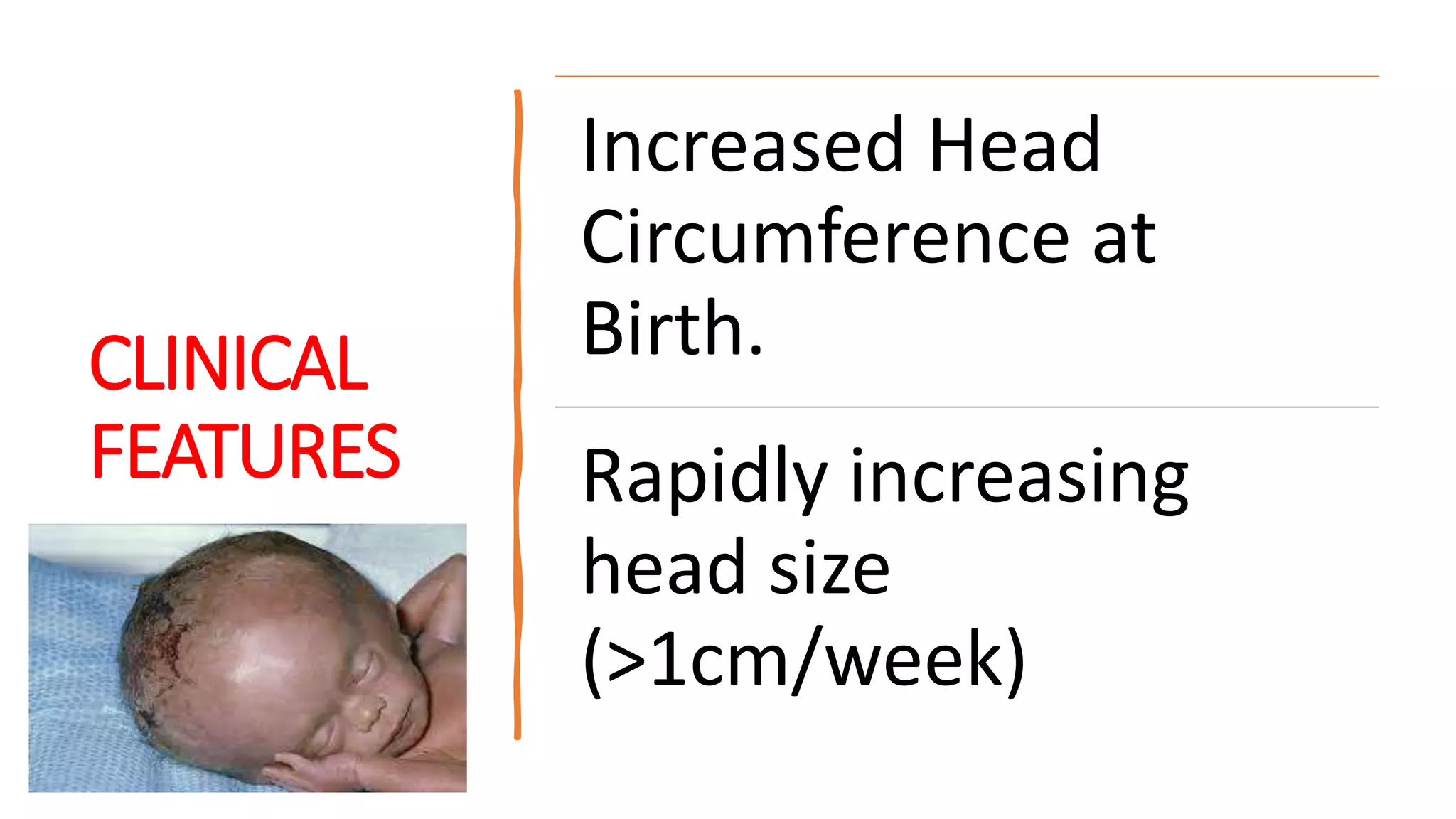
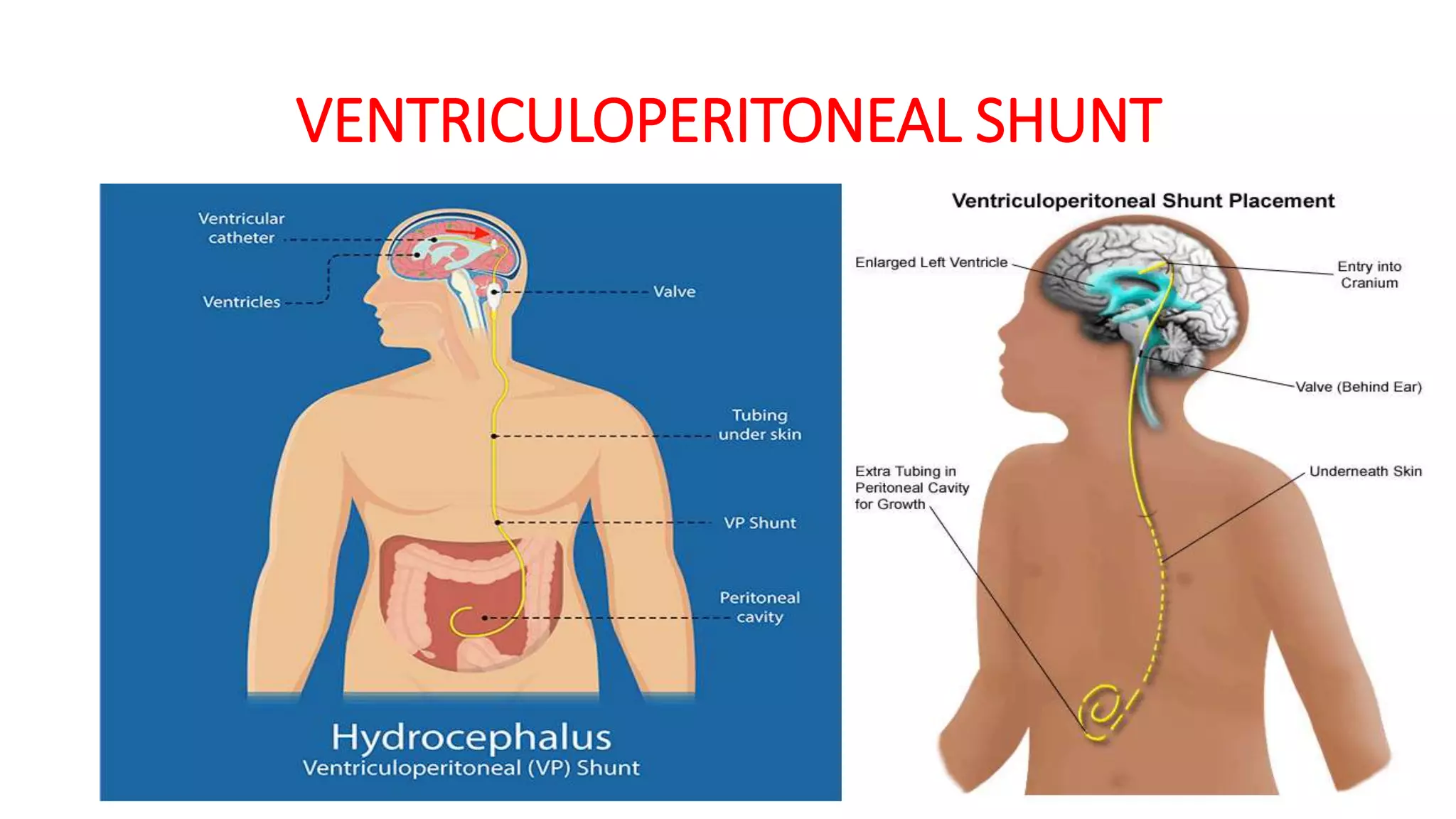
HYDROCEPHALUS
Dr Kaushik Barot
 Total volume of CSF
 125-150 ml (1.5-
2ml/kg) in
adults,50ml in
infants at any time.
 Ultrafiltration
 500 ml per day,
 25ml/hour
 Children-3ml/kg
 Infants-4ml/kg
 ICP is pulsatile
 Newborn < 5mm
of Hg
 Infants-6-15 mm
 Children-10-15
mm
WHAT
IS
HDROCEPHALUS ?
Increasedventricular
sizedue toincreasein
volume of CSF due to
either
– Increased
production
– Obstruction
– Impaired
absorption
DON’T CONFUSE !
HYDROCEPHALUS HYDRANCEPHALY
TYPES OF HYDROCEPHALUS
1) COMMUNICATING
OR
NON-OBSTRUCTIVE
2) NON-COMMUNICATING
or
OBSTRUCTIVE
3) MIXED
NON-COMMUNICATING
Or OBSTRUCTIVE
ACQUIRED
CONGENITAL
1) CONGENITAL ANOMALIES
• Aqueductalstenosis(xlinked)
• Arnold Chiarimalformation
• Dandy walkermalformation
• Vein of Galenmalformation
• Spinabifida
• AVmalformation
2)CONGENITALINFECTIONS
• Toxoplasmosis-aqueductalstenosis
• Zikavirus
INFECTIONS VENTRICULARHEMORRHAGE
• Prematurity
• AVmalformation
• Meningitis
• Mumps
SPACEOCCUPYINGLESION
• Intra‐ventriculartumor
• Posteriorfossatumor
COMMUNICATING or
NON-OBSTRUCTIVE
INFECTIONS
OVERPRODUCTION
UNDERABSORPTION
 CONGENITALDEFICIENCYOF
ARACHNOID GRANULATION
 POSTHEMORRHAGIC
 INFLAMMATION asinmeningitis
 CHOROID PLEXUS PAPILLOMA
 CHOROID PLEXUS CARCINOMA
 CHOROID PLEXUS
INFLAMMATION
HEMORRHAGE
 CONGENITAL INFECTIONS
 MENINGITIS (PYO OR
TBME)
 SUBARACHNOID
HEMORRHAGE
 TRAUMA
 Achondroplasia
 Normal pressure
hydrocephalus
 Hydrocephalus ex vacuo
OTHERS
DEFECTSIN SUBAROCHNOID
SPACE
INFILTRATES
FOR PRACTICAL'S:- COMMON CAUSES
PATHOPHYSIOLOGY
Obstruction to CSFflow
Reversal of ventricular fluid into periventricularwhite
matter
Demyelination and progressive gliosis
Damageto periventricularwhitematterand latergray
matter
Risk of Herniation
CLINICAL
FEATURES
Increased Head
Circumference at
Birth.
Rapidly increasing
head size
(>1cm/week)
CLINICAL FEATURES
• Neonates and infants
• Irritability
• Poor appetite, Failure to thrive
• Vomiting
• Poor head control
• Tense fontanelle, Delayed fusion of sutures
• Dilated scalp veins
• Sun Setting sign (visible sclera above cornea)
• Macewans or crack pot sign positive (>1 yr.
of age)
• Transillumination may be positive
SUN SETTING SIGN CRACKPOT (MACEWEN’S SIGN
TRANSILLUMINATION
• Torch with rubber rim in dark
room.
• Rim of lucency >2-2.5 cm in frontal
region
• >1cm in occipital region
• POSITIVE in
1) Hydrocephalus
2) Hydrancephaly
3) Porencephaly
4) Subdural effusion
5) Subudral hematoma
CLINICAL FEATURES
• Older children
• Sign S/S raised ICT
• Headache, worst in the morning
• Nausea and vomiting
• Blurred vision
• Personality and behavioral
disturbances
• Gait abnormalities
• Drowsiness/depressed level of
consciousness
• Seizures
• Cushings triad:- (bradycardia,
irregular respiration, wide pulse
pressure)
EXAMINATION
Open squamoparietal suture beyond one month is
an early sign
• Serial HC measurement/HC more than 2 std. deviation
• Papilledema
• Abducens palsy
• Pyramidal tract lesions ( lower extremities )
• Crackpot sign positive
FEW
MALFORMATIONS
AHEAD !
POSTERIOR FOSSA
CONTENTS
• CEREBELLUM
• BRAIN STEM
• OCCIPITAL LOBE
• 4th VENTRICLE
CHIARIMALFORMATION or
previously k/a Arnold Chiari malformation
• CEREBELLAR
TONSILS
TYPE - I
• CEREBELLAR
TONSILS
• VERMIS
• BRAIN STEM
TYPE-II TYPE -III
Per mont
DOWNWARDS DISPLACMENT towards the
foramen magnum OF THE………
CHIARIMALFORMATION
TYPE 1
• Seen inadolescence
• Not associated with
Hydrocephalus
• Headache,Neckpain
• Progressivespasticity
• Associated with
Syringomyelia
TYPE 2
• Seen in infants
• Progressivehydrocephalus
Weak cry, stridor, apnea
• Abnormality ofgait, Spasticity
• Incoordination
• Associated with Myelo‐meningocele,
Spina bifida
DANDY WALKER MALFORMATION
• Failureof development of roof
of4th ventricle
• Cysticdilatationof 4thventricle
(Blake's pouch)
• Cerebellarhypoplasia
• Ass.Anomalies‐absenceof
corpuscallosum
DANDY WALKERMALFORMATION
Clinical features
• Increasing headsize
• Prominentocciput
• Cerebellarataxia
• Delayed motor &
cognitivedevelopment
• Trans‐illumination
VEIN OF GALEN ANEURYSMAL MALFORMATIONS
• VGAM consist of a
tangled mass of dilated
vessels supplied by an
enlarged artery.
• Hydrocephalus, High
output cardiac failure.
HOW TO REMEMBER !
NORMAL PRESURE HYDROCEPHALUS
Rare in children
• Gait disturbance
• Dementia
• Urinary incontinence
HAKIMS or Adams triad consists of:-
HYDROCEPHALUS EX VACUO
• EX-out of
Vacuo-space
• Enlargement of cerebral
ventricles & subarachnoid
spaces, caused by
encephalic volume loss.
• NOT a true hydrocephalus
• Increases in CSF
volume without
increased CSF pressure
DO ALL HYDROCEPHALUS PROGRESS ?...NO
ARRESTED HYDROCEPHALUS
• A large proportion of
congenital and acquired
hydrocephalus may
undergo spontaneous
arrest.
• No surgical intervention
needed.
INVESTIGATIONS
X‐RAYSKULL
Separated sutures
Silver beaten appearance
Shallow orbit
ULTRASOUND BRAIN
Screening if AF is open.
Cheap, nonionizing, can be done at
bedside.
Detects IVH in preterm.
Serial monitoring of ventricular size.
CTSCAN
Useful when AF is closed
WATCH FOR
1) Ventricle size
2) Cortical mantle
3) Periventricular ooze
4) Associated malformation
5) EVANS ratio (lateral
ventricular width:
hemispheric width >33%-
needs surgery)
MRI BRAIN
•Dilated ventricles
•Useful when
suspecting posterior
fossa or white matter
lesions.
TREATMENT
•Goals of therapy is to decrease
ICT to same limits preserving
brain tissue.
•Mild /Arrested cases managed
medically.
•Severe cases with
cortical mantle < 1 cm
optic atrophy
associated anomalies
Won’t benefit from surgery.
•Moderate cases should be
operated early.
MEDICAL
TREATMENT
Reduction of CSFproduction
•Acetazolamide 50-100mg/kg/day.
w/f metabolic acidosis
•Oral glycerol 1ml/kg/dose 8 hourly
•Mannitol for emergency (5ml/kg
f/b 2ml/kg qds x 2 days)
•Furosemide 0.5-2mg/kg/day
INDICATIONS
FOR
SURGERY
•Progressive thinning of
cortical mantle despite
medical therapy
•Cortical mantle less then 2-
2.5 cm especially in infancy
•Papilledema on fundoscopy
•Periventricular ooze on CT
scan
•Evans ratio > 33 %
SURGICAL
TREATMENT
 Diversion of CSFto extra
cranialsites using shunts
 Ventriculo peritoneal
 Ventriculo atrial
 Ventriculo azygous
 Theco peritoneal
 Endoscopic third
ventriculostomy
VENTRICULOPERITONEAL SHUNT
TYPESOF VPSHUNT
1) Chabra’s
2) Pudenz – Hakin
3) Splitz – Holter valve
TREATMENT
•Opening of ventricular
system into subarachnoid
space via lamina terminalis.
Ventriculostomy
•TB meningitis – ATT
•Pyogenic Meningitis ‐
Antibiotics
Treatment of cause :
EXTERNAL VENTRICULAR DRAIN DEVICE
COMPLICATIONS
OF VPSHUNT
Blockage
Infection
Shunt dependence
Slit Ventricle Syndrome
Migration of tube
Intestinal obstruction
Peritonitis
Arrhythmias
PROGNOSIS
•Untreated – 50 % mortality in
severe hydrocephalus
•Medical management- 30 %
improvement in ventriculomegaly
•VP shunt- 60 % improvement in
ventriculomegaly
44 % in fundus
•70% - neurologically impaired
Hydrocephalus

Hydrocephalus

Editor's Notes

  • #4 Intracranial pressure refers to the pressure of CSF within the cranium. It is normally pulsatile and less than 5 mm Hg in newborns, 6-15 mm Hg in infants and 10-15 mm Hg in older children. Intracranial pressure is considered severely elevated, if raised above 40 mm Hg.
  • #9 : When the narrowing near the base of the spine 
  • #12 Central transtentorial herniation is common in children with progressive hydrocephalus  to protrude through an abnormal body opening
  • #15 > 2 cm transillumination is positive
  • #40 Shunt dictionary meaning- to move something from one place to other