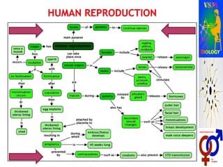
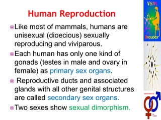
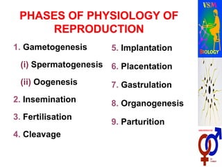
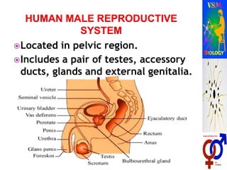
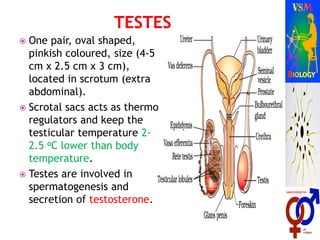
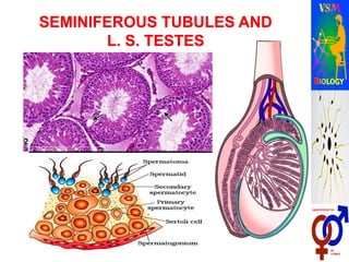
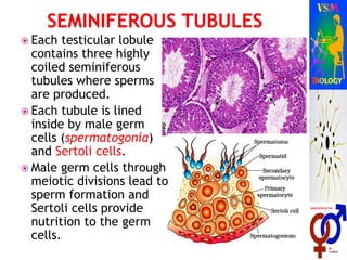
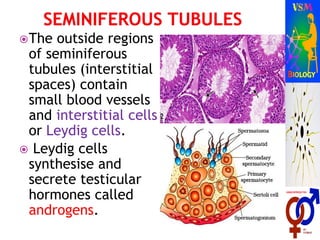
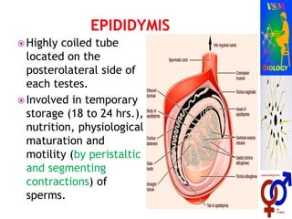
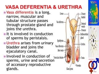
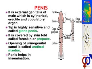
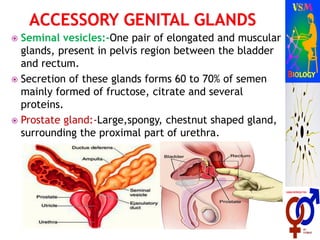
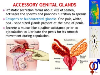
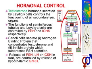
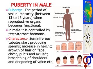
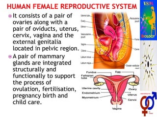
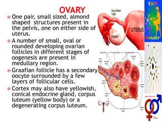
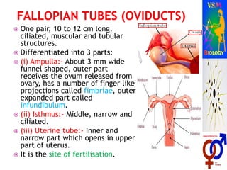
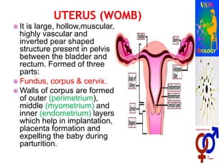
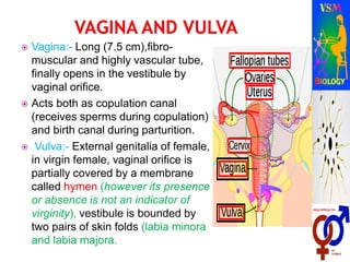
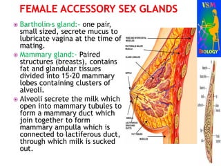
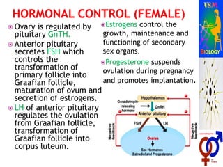
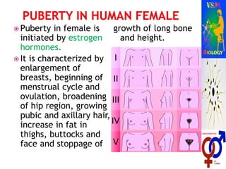
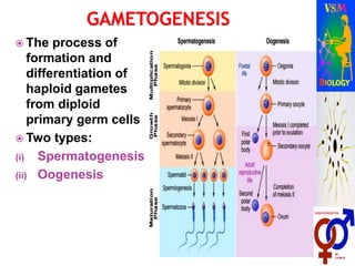
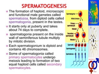
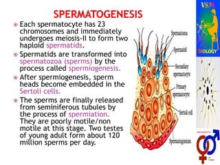
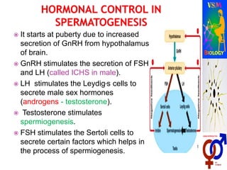
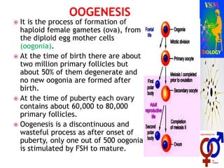
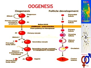
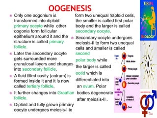
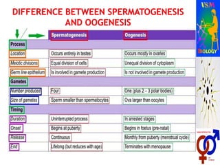
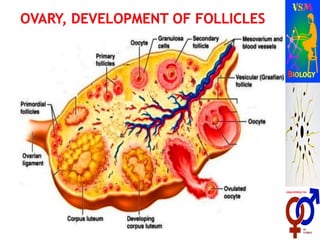
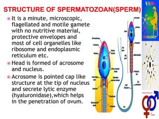
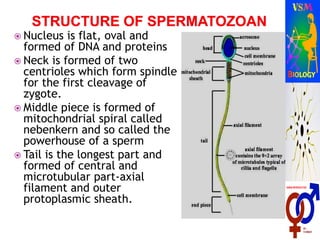
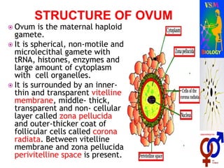
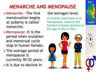
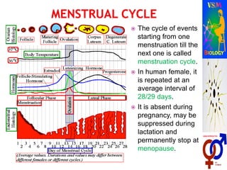
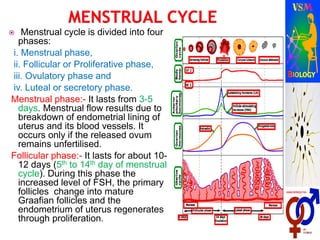
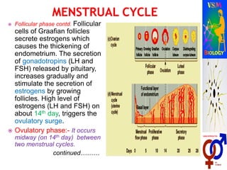
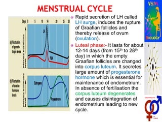
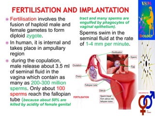
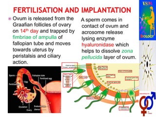
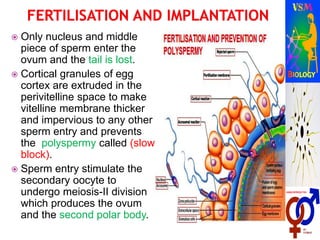
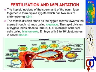
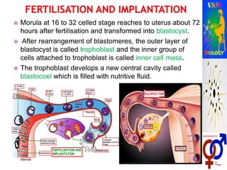
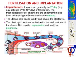
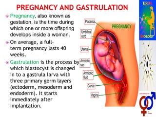
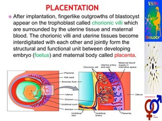
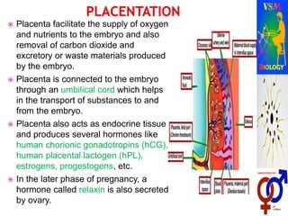
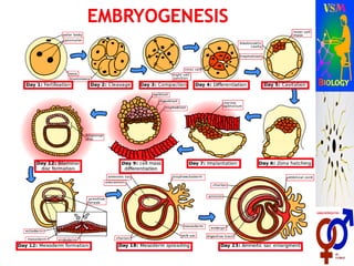
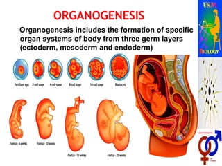
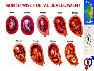
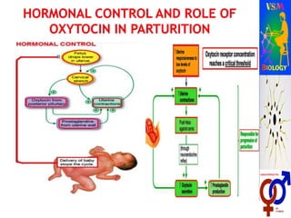
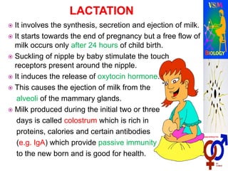
By:- V. S. Malik
Biology Lecturer (Retd.)
Like most of mammals, humans are
unisexual (dioecious) sexually
reproducing and viviparous.
Each human has only one kind of
gonads (testes in male and ovary in
female) as primary sex organs.
 Reproductive ducts and associated
glands with all other genital structures
are called secondary sex organs.
Two sexes show sexual dimorphism.
1. Gametogenesis
(i) Spermatogenesis
(ii) Oogenesis
2. Insemination
3. Fertilisation
4. Cleavage
5. Implantation
6. Placentation
7. Gastrulation
8. Organogenesis
9. Parturition
Located in pelvic region.
Includes a pair of testes, accessory
ducts, glands and external genitalia.
 One pair, oval shaped,
pinkish coloured, size (4-5
cm x 2.5 cm x 3 cm),
located in scrotum (extra
abdominal).
 Scrotal sacs acts as thermo
regulators and keep the
testicular temperature 2-
2.5 oC lower than body
temperature.
 Testes are involved in
spermatogenesis and
secretion of testosterone.
 Each testicular lobule
contains three highly
coiled seminiferous
tubules where sperms
are produced.
 Each tubule is lined
inside by male germ
cells (spermatogonia)
and Sertoli cells.
 Male germ cells through
meiotic divisions lead to
sperm formation and
Sertoli cells provide
nutrition to the germ
cells.
The outside regions
of seminiferous
tubules (interstitial
spaces) contain
small blood vessels
and interstitial cells
or Leydig cells.
 Leydig cells
synthesise and
secrete testicular
hormones called
androgens.
 Highly coiled tube
located on the
posterolateral side of
each testes.
 Involved in temporary
storage (18 to 24 hrs.),
nutrition, physiological
maturation and
motility (by peristaltic
and segmenting
contractions) of
sperms.
 Vasa differentia is a long,
narrow, muscular and
tubular structure passes
through prostate gland and
joins the urethra.
 It is involved in conduction
of sperms by peristalsis.
 Urethra arises from urinary
bladder and joins the
ejaculatory canal.
 Involved in conduction of
sperms, urine and secretion
of accessory reproductive
glands.
 It is external genitalia of
male which is cylindrical,
erectile and copulatory
organ.
 Tip is highly sensitive and
called glans penis.
 It is covered by skin fold
called foreskin or prepuce.
 Opening of urinogenital
canal is called urethral
maetus.
 Penis helps in
insemination.
 Seminal vesicles:-One pair of elongated and muscular
glands, present in pelvis region between the bladder
and rectum.
 Secretion of these glands forms 60 to 70% of semen
mainly formed of fructose, citrate and several
proteins.
 Prostate gland:-Large,spongy, chestnut shaped gland,
surrounding the proximal part of urethra.
 Prostatic secretion forms about 20% of semen,
activates the sperms and provides nutrition to sperms.
 Cowper's or Bulbourethral glands:- One pair, white,
pea - seed sized glands present at the base of penis.
 Secrete a mucus-like alkaline substance prior to
ejaculation to lubricate the penis for its smooth
movement during copulation.
 Testosterone hormone secreted
by Leydig,s cells controls the
functioning of all secondary sex
organs.
 The functions of seminiferous
tubules and Leydig,s cells are
controlled by FSH and ICHS
respectively.
 Sertoli cells secrete (i) Androgen
Binding Protein that
concentrates testosterone and
(ii) Inhibin protein which
suppresses FSH secretion.
 Release of FSH, LH or ICSH in
turn, are controlled by release of
hypothalamic GnRH.
 Puberty:- The period of
sexual maturity (between
13 to 16 years) when
reproductive organs
becomes functional.
 In male it is controlled by
testosterone hormone.
 Characters:- Seminiferous
tubules start producing
sperms; increase in height;
growth of hair on face,
chest, pubis and axillae;
broadening of shoulders
and deepening of voice etc.
 It consists of a pair of
ovaries along with a
pair of oviducts, uterus,
cervix, vagina and the
external genitalia
located in pelvic region.
 A pair of mammary
glands are integrated
structurally and
functionally to support
the process of
ovulation, fertilisation,
pregnancy birth and
child care.
 One pair, small sized, almond
shaped structures present in
the pelvis, one on either side of
uterus.
 A number of small, oval or
rounded developing ovarian
follicles in different stages of
oogenesis are present in
medullary region.
 Graafian follicle has a secondary
oocyte surrounded by a few
layers of follicular cells.
 Cortex may also have yellowish,
conical endocrine gland, corpus
luteum (yellow body) or a
degenerating corpus luteum.
.
 One pair, 10 to 12 cm long,
ciliated, muscular and tubular
structures.
 Differentiated into 3 parts:
 (i) Ampulla:- About 3 mm wide
funnel shaped, outer part
receives the ovum released from
ovary, has a number of finger like
projections called fimbriae, outer
expanded part called
infundibulum.
 (ii) Isthmus:- Middle, narrow and
ciliated.
 (iii) Uterine tube:- Inner and
narrow part which opens in upper
part of uterus.
 It is the site of fertilisation.
 It is large, hollow,muscular,
highly vascular and
inverted pear shaped
structure present in pelvis
between the bladder and
rectum. Formed of three
parts:
 Fundus, corpus & cervix.
 Walls of corpus are formed
of outer (perimetrium),
middle (myometrium) and
inner (endometrium) layers
which help in implantation,
placenta formation and
expelling the baby during
parturition.
 Vagina:- Long (7.5 cm),fibro-
muscular and highly vascular tube,
finally opens in the vestibule by
vaginal orifice.
 Acts both as copulation canal
(receives sperms during copulation)
and birth canal during parturition.
 Vulva:- External genitalia of female,
in virgin female, vaginal orifice is
partially covered by a membrane
called hymen (however its presence
or absence is not an indicator of
virginity), vestibule is bounded by
two pairs of skin folds (labia minora
and labia majora.
 Bartholin,s gland:- one pair,
small sized, secrete mucus to
lubricate vagina at the time of
mating.
 Mammary gland:- Paired
structures (breasts), contains
fat and glandular tissues
divided into 15-20 mammary
lobes containing clusters of
alveoli.
 Alveoli secrete the milk which
open into mammary tubules to
form a mammary duct which
join together to form
mammary ampulla which is
connected to lactiferous duct,
through which milk is sucked
out.
 Ovary is regulated by
pituitary GnTH.
 Anterior pituitary
secretes FSH which
controls the
transformation of
primary follicle into
Graafian follicle,
maturation of ovum and
secretion of estrogens.
 LH of anterior pituitary
regulates the ovulation
from Graafian follicle,
transformation of
Graafian follicle into
corpus luteum.
Estrogens control the
growth, maintenance and
functioning of secondary
sex organs.
Progesterone suspends
ovulation during pregnancy
and promotes implantation.
 Puberty in female is
initiated by estrogen
hormones.
 It is characterized by
enlargement of
breasts, beginning of
menstrual cycle and
ovulation, broadening
of hip region, growing
pubic and axillary hair,
increase in fat in
thighs, buttocks and
face and stoppage of
growth of long bone
and height.
 The process of
formation and
differentiation of
haploid gametes
from diploid
primary germ cells
 Two types:
(i) Spermatogenesis
(ii) Oogenesis
 The formation of haploid, microscopic
and functional male gametes called
spermatozoa, from diploid cells called
spermatogonia, present in the testes.
 It starts only at puberty and takes
about 74 days to complete.
 spermatogonia present on the inside
wall of seminiferous tubule multiply
by mitotic division.
 Each spermatogonium is diploid and
contains 46 chromosomes.
 Some of spermatogonia called
primary spermatocytes undergo
meiosis leading to formation of two
equal haploid cells called secondary
spermatocytes.
 Each spermatocyte has 23
chromosomes and immediately
undergoes meiosis-II to form two
haploid spermatids.
 Spermatids are transformed into
spermatozoa (sperms) by the
process called spermiogenesis.
 After spermiogenesis, sperm
heads become embedded in the
Sertoli cells.
 The sperms are finally released
from seminiferous tubules by
the process of spermiation.
They are poorly motile/non
motile at this stage. Two testes
of young adult form about 120
million sperms per day.
 It starts at puberty due to increased
secretion of GnRH from hypothalamus
of brain.
 GnRH stimulates the secretion of FSH
and LH (called ICHS in male).
 LH stimulates the Leydig,s cells to
secrete male sex hormones
(androgens - testosterone).
 Testosterone stimulates
spermiogenesis.
 FSH stimulates the Sertoli cells to
secrete certain factors which helps in
the process of spermiogenesis.
 It is the process of formation of
haploid female gametes (ova), from
the diploid egg mother cells
(oogonia).
 At the time of birth there are about
two million primary follicles but
about 50% of them degenerate and
no new oogonia are formed after
birth.
 At the time of puberty each ovary
contains about 60,000 to 80,000
primary follicles.
 Oogenesis is a discontinuous and
wasteful process as after onset of
puberty, only one out of 500 oogonia
is stimulated by FSH to mature.
 Only one oogonium is
transformed into diploid
primary oocyte while other
oogonia form follicular
epithelium around it and the
structure is called primary
follicle.
 Later the secondary oocyte
gets surrounded more
granulosal layers and changes
into secondary follicle.
 A fluid filled cavity (antrum) is
formed inside it and it is now
called tertiary follicle.
 It further changes into Graafian
follicle.
 Diploid and fully grown primary
oocyte undergoes meiosis-I to
form two unequal haploid cells,
the smaller is called first polar
body and the larger is called
secondary oocyte.
 Secondary oocyte undergoes
meiosis-II to form two unequal
cells and smaller is called
second
polar body while
the larger is called
ootid which is
differentiated into
an ovum. Polar
bodies degenerate
after meiosis-II .
 It is a minute, microscopic,
flagellated and motile gamete
with no nutritive material,
protective envelopes and
most of cell organelles like
ribosome and endoplasmic
reticulum etc.
 Head is formed of acrosome
and nucleus.
 Acrosome is pointed cap like
structure at the tip of nucleus
and secrete lytic enzyme
(hyaluronidase),which helps
in the penetration of ovum.
 Nucleus is flat, oval and
formed of DNA and proteins
 Neck is formed of two
centrioles which form spindle
for the first cleavage of
zygote.
 Middle piece is formed of
mitochondrial spiral called
nebenkern and so called the
powerhouse of a sperm
 Tail is the longest part and
formed of central and
microtubular part-axial
filament and outer
protoplasmic sheath.
 Ovum is the maternal haploid
gamete.
 It is spherical, non-motile and
microlecithal gamete with
tRNA, histones, enzymes and
large amount of cytoplasm
with cell organelles.
 It is surrounded by an inner-
thin and transparent vitelline
membrane, middle- thick,
transparent and non- cellular
layer called zona pellucida
and outer-thicker coat of
follicular cells called corona
radiata. Between vitelline
membrane and zona pellucida
perivitelline space is present.
 Menarche:- The first
menstruation begins
at puberty is called
menarche.
 Menopause:-It is the
period when ovulation
and menstrual cycle
stop in human female.
 The average period of
menopause is
currently 50-52 years.
 It is due to decline in
the estrogen level.
(In human male there is no
menopause, however the
function of testes declines at
the age of 40s or 50s.)
 The cycle of events
starting from one
menstruation till the
next one is called
menstruation cycle.
 In human female, it
is repeated at an
average interval of
28/29 days.
 It is absent during
pregnancy, may be
suppressed during
lactation and
permanently stop at
menopause.
 Menstrual cycle is divided into four
phases:
i. Menstrual phase,
ii. Follicular or Proliferative phase,
iii. Ovulatory phase and
iv. Luteal or secretory phase.
Menstrual phase:- It lasts from 3-5
days. Menstrual flow results due to
breakdown of endometrial lining of
uterus and its blood vessels. It
occurs only if the released ovum
remains unfertilised.
Follicular phase:- It lasts for about 10-
12 days (5th to 14th day of menstrual
cycle). During this phase the
increased level of FSH, the primary
follicles change into mature
Graafian follicles and the
endometrium of uterus regenerates
through proliferation.
 Follicular phase contd. Follicular
cells of Graafian follicles
secrete estrogens which
causes the thickening of
endometrium. The secretion
of gonadotropins (LH and
FSH) released by pituitary,
increases gradually and
stimulate the secretion of
estrogens by growing
follicles. High level of
estrogens (LH and FSH) on
about 14th day, triggers the
ovulatory surge.
 Ovulatory phase:- It occurs
midway (on 14th day) between
two menstrual cycles.
continued………
 Rapid secretion of LH called
LH surge, induces the rupture
of Graafian follicles and
thereby release of ovum
(ovulation).
 Luteal phase:- It lasts for about
12-14 days (from 15th to 28th
day) in which the empty
Graafian follicles are changed
into corpus luteum. It secretes
large amount of progesterone
hormone which is essential for
maintenance of endometrium.
In absence of fertilisation the
corpus luteum degenerates
and causes disintegration of
endometrium leading to new
cycle.
 Fertilisation involves the
fusion of haploid male and
female gametes to form
diploid zygote.
 In human, it is internal and
takes place in ampullary
region
 during the copulation,
male release about 3.5 ml
of seminal fluid in the
vagina which contain as
many as 200-300 million
sperms. Only about 100
sperms reach the fallopian
tube (because about 50% are
killed by acidity of female genital
tract and many sperms are
engulfed by phagocytes of
vaginal epithelium).
Sperms swim in the
seminal fluid at the rate
of 1-4 mm per minute.
 Ovum is released from the
Graafian follicles of ovary
on 14th day and trapped by
fimbriae of ampulla of
fallopian tube and moves
towards uterus by
peristalsis and ciliary
action.
A sperm comes in
contact of ovum and
acrosome release
lysing enzyme
hyaluronidase which
helps to dissolve zona
pellucida layer of ovum.
 Only nucleus and middle
piece of sperm enter the
ovum and the tail is lost.
 Cortical granules of egg
cortex are extruded in the
perivitelline space to make
vitelline membrane thicker
and impervious to any other
sperm entry and prevents
the polyspermy called (slow
block).
 Sperm entry stimulate the
secondary oocyte to
undergo meiosis-II division
which produces the ovum
and the second polar body.
 The haploid nucleus of the sperm and of the ovum fuse
together to form diploid zygote which has two sets of
chromosomes (2n).
 The mitotic division starts as the zygote moves towards the
uterus through isthmus called cleavage. The rapid division
of zygote takes place to form 2, 4, 8,16 hollow, spherical
cells called blastomeres. Embryo with 8 to 16 blastomeres
is called morula.
 Morula at 16 to 32 celled stage reaches to uterus about 72
hours after fertilisation and transformed into blastocyst.
 After rearrangement of blastomeres, the outer layer of
blastocyst is called trophoblast and the inner group of
cells attached to trophoblast is called inner cell mass.
 The trophoblast develops a new central cavity called
blastocoel which is filled with nutritive fluid.
 Implantation:- It may occur generally on 7th day (any
day between 6th to 10th day) of fertilisation. The
trophoblast layer get attached to the endometrium and
inner cell mass get differentiated as the embryo.
 The uterine cells divide rapidly and covers the blastocyst.
 The blastocyst becomes embedded in the endometrium of
the uterus. This is called implantation and leads to
pregnancy.
 Pregnancy, also known as
gestation, is the time during
which one or more offspring
develops inside a woman.
 On average, a full-
term pregnancy lasts 40
weeks.
 Gastrulation is the process by
which blastocyst is changed
in to a gastrula larva with
three primary germ layers
(ectoderm, mesoderm and
endoderm). It starts
immediately after
implantation.
 After implantation, fingerlike outgrowths of blastocyst
appear on the trophoblast called chorionic villi which
are surrounded by the uterine tissue and maternal
blood. The chorionic villi and uterine tissues become
interdigitated with each other and jointly form the
structural and functional unit between developing
embryo (foetus) and maternal body called placenta.
 Placenta facilitate the supply of oxygen
and nutrients to the embryo and also
removal of carbon dioxide and
excretory or waste materials produced
by the embryo.
 Placenta is connected to the embryo
through an umbilical cord which helps
in the transport of substances to and
from the embryo.
 Placenta also acts as endocrine tissue
and produces several hormones like
human chorionic gonadotropins (hCG),
human placental lactogen (hPL),
estrogens, progestogens, etc.
 In the later phase of pregnancy, a
hormone called relaxin is also secreted
by ovary.
.
Organogenesis includes the formation of specific
organ systems of body from three germ layers
(ectoderm, mesoderm and endoderm)
STAGE Embryo/
Foetus
Important features
1 Month Embryo consists of two layers of cells from which
all organs and body parts will develop. Embryo's
heart begins to beat; fore and hind limb buds liver
and pancreatic buds are formed.
2 Months Embryo is now about the size of a kidney bean and
is constantly moving. This has distinct, slightly
webbed fingers. Limbs become paddle-like. Nasal
pits, pinna tubercles and eyelids established.
3 Months By now the foetus is around 7 to 8 cm long and weighs
about the same as half a banana. Its tiny, unique
fingerprints are now in place. Major organ systems are
established; external genitalia and limbs well developed.
4 Months Foetus is now about 13cm long and weighs 140g.
Its skeleton is starting to harden from rubbery
cartilage to bone. Medullated nerve fibres
formation begins.
5 Months Eyebrows and eyelids are now in place.
Foetus would now be more than 27 cm long.
First movement of foetus is noticed. Eruption
of hair on head begins.
6 Months Foetus weighs about 660 g. Its wrinkled skin is
starting to smooth out as he/she puts on baby
fat. Appearance of fine hair on whole body.
7 Months By now, foetus is about 35-40 cm long. It can
open and close its eyes and responds to all
sounds. If born prematurely, the baby would
be likely to survive.
8 Months Foetus now weighs about 2.2 kg. Its layers of
fat are filling him out, making it rounder, and
its lungs are well developed. Testes descend
in scrotal sacs.
9 Months Foetus is fully developed and is ready for
birth. At birth, the average baby is about
51cm long from head to toe and weighs an
average of 2.9kg.
 It is the expelling of fully developed
foetus (baby) from the mother's uterus
after the gestation period (about 280
days or 40 weeks) after the last
menstruation.
 It is induced by complex
neuroendocrine mechanism, which is
triggered by fully formed foetus and
the placenta called foetal ejection
reflex.
 This triggers release of oxytocin from
the mother's posterior pituitary.
 Oxytocin causes the forceful
contraction of smooth muscles of
myometrium, called labour pains.
 Labour pains pushes the baby
gradually out through the dilated
cervix (caused by relaxin) and
vagina, with the head foremost.
 Uterine contraction stimulates
further secretion of oxytocin
resulting in stronger and stronger
contractions.
 The stimulatory reflex between the
uterine contraction and oxytocin
secretion continues resulting to
expulsion of baby out of the uterus
through the birth canal.
 Soon after the birth of baby, the
placenta is also expelled out of the
uterus.
 It involves the synthesis, secretion and ejection of milk.
 It starts towards the end of pregnancy but a free flow of
milk occurs only after 24 hours of child birth.
 Suckling of nipple by baby stimulate the touch
receptors present around the nipple.
 It induces the release of oxytocin hormone.
 This causes the ejection of milk from the
alveoli of the mammary glands.
 Milk produced during the initial two or three
days is called colostrum which is rich in
proteins, calories and certain antibodies
(e.g. IgA) which provide passive immunity
to the new born and is good for health.
Human

Human

  • 1.
    By:- V. S.Malik Biology Lecturer (Retd.)
  • 3.
    Like most ofmammals, humans are unisexual (dioecious) sexually reproducing and viviparous. Each human has only one kind of gonads (testes in male and ovary in female) as primary sex organs.  Reproductive ducts and associated glands with all other genital structures are called secondary sex organs. Two sexes show sexual dimorphism.
  • 4.
    1. Gametogenesis (i) Spermatogenesis (ii)Oogenesis 2. Insemination 3. Fertilisation 4. Cleavage 5. Implantation 6. Placentation 7. Gastrulation 8. Organogenesis 9. Parturition
  • 5.
    Located in pelvicregion. Includes a pair of testes, accessory ducts, glands and external genitalia.
  • 6.
     One pair,oval shaped, pinkish coloured, size (4-5 cm x 2.5 cm x 3 cm), located in scrotum (extra abdominal).  Scrotal sacs acts as thermo regulators and keep the testicular temperature 2- 2.5 oC lower than body temperature.  Testes are involved in spermatogenesis and secretion of testosterone.
  • 8.
     Each testicularlobule contains three highly coiled seminiferous tubules where sperms are produced.  Each tubule is lined inside by male germ cells (spermatogonia) and Sertoli cells.  Male germ cells through meiotic divisions lead to sperm formation and Sertoli cells provide nutrition to the germ cells.
  • 9.
    The outside regions ofseminiferous tubules (interstitial spaces) contain small blood vessels and interstitial cells or Leydig cells.  Leydig cells synthesise and secrete testicular hormones called androgens.
  • 10.
     Highly coiledtube located on the posterolateral side of each testes.  Involved in temporary storage (18 to 24 hrs.), nutrition, physiological maturation and motility (by peristaltic and segmenting contractions) of sperms.
  • 11.
     Vasa differentiais a long, narrow, muscular and tubular structure passes through prostate gland and joins the urethra.  It is involved in conduction of sperms by peristalsis.  Urethra arises from urinary bladder and joins the ejaculatory canal.  Involved in conduction of sperms, urine and secretion of accessory reproductive glands.
  • 12.
     It isexternal genitalia of male which is cylindrical, erectile and copulatory organ.  Tip is highly sensitive and called glans penis.  It is covered by skin fold called foreskin or prepuce.  Opening of urinogenital canal is called urethral maetus.  Penis helps in insemination.
  • 13.
     Seminal vesicles:-Onepair of elongated and muscular glands, present in pelvis region between the bladder and rectum.  Secretion of these glands forms 60 to 70% of semen mainly formed of fructose, citrate and several proteins.  Prostate gland:-Large,spongy, chestnut shaped gland, surrounding the proximal part of urethra.
  • 14.
     Prostatic secretionforms about 20% of semen, activates the sperms and provides nutrition to sperms.  Cowper's or Bulbourethral glands:- One pair, white, pea - seed sized glands present at the base of penis.  Secrete a mucus-like alkaline substance prior to ejaculation to lubricate the penis for its smooth movement during copulation.
  • 15.
     Testosterone hormonesecreted by Leydig,s cells controls the functioning of all secondary sex organs.  The functions of seminiferous tubules and Leydig,s cells are controlled by FSH and ICHS respectively.  Sertoli cells secrete (i) Androgen Binding Protein that concentrates testosterone and (ii) Inhibin protein which suppresses FSH secretion.  Release of FSH, LH or ICSH in turn, are controlled by release of hypothalamic GnRH.
  • 16.
     Puberty:- Theperiod of sexual maturity (between 13 to 16 years) when reproductive organs becomes functional.  In male it is controlled by testosterone hormone.  Characters:- Seminiferous tubules start producing sperms; increase in height; growth of hair on face, chest, pubis and axillae; broadening of shoulders and deepening of voice etc.
  • 17.
     It consistsof a pair of ovaries along with a pair of oviducts, uterus, cervix, vagina and the external genitalia located in pelvic region.  A pair of mammary glands are integrated structurally and functionally to support the process of ovulation, fertilisation, pregnancy birth and child care.
  • 18.
     One pair,small sized, almond shaped structures present in the pelvis, one on either side of uterus.  A number of small, oval or rounded developing ovarian follicles in different stages of oogenesis are present in medullary region.  Graafian follicle has a secondary oocyte surrounded by a few layers of follicular cells.  Cortex may also have yellowish, conical endocrine gland, corpus luteum (yellow body) or a degenerating corpus luteum. .
  • 19.
     One pair,10 to 12 cm long, ciliated, muscular and tubular structures.  Differentiated into 3 parts:  (i) Ampulla:- About 3 mm wide funnel shaped, outer part receives the ovum released from ovary, has a number of finger like projections called fimbriae, outer expanded part called infundibulum.  (ii) Isthmus:- Middle, narrow and ciliated.  (iii) Uterine tube:- Inner and narrow part which opens in upper part of uterus.  It is the site of fertilisation.
  • 20.
     It islarge, hollow,muscular, highly vascular and inverted pear shaped structure present in pelvis between the bladder and rectum. Formed of three parts:  Fundus, corpus & cervix.  Walls of corpus are formed of outer (perimetrium), middle (myometrium) and inner (endometrium) layers which help in implantation, placenta formation and expelling the baby during parturition.
  • 21.
     Vagina:- Long(7.5 cm),fibro- muscular and highly vascular tube, finally opens in the vestibule by vaginal orifice.  Acts both as copulation canal (receives sperms during copulation) and birth canal during parturition.  Vulva:- External genitalia of female, in virgin female, vaginal orifice is partially covered by a membrane called hymen (however its presence or absence is not an indicator of virginity), vestibule is bounded by two pairs of skin folds (labia minora and labia majora.
  • 22.
     Bartholin,s gland:-one pair, small sized, secrete mucus to lubricate vagina at the time of mating.  Mammary gland:- Paired structures (breasts), contains fat and glandular tissues divided into 15-20 mammary lobes containing clusters of alveoli.  Alveoli secrete the milk which open into mammary tubules to form a mammary duct which join together to form mammary ampulla which is connected to lactiferous duct, through which milk is sucked out.
  • 23.
     Ovary isregulated by pituitary GnTH.  Anterior pituitary secretes FSH which controls the transformation of primary follicle into Graafian follicle, maturation of ovum and secretion of estrogens.  LH of anterior pituitary regulates the ovulation from Graafian follicle, transformation of Graafian follicle into corpus luteum. Estrogens control the growth, maintenance and functioning of secondary sex organs. Progesterone suspends ovulation during pregnancy and promotes implantation.
  • 24.
     Puberty infemale is initiated by estrogen hormones.  It is characterized by enlargement of breasts, beginning of menstrual cycle and ovulation, broadening of hip region, growing pubic and axillary hair, increase in fat in thighs, buttocks and face and stoppage of growth of long bone and height.
  • 25.
     The processof formation and differentiation of haploid gametes from diploid primary germ cells  Two types: (i) Spermatogenesis (ii) Oogenesis
  • 26.
     The formationof haploid, microscopic and functional male gametes called spermatozoa, from diploid cells called spermatogonia, present in the testes.  It starts only at puberty and takes about 74 days to complete.  spermatogonia present on the inside wall of seminiferous tubule multiply by mitotic division.  Each spermatogonium is diploid and contains 46 chromosomes.  Some of spermatogonia called primary spermatocytes undergo meiosis leading to formation of two equal haploid cells called secondary spermatocytes.
  • 27.
     Each spermatocytehas 23 chromosomes and immediately undergoes meiosis-II to form two haploid spermatids.  Spermatids are transformed into spermatozoa (sperms) by the process called spermiogenesis.  After spermiogenesis, sperm heads become embedded in the Sertoli cells.  The sperms are finally released from seminiferous tubules by the process of spermiation. They are poorly motile/non motile at this stage. Two testes of young adult form about 120 million sperms per day.
  • 28.
     It startsat puberty due to increased secretion of GnRH from hypothalamus of brain.  GnRH stimulates the secretion of FSH and LH (called ICHS in male).  LH stimulates the Leydig,s cells to secrete male sex hormones (androgens - testosterone).  Testosterone stimulates spermiogenesis.  FSH stimulates the Sertoli cells to secrete certain factors which helps in the process of spermiogenesis.
  • 29.
     It isthe process of formation of haploid female gametes (ova), from the diploid egg mother cells (oogonia).  At the time of birth there are about two million primary follicles but about 50% of them degenerate and no new oogonia are formed after birth.  At the time of puberty each ovary contains about 60,000 to 80,000 primary follicles.  Oogenesis is a discontinuous and wasteful process as after onset of puberty, only one out of 500 oogonia is stimulated by FSH to mature.
  • 31.
     Only oneoogonium is transformed into diploid primary oocyte while other oogonia form follicular epithelium around it and the structure is called primary follicle.  Later the secondary oocyte gets surrounded more granulosal layers and changes into secondary follicle.  A fluid filled cavity (antrum) is formed inside it and it is now called tertiary follicle.  It further changes into Graafian follicle.  Diploid and fully grown primary oocyte undergoes meiosis-I to form two unequal haploid cells, the smaller is called first polar body and the larger is called secondary oocyte.  Secondary oocyte undergoes meiosis-II to form two unequal cells and smaller is called second polar body while the larger is called ootid which is differentiated into an ovum. Polar bodies degenerate after meiosis-II .
  • 34.
     It isa minute, microscopic, flagellated and motile gamete with no nutritive material, protective envelopes and most of cell organelles like ribosome and endoplasmic reticulum etc.  Head is formed of acrosome and nucleus.  Acrosome is pointed cap like structure at the tip of nucleus and secrete lytic enzyme (hyaluronidase),which helps in the penetration of ovum.
  • 35.
     Nucleus isflat, oval and formed of DNA and proteins  Neck is formed of two centrioles which form spindle for the first cleavage of zygote.  Middle piece is formed of mitochondrial spiral called nebenkern and so called the powerhouse of a sperm  Tail is the longest part and formed of central and microtubular part-axial filament and outer protoplasmic sheath.
  • 36.
     Ovum isthe maternal haploid gamete.  It is spherical, non-motile and microlecithal gamete with tRNA, histones, enzymes and large amount of cytoplasm with cell organelles.  It is surrounded by an inner- thin and transparent vitelline membrane, middle- thick, transparent and non- cellular layer called zona pellucida and outer-thicker coat of follicular cells called corona radiata. Between vitelline membrane and zona pellucida perivitelline space is present.
  • 37.
     Menarche:- Thefirst menstruation begins at puberty is called menarche.  Menopause:-It is the period when ovulation and menstrual cycle stop in human female.  The average period of menopause is currently 50-52 years.  It is due to decline in the estrogen level. (In human male there is no menopause, however the function of testes declines at the age of 40s or 50s.)
  • 38.
     The cycleof events starting from one menstruation till the next one is called menstruation cycle.  In human female, it is repeated at an average interval of 28/29 days.  It is absent during pregnancy, may be suppressed during lactation and permanently stop at menopause.
  • 39.
     Menstrual cycleis divided into four phases: i. Menstrual phase, ii. Follicular or Proliferative phase, iii. Ovulatory phase and iv. Luteal or secretory phase. Menstrual phase:- It lasts from 3-5 days. Menstrual flow results due to breakdown of endometrial lining of uterus and its blood vessels. It occurs only if the released ovum remains unfertilised. Follicular phase:- It lasts for about 10- 12 days (5th to 14th day of menstrual cycle). During this phase the increased level of FSH, the primary follicles change into mature Graafian follicles and the endometrium of uterus regenerates through proliferation.
  • 40.
     Follicular phasecontd. Follicular cells of Graafian follicles secrete estrogens which causes the thickening of endometrium. The secretion of gonadotropins (LH and FSH) released by pituitary, increases gradually and stimulate the secretion of estrogens by growing follicles. High level of estrogens (LH and FSH) on about 14th day, triggers the ovulatory surge.  Ovulatory phase:- It occurs midway (on 14th day) between two menstrual cycles. continued………
  • 41.
     Rapid secretionof LH called LH surge, induces the rupture of Graafian follicles and thereby release of ovum (ovulation).  Luteal phase:- It lasts for about 12-14 days (from 15th to 28th day) in which the empty Graafian follicles are changed into corpus luteum. It secretes large amount of progesterone hormone which is essential for maintenance of endometrium. In absence of fertilisation the corpus luteum degenerates and causes disintegration of endometrium leading to new cycle.
  • 42.
     Fertilisation involvesthe fusion of haploid male and female gametes to form diploid zygote.  In human, it is internal and takes place in ampullary region  during the copulation, male release about 3.5 ml of seminal fluid in the vagina which contain as many as 200-300 million sperms. Only about 100 sperms reach the fallopian tube (because about 50% are killed by acidity of female genital tract and many sperms are engulfed by phagocytes of vaginal epithelium). Sperms swim in the seminal fluid at the rate of 1-4 mm per minute.
  • 43.
     Ovum isreleased from the Graafian follicles of ovary on 14th day and trapped by fimbriae of ampulla of fallopian tube and moves towards uterus by peristalsis and ciliary action. A sperm comes in contact of ovum and acrosome release lysing enzyme hyaluronidase which helps to dissolve zona pellucida layer of ovum.
  • 44.
     Only nucleusand middle piece of sperm enter the ovum and the tail is lost.  Cortical granules of egg cortex are extruded in the perivitelline space to make vitelline membrane thicker and impervious to any other sperm entry and prevents the polyspermy called (slow block).  Sperm entry stimulate the secondary oocyte to undergo meiosis-II division which produces the ovum and the second polar body.
  • 45.
     The haploidnucleus of the sperm and of the ovum fuse together to form diploid zygote which has two sets of chromosomes (2n).  The mitotic division starts as the zygote moves towards the uterus through isthmus called cleavage. The rapid division of zygote takes place to form 2, 4, 8,16 hollow, spherical cells called blastomeres. Embryo with 8 to 16 blastomeres is called morula.
  • 46.
     Morula at16 to 32 celled stage reaches to uterus about 72 hours after fertilisation and transformed into blastocyst.  After rearrangement of blastomeres, the outer layer of blastocyst is called trophoblast and the inner group of cells attached to trophoblast is called inner cell mass.  The trophoblast develops a new central cavity called blastocoel which is filled with nutritive fluid.
  • 47.
     Implantation:- Itmay occur generally on 7th day (any day between 6th to 10th day) of fertilisation. The trophoblast layer get attached to the endometrium and inner cell mass get differentiated as the embryo.  The uterine cells divide rapidly and covers the blastocyst.  The blastocyst becomes embedded in the endometrium of the uterus. This is called implantation and leads to pregnancy.
  • 48.
     Pregnancy, alsoknown as gestation, is the time during which one or more offspring develops inside a woman.  On average, a full- term pregnancy lasts 40 weeks.  Gastrulation is the process by which blastocyst is changed in to a gastrula larva with three primary germ layers (ectoderm, mesoderm and endoderm). It starts immediately after implantation.
  • 49.
     After implantation,fingerlike outgrowths of blastocyst appear on the trophoblast called chorionic villi which are surrounded by the uterine tissue and maternal blood. The chorionic villi and uterine tissues become interdigitated with each other and jointly form the structural and functional unit between developing embryo (foetus) and maternal body called placenta.
  • 50.
     Placenta facilitatethe supply of oxygen and nutrients to the embryo and also removal of carbon dioxide and excretory or waste materials produced by the embryo.  Placenta is connected to the embryo through an umbilical cord which helps in the transport of substances to and from the embryo.  Placenta also acts as endocrine tissue and produces several hormones like human chorionic gonadotropins (hCG), human placental lactogen (hPL), estrogens, progestogens, etc.  In the later phase of pregnancy, a hormone called relaxin is also secreted by ovary.
  • 51.
  • 52.
    Organogenesis includes theformation of specific organ systems of body from three germ layers (ectoderm, mesoderm and endoderm)
  • 54.
    STAGE Embryo/ Foetus Important features 1Month Embryo consists of two layers of cells from which all organs and body parts will develop. Embryo's heart begins to beat; fore and hind limb buds liver and pancreatic buds are formed. 2 Months Embryo is now about the size of a kidney bean and is constantly moving. This has distinct, slightly webbed fingers. Limbs become paddle-like. Nasal pits, pinna tubercles and eyelids established. 3 Months By now the foetus is around 7 to 8 cm long and weighs about the same as half a banana. Its tiny, unique fingerprints are now in place. Major organ systems are established; external genitalia and limbs well developed. 4 Months Foetus is now about 13cm long and weighs 140g. Its skeleton is starting to harden from rubbery cartilage to bone. Medullated nerve fibres formation begins.
  • 55.
    5 Months Eyebrowsand eyelids are now in place. Foetus would now be more than 27 cm long. First movement of foetus is noticed. Eruption of hair on head begins. 6 Months Foetus weighs about 660 g. Its wrinkled skin is starting to smooth out as he/she puts on baby fat. Appearance of fine hair on whole body. 7 Months By now, foetus is about 35-40 cm long. It can open and close its eyes and responds to all sounds. If born prematurely, the baby would be likely to survive. 8 Months Foetus now weighs about 2.2 kg. Its layers of fat are filling him out, making it rounder, and its lungs are well developed. Testes descend in scrotal sacs. 9 Months Foetus is fully developed and is ready for birth. At birth, the average baby is about 51cm long from head to toe and weighs an average of 2.9kg.
  • 56.
     It isthe expelling of fully developed foetus (baby) from the mother's uterus after the gestation period (about 280 days or 40 weeks) after the last menstruation.  It is induced by complex neuroendocrine mechanism, which is triggered by fully formed foetus and the placenta called foetal ejection reflex.  This triggers release of oxytocin from the mother's posterior pituitary.  Oxytocin causes the forceful contraction of smooth muscles of myometrium, called labour pains.
  • 57.
     Labour painspushes the baby gradually out through the dilated cervix (caused by relaxin) and vagina, with the head foremost.  Uterine contraction stimulates further secretion of oxytocin resulting in stronger and stronger contractions.  The stimulatory reflex between the uterine contraction and oxytocin secretion continues resulting to expulsion of baby out of the uterus through the birth canal.  Soon after the birth of baby, the placenta is also expelled out of the uterus.
  • 59.
     It involvesthe synthesis, secretion and ejection of milk.  It starts towards the end of pregnancy but a free flow of milk occurs only after 24 hours of child birth.  Suckling of nipple by baby stimulate the touch receptors present around the nipple.  It induces the release of oxytocin hormone.  This causes the ejection of milk from the alveoli of the mammary glands.  Milk produced during the initial two or three days is called colostrum which is rich in proteins, calories and certain antibodies (e.g. IgA) which provide passive immunity to the new born and is good for health.