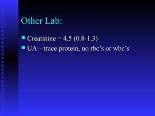
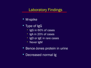
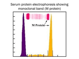
Multiple Myeloma
Alan Johns, M.D.
Kristine Krafts, M.D.
Richard – A Case Study
Richard S.
 51 year old male presented 8/95 with vague51 year old male presented 8/95 with vague
epigastric distress and weight loss of 5 lbs.epigastric distress and weight loss of 5 lbs.
 Denies fevers, chills or back painDenies fevers, chills or back pain
 PMH – neg.PMH – neg.
 Meds – noneMeds – none
 Smoking, alcohol – noneSmoking, alcohol – none
 Works as a carpenterWorks as a carpenter
 Physical Exam: BP= 134/84, Pulse = 70Physical Exam: BP= 134/84, Pulse = 70
Temp = 98.4 degreesTemp = 98.4 degrees
HEENT – negHEENT – neg
Neck – no lymphadenophyNeck – no lymphadenophy
Lungs – clear, Heart – no gallop or murmurLungs – clear, Heart – no gallop or murmur
Abdomen – nontender, no organomegalyAbdomen – nontender, no organomegaly
Rectal – normal, stool hemoccult negativeRectal – normal, stool hemoccult negative
Extremities – no edema or deformities, noExtremities – no edema or deformities, no
Initial Lab:
 Hemoglobin = 6.7Hemoglobin = 6.7
 WBC = 8,300WBC = 8,300
 Plts. = 166,000Plts. = 166,000
 MCV = 94.5 (82-99)MCV = 94.5 (82-99)
 RDW = 13.0 (11.0-15.0)RDW = 13.0 (11.0-15.0)
 Reticulocyte Count = 0.9% (0.4-1.8)Reticulocyte Count = 0.9% (0.4-1.8)
 Hemoccult (stool) – negative for bloodHemoccult (stool) – negative for blood
Other Lab:
 Creatinine = 4.5 (0.8-1.3)Creatinine = 4.5 (0.8-1.3)
 UA – trace protein, no rbc’s or wbc’sUA – trace protein, no rbc’s or wbc’s
Problem List:
 1) Severe anemia1) Severe anemia
 2) Acute renal failure with proteinurea2) Acute renal failure with proteinurea
 3) Epigastric distress3) Epigastric distress
 4) Weight loss4) Weight loss
Bone Marrow
 Diagnosis – Multiple MyelomaDiagnosis – Multiple Myeloma
Multiple Myeloma
• monoclonal plasma cell proliferation
• monoclonal gammopathy
• decreased normal immunoglobulins
• osteolytic lesions
Things You Must Know
• M-spike
• Type of IgG
• IgG in 60% of cases
• IgA in 20% of cases
• IgD or IgE in rare cases
• Never IgM
• Bence-Jones protein in urine
• Decreased normal Ig
Laboratory Findings
Normal serum protein electrophoresis
Normal serum protein electrophoresisSerum protein electrophoresis showing
monoclonal band (M protein)
• Blood: anemia, rouleaux
• Marrow: plasma cells, amyloid
Morphology
Multiple Myeloma
Multiple Myeloma
Myeloma, mature type
Myeloma, intermediate type
Myeloma, plasmablastic type
Flame cells
Russell bodies
Dutcher body and Mott cell
Rouleaux
Amyloid
• Solitary plasmacytoma
• Plasma cell leukemia
• Waldenström macroglobulinemia
• Lymphoplasmacytoid lymphoma
• IgM
• Hyperviscosity syndrome
• MGUS (Monoclonal gammopathy of
undetermined significance)
• Small M spike with no myeloma symptoms
• Occasionally transforms into myeloma
OTHER PLASMA CELL TUMORS
Richard
 Serum protein electrophoresis:Serum protein electrophoresis:
 Serum immunoelectrophoresisSerum immunoelectrophoresis
 Urine immunoelectrophoresisUrine immunoelectrophoresis
Biology of Normal Plasma Cells
Plasmablasts in lymph nodes (IgM)Plasmablasts in lymph nodes (IgM)
Activated B cells in bone marrow (IgG, IgA)Activated B cells in bone marrow (IgG, IgA)
Differentiate into plasma cells (small inDifferentiate into plasma cells (small in
number, well-differentiated, characteristicnumber, well-differentiated, characteristic
phenotype, die by apoptosis)phenotype, die by apoptosis)
Biology of Malignant Plasma
Cells
 Plasmablasts in lymph nodesPlasmablasts in lymph nodes
 Plasmablasts in bone marrow (IgG, IgA)Plasmablasts in bone marrow (IgG, IgA)
 Plasmablasts do not differentiate intoPlasmablasts do not differentiate into
plasma cells, continue to proliferate andplasma cells, continue to proliferate and
accumulate in marrow, produce largeaccumulate in marrow, produce large
amounts of immunoglobulins, normal deathamounts of immunoglobulins, normal death
of cells doesn’t occur, crowds out otherof cells doesn’t occur, crowds out other
cells – rbc precursors. Suppress antibodycells – rbc precursors. Suppress antibody
Interleukin – 6
 Essential for survival and growth ofEssential for survival and growth of
myeloma cellsmyeloma cells
 Growth factor for myeloma cellsGrowth factor for myeloma cells
 Also promotes survival of myeloma cells byAlso promotes survival of myeloma cells by
preventing spontaneous apoptosis.preventing spontaneous apoptosis.
 Increased levels in myeloma patientsIncreased levels in myeloma patients
Clinical Features
 80% of patients present with bone pain80% of patients present with bone pain
(low back, pelvis, or ribs). Pain is(low back, pelvis, or ribs). Pain is
associated with multiple lytic bone lesions.associated with multiple lytic bone lesions.
• Bruising or bleeding from decreasedBruising or bleeding from decreased
plateletsplatelets
• Infections from decreased levels of normalInfections from decreased levels of normal
immunoglobulinsimmunoglobulins
Clinical Features – con’t
 Hypercalcemia from bone destructionHypercalcemia from bone destruction
 50% of patients present with renal failure50% of patients present with renal failure
 Hyperviscosity syndrome – caused by largeHyperviscosity syndrome – caused by large
amounts of circulating immunoglobulinsamounts of circulating immunoglobulins
causing purpura, confusion, decreasedcausing purpura, confusion, decreased
visionvision
 Major causes of death – infection, renalMajor causes of death – infection, renal
failurefailure
 Classic triad – anemia, bone pain, renalClassic triad – anemia, bone pain, renal
Criteria for Diagnosis
1) Bone marrow with >20% plasma cells OR1) Bone marrow with >20% plasma cells OR
2) Plasmacytoma plus one of the following:2) Plasmacytoma plus one of the following:
monoclonal protein in serum > 3 g/dlmonoclonal protein in serum > 3 g/dl
monoclonal protein in urinemonoclonal protein in urine
lytic lesionslytic lesions
3) Usual clinical features of myeloma3) Usual clinical features of myeloma
4) Exclude connective tissue diseases, chronic4) Exclude connective tissue diseases, chronic
infections, carcinoma, lymphoma, leukemiainfections, carcinoma, lymphoma, leukemia
Therapy
 Conventional Dose ChemotherapyConventional Dose Chemotherapy
Classic combination – melphalan andClassic combination – melphalan and
prednisone (1962)prednisone (1962)
Complete Remission - < 5%Complete Remission - < 5%
Median Survival – 3 yearsMedian Survival – 3 years
Conventional Chemo-con’t
VAD – vincristine, doxyrubicin andVAD – vincristine, doxyrubicin and
dexamethasonedexamethasone
VAMP – vincristine, doxyrubicin andVAMP – vincristine, doxyrubicin and
methyprednisolonemethyprednisolone
Did not prolong survival more than otherDid not prolong survival more than other
regimensregimens
Excessive morbidity and mortality fromExcessive morbidity and mortality from
prolonged myelosupressionprolonged myelosupression
Autologous Peripheral Blood
Stem Cell Transplant - PBSC
 Hematopoietic stem cells from peripheralHematopoietic stem cells from peripheral
bloodblood
 Growth factors are given afterGrowth factors are given after
transplantationtransplantation
 Safe – 1-2% death rate from the transplantSafe – 1-2% death rate from the transplant
 Problem – contamination of the autologousProblem – contamination of the autologous
graft by myeloma cellsgraft by myeloma cells
High-Dose Therapy with Stem-cell
Transplant (1992)
Melphalan in high doses can induce completeMelphalan in high doses can induce complete
remissions in 20-30%. Death fromremissions in 20-30%. Death from
treatment alone is 10-30%.treatment alone is 10-30%.
Stem-cell transplant after high doseStem-cell transplant after high dose
Melphalan (with or without radiation) canMelphalan (with or without radiation) can
produce a 30-50% complete remission inproduce a 30-50% complete remission in
newly diagnosed patients. Problems – onlynewly diagnosed patients. Problems – only
58% of patients over 60 could tolerate.58% of patients over 60 could tolerate.
Single vs. double autologous
stem-cell transplantation
(2003)
New Agents
 ThalidomideThalidomide
First used with advanced and refractoryFirst used with advanced and refractory
myeloma (2001)myeloma (2001)
Now used for newly diagnosed disease inNow used for newly diagnosed disease in
combination with high-dose melphalan andcombination with high-dose melphalan and
double stem-cell transplant (2005)double stem-cell transplant (2005)
Copyright ©2004 American Society of Hematology. Copyright restrictions may apply.
Barlogie, B. et al. Blood 2004;103:20-32
Figure 3. Thalidomide in advanced and refractory myeloma
 Bortezomib (Velcade)Bortezomib (Velcade)
Proteasome inhibitorProteasome inhibitor
Copyright ©2004 American Society of Hematology. Copyright restrictions may apply.
Barlogie, B. et al. Blood 2004;103:20-32
Figure 6. PS 341 (Velcade) plus thalidomide for posttransplantation relapse in 46 patients
New Supportive Therapies
 Biphosphonates – inhibit bone resorption,Biphosphonates – inhibit bone resorption,
treats bone lesions and hypercalcemia.treats bone lesions and hypercalcemia.
 Erythropoietin – helps anemia andErythropoietin – helps anemia and
decreases need for transfusions.decreases need for transfusions.
Future Approaches
 Interleukin-2, Interleukin-4, InterferonInterleukin-2, Interleukin-4, Interferon
gamma- pilot studies show no benefit.gamma- pilot studies show no benefit.
 Anti-interleukin-6Anti-interleukin-6
Initial studies produced some effect but noInitial studies produced some effect but no
lasting benefit.lasting benefit.
Further trials underwayFurther trials underway
Future approaches – con’t
 ImmunotherapyImmunotherapy
Monoclonal immunoglobulins in anMonoclonal immunoglobulins in an
individual patient may have a tumor-individual patient may have a tumor-
specific antigen.specific antigen.
T-cells seem to recognize the idiotypes ofT-cells seem to recognize the idiotypes of
the patients myeloma protein.the patients myeloma protein.
IgG from patient transferred to a boneIgG from patient transferred to a bone
marrow donor then patient receivedmarrow donor then patient received
transplant. Two years later patient hastransplant. Two years later patient has
remained well with minimal M component.remained well with minimal M component.
Prognosis
 15 % die within 3 months of diagnosis15 % die within 3 months of diagnosis
 Subsequent death rate 15% per yearSubsequent death rate 15% per year
 Causes of death- marrow replacement withCauses of death- marrow replacement with
pancytopenia (16%), renal failure (10%),pancytopenia (16%), renal failure (10%),
sepsis (14%), acute leukemia (5%), othersepsis (14%), acute leukemia (5%), other
chronic illnesses unrelated to myelomachronic illnesses unrelated to myeloma
(23%)(23%)
Richards’ Treatment
 8/95 Melphalan and Prednisone cycles8/95 Melphalan and Prednisone cycles
started. M = 6.6 g%started. M = 6.6 g%
 9/95 M = 3.96 Creatinine = 1.29/95 M = 3.96 Creatinine = 1.2
 4/96 M = 1.954/96 M = 1.95
 5/96 M = 2.4 Bone Marrow 7% plasma5/96 M = 2.4 Bone Marrow 7% plasma
cellscells
 8/96 M = 1.858/96 M = 1.85
Richards’ Treatment Cont’
 2/97 M = 2.52/97 M = 2.5
 4/97 M = 4.05 Bone Marrow shows 44%4/97 M = 4.05 Bone Marrow shows 44%
plasma cellsplasma cells
 5/97 VAD started5/97 VAD started
 7/97 M = 3.87/97 M = 3.8
 8/97 Bone lesions noted pelvis and femur8/97 Bone lesions noted pelvis and femur
 1/98 M = 3.9 Bone marrow shows 20%1/98 M = 3.9 Bone marrow shows 20%
plasma cellsplasma cells
Richards’ Treatment Cont’
 4/98 M = 5.24/98 M = 5.2
Allogenic bone marrow transplant afterAllogenic bone marrow transplant after
Cytoxan and whole body radiation at MayoCytoxan and whole body radiation at Mayo
Clinic (brother was donor)Clinic (brother was donor)
Post-transplant renal failure andPost-transplant renal failure and
pulmonary hemorrhage.pulmonary hemorrhage.
Died 6/6/98Died 6/6/98

Histopics

  • 1.
    Multiple Myeloma Alan Johns,M.D. Kristine Krafts, M.D.
  • 2.
    Richard – ACase Study
  • 3.
    Richard S.  51year old male presented 8/95 with vague51 year old male presented 8/95 with vague epigastric distress and weight loss of 5 lbs.epigastric distress and weight loss of 5 lbs.  Denies fevers, chills or back painDenies fevers, chills or back pain  PMH – neg.PMH – neg.  Meds – noneMeds – none  Smoking, alcohol – noneSmoking, alcohol – none  Works as a carpenterWorks as a carpenter
  • 4.
     Physical Exam:BP= 134/84, Pulse = 70Physical Exam: BP= 134/84, Pulse = 70 Temp = 98.4 degreesTemp = 98.4 degrees HEENT – negHEENT – neg Neck – no lymphadenophyNeck – no lymphadenophy Lungs – clear, Heart – no gallop or murmurLungs – clear, Heart – no gallop or murmur Abdomen – nontender, no organomegalyAbdomen – nontender, no organomegaly Rectal – normal, stool hemoccult negativeRectal – normal, stool hemoccult negative Extremities – no edema or deformities, noExtremities – no edema or deformities, no
  • 5.
    Initial Lab:  Hemoglobin= 6.7Hemoglobin = 6.7  WBC = 8,300WBC = 8,300  Plts. = 166,000Plts. = 166,000  MCV = 94.5 (82-99)MCV = 94.5 (82-99)  RDW = 13.0 (11.0-15.0)RDW = 13.0 (11.0-15.0)  Reticulocyte Count = 0.9% (0.4-1.8)Reticulocyte Count = 0.9% (0.4-1.8)  Hemoccult (stool) – negative for bloodHemoccult (stool) – negative for blood
  • 6.
    Other Lab:  Creatinine= 4.5 (0.8-1.3)Creatinine = 4.5 (0.8-1.3)  UA – trace protein, no rbc’s or wbc’sUA – trace protein, no rbc’s or wbc’s
  • 7.
    Problem List:  1)Severe anemia1) Severe anemia  2) Acute renal failure with proteinurea2) Acute renal failure with proteinurea  3) Epigastric distress3) Epigastric distress  4) Weight loss4) Weight loss
  • 8.
  • 12.
     Diagnosis –Multiple MyelomaDiagnosis – Multiple Myeloma
  • 13.
    Multiple Myeloma • monoclonalplasma cell proliferation • monoclonal gammopathy • decreased normal immunoglobulins • osteolytic lesions Things You Must Know
  • 14.
    • M-spike • Typeof IgG • IgG in 60% of cases • IgA in 20% of cases • IgD or IgE in rare cases • Never IgM • Bence-Jones protein in urine • Decreased normal Ig Laboratory Findings
  • 15.
    Normal serum proteinelectrophoresis Normal serum protein electrophoresis
  • 16.
    Normal serum proteinelectrophoresisSerum protein electrophoresis showing monoclonal band (M protein)
  • 17.
    • Blood: anemia,rouleaux • Marrow: plasma cells, amyloid Morphology
  • 18.
  • 19.
  • 20.
  • 21.
  • 22.
  • 23.
  • 24.
  • 25.
  • 26.
  • 27.
  • 28.
    • Solitary plasmacytoma •Plasma cell leukemia • Waldenström macroglobulinemia • Lymphoplasmacytoid lymphoma • IgM • Hyperviscosity syndrome • MGUS (Monoclonal gammopathy of undetermined significance) • Small M spike with no myeloma symptoms • Occasionally transforms into myeloma OTHER PLASMA CELL TUMORS
  • 29.
    Richard  Serum proteinelectrophoresis:Serum protein electrophoresis:  Serum immunoelectrophoresisSerum immunoelectrophoresis  Urine immunoelectrophoresisUrine immunoelectrophoresis
  • 33.
    Biology of NormalPlasma Cells Plasmablasts in lymph nodes (IgM)Plasmablasts in lymph nodes (IgM) Activated B cells in bone marrow (IgG, IgA)Activated B cells in bone marrow (IgG, IgA) Differentiate into plasma cells (small inDifferentiate into plasma cells (small in number, well-differentiated, characteristicnumber, well-differentiated, characteristic phenotype, die by apoptosis)phenotype, die by apoptosis)
  • 34.
    Biology of MalignantPlasma Cells  Plasmablasts in lymph nodesPlasmablasts in lymph nodes  Plasmablasts in bone marrow (IgG, IgA)Plasmablasts in bone marrow (IgG, IgA)  Plasmablasts do not differentiate intoPlasmablasts do not differentiate into plasma cells, continue to proliferate andplasma cells, continue to proliferate and accumulate in marrow, produce largeaccumulate in marrow, produce large amounts of immunoglobulins, normal deathamounts of immunoglobulins, normal death of cells doesn’t occur, crowds out otherof cells doesn’t occur, crowds out other cells – rbc precursors. Suppress antibodycells – rbc precursors. Suppress antibody
  • 36.
    Interleukin – 6 Essential for survival and growth ofEssential for survival and growth of myeloma cellsmyeloma cells  Growth factor for myeloma cellsGrowth factor for myeloma cells  Also promotes survival of myeloma cells byAlso promotes survival of myeloma cells by preventing spontaneous apoptosis.preventing spontaneous apoptosis.  Increased levels in myeloma patientsIncreased levels in myeloma patients
  • 37.
    Clinical Features  80%of patients present with bone pain80% of patients present with bone pain (low back, pelvis, or ribs). Pain is(low back, pelvis, or ribs). Pain is associated with multiple lytic bone lesions.associated with multiple lytic bone lesions. • Bruising or bleeding from decreasedBruising or bleeding from decreased plateletsplatelets • Infections from decreased levels of normalInfections from decreased levels of normal immunoglobulinsimmunoglobulins
  • 40.
    Clinical Features –con’t  Hypercalcemia from bone destructionHypercalcemia from bone destruction  50% of patients present with renal failure50% of patients present with renal failure  Hyperviscosity syndrome – caused by largeHyperviscosity syndrome – caused by large amounts of circulating immunoglobulinsamounts of circulating immunoglobulins causing purpura, confusion, decreasedcausing purpura, confusion, decreased visionvision  Major causes of death – infection, renalMajor causes of death – infection, renal failurefailure  Classic triad – anemia, bone pain, renalClassic triad – anemia, bone pain, renal
  • 41.
    Criteria for Diagnosis 1)Bone marrow with >20% plasma cells OR1) Bone marrow with >20% plasma cells OR 2) Plasmacytoma plus one of the following:2) Plasmacytoma plus one of the following: monoclonal protein in serum > 3 g/dlmonoclonal protein in serum > 3 g/dl monoclonal protein in urinemonoclonal protein in urine lytic lesionslytic lesions 3) Usual clinical features of myeloma3) Usual clinical features of myeloma 4) Exclude connective tissue diseases, chronic4) Exclude connective tissue diseases, chronic infections, carcinoma, lymphoma, leukemiainfections, carcinoma, lymphoma, leukemia
  • 43.
    Therapy  Conventional DoseChemotherapyConventional Dose Chemotherapy Classic combination – melphalan andClassic combination – melphalan and prednisone (1962)prednisone (1962) Complete Remission - < 5%Complete Remission - < 5% Median Survival – 3 yearsMedian Survival – 3 years
  • 44.
    Conventional Chemo-con’t VAD –vincristine, doxyrubicin andVAD – vincristine, doxyrubicin and dexamethasonedexamethasone VAMP – vincristine, doxyrubicin andVAMP – vincristine, doxyrubicin and methyprednisolonemethyprednisolone Did not prolong survival more than otherDid not prolong survival more than other regimensregimens Excessive morbidity and mortality fromExcessive morbidity and mortality from prolonged myelosupressionprolonged myelosupression
  • 45.
    Autologous Peripheral Blood StemCell Transplant - PBSC  Hematopoietic stem cells from peripheralHematopoietic stem cells from peripheral bloodblood  Growth factors are given afterGrowth factors are given after transplantationtransplantation  Safe – 1-2% death rate from the transplantSafe – 1-2% death rate from the transplant  Problem – contamination of the autologousProblem – contamination of the autologous graft by myeloma cellsgraft by myeloma cells
  • 46.
    High-Dose Therapy withStem-cell Transplant (1992) Melphalan in high doses can induce completeMelphalan in high doses can induce complete remissions in 20-30%. Death fromremissions in 20-30%. Death from treatment alone is 10-30%.treatment alone is 10-30%. Stem-cell transplant after high doseStem-cell transplant after high dose Melphalan (with or without radiation) canMelphalan (with or without radiation) can produce a 30-50% complete remission inproduce a 30-50% complete remission in newly diagnosed patients. Problems – onlynewly diagnosed patients. Problems – only 58% of patients over 60 could tolerate.58% of patients over 60 could tolerate.
  • 47.
    Single vs. doubleautologous stem-cell transplantation (2003)
  • 48.
    New Agents  ThalidomideThalidomide Firstused with advanced and refractoryFirst used with advanced and refractory myeloma (2001)myeloma (2001) Now used for newly diagnosed disease inNow used for newly diagnosed disease in combination with high-dose melphalan andcombination with high-dose melphalan and double stem-cell transplant (2005)double stem-cell transplant (2005)
  • 49.
    Copyright ©2004 AmericanSociety of Hematology. Copyright restrictions may apply. Barlogie, B. et al. Blood 2004;103:20-32 Figure 3. Thalidomide in advanced and refractory myeloma
  • 50.
     Bortezomib (Velcade)Bortezomib(Velcade) Proteasome inhibitorProteasome inhibitor
  • 51.
    Copyright ©2004 AmericanSociety of Hematology. Copyright restrictions may apply. Barlogie, B. et al. Blood 2004;103:20-32 Figure 6. PS 341 (Velcade) plus thalidomide for posttransplantation relapse in 46 patients
  • 52.
    New Supportive Therapies Biphosphonates – inhibit bone resorption,Biphosphonates – inhibit bone resorption, treats bone lesions and hypercalcemia.treats bone lesions and hypercalcemia.  Erythropoietin – helps anemia andErythropoietin – helps anemia and decreases need for transfusions.decreases need for transfusions.
  • 53.
    Future Approaches  Interleukin-2,Interleukin-4, InterferonInterleukin-2, Interleukin-4, Interferon gamma- pilot studies show no benefit.gamma- pilot studies show no benefit.  Anti-interleukin-6Anti-interleukin-6 Initial studies produced some effect but noInitial studies produced some effect but no lasting benefit.lasting benefit. Further trials underwayFurther trials underway
  • 54.
    Future approaches –con’t  ImmunotherapyImmunotherapy Monoclonal immunoglobulins in anMonoclonal immunoglobulins in an individual patient may have a tumor-individual patient may have a tumor- specific antigen.specific antigen. T-cells seem to recognize the idiotypes ofT-cells seem to recognize the idiotypes of the patients myeloma protein.the patients myeloma protein. IgG from patient transferred to a boneIgG from patient transferred to a bone marrow donor then patient receivedmarrow donor then patient received transplant. Two years later patient hastransplant. Two years later patient has remained well with minimal M component.remained well with minimal M component.
  • 55.
    Prognosis  15 %die within 3 months of diagnosis15 % die within 3 months of diagnosis  Subsequent death rate 15% per yearSubsequent death rate 15% per year  Causes of death- marrow replacement withCauses of death- marrow replacement with pancytopenia (16%), renal failure (10%),pancytopenia (16%), renal failure (10%), sepsis (14%), acute leukemia (5%), othersepsis (14%), acute leukemia (5%), other chronic illnesses unrelated to myelomachronic illnesses unrelated to myeloma (23%)(23%)
  • 56.
    Richards’ Treatment  8/95Melphalan and Prednisone cycles8/95 Melphalan and Prednisone cycles started. M = 6.6 g%started. M = 6.6 g%  9/95 M = 3.96 Creatinine = 1.29/95 M = 3.96 Creatinine = 1.2  4/96 M = 1.954/96 M = 1.95  5/96 M = 2.4 Bone Marrow 7% plasma5/96 M = 2.4 Bone Marrow 7% plasma cellscells  8/96 M = 1.858/96 M = 1.85
  • 57.
    Richards’ Treatment Cont’ 2/97 M = 2.52/97 M = 2.5  4/97 M = 4.05 Bone Marrow shows 44%4/97 M = 4.05 Bone Marrow shows 44% plasma cellsplasma cells  5/97 VAD started5/97 VAD started  7/97 M = 3.87/97 M = 3.8  8/97 Bone lesions noted pelvis and femur8/97 Bone lesions noted pelvis and femur  1/98 M = 3.9 Bone marrow shows 20%1/98 M = 3.9 Bone marrow shows 20% plasma cellsplasma cells
  • 58.
    Richards’ Treatment Cont’ 4/98 M = 5.24/98 M = 5.2 Allogenic bone marrow transplant afterAllogenic bone marrow transplant after Cytoxan and whole body radiation at MayoCytoxan and whole body radiation at Mayo Clinic (brother was donor)Clinic (brother was donor) Post-transplant renal failure andPost-transplant renal failure and pulmonary hemorrhage.pulmonary hemorrhage. Died 6/6/98Died 6/6/98