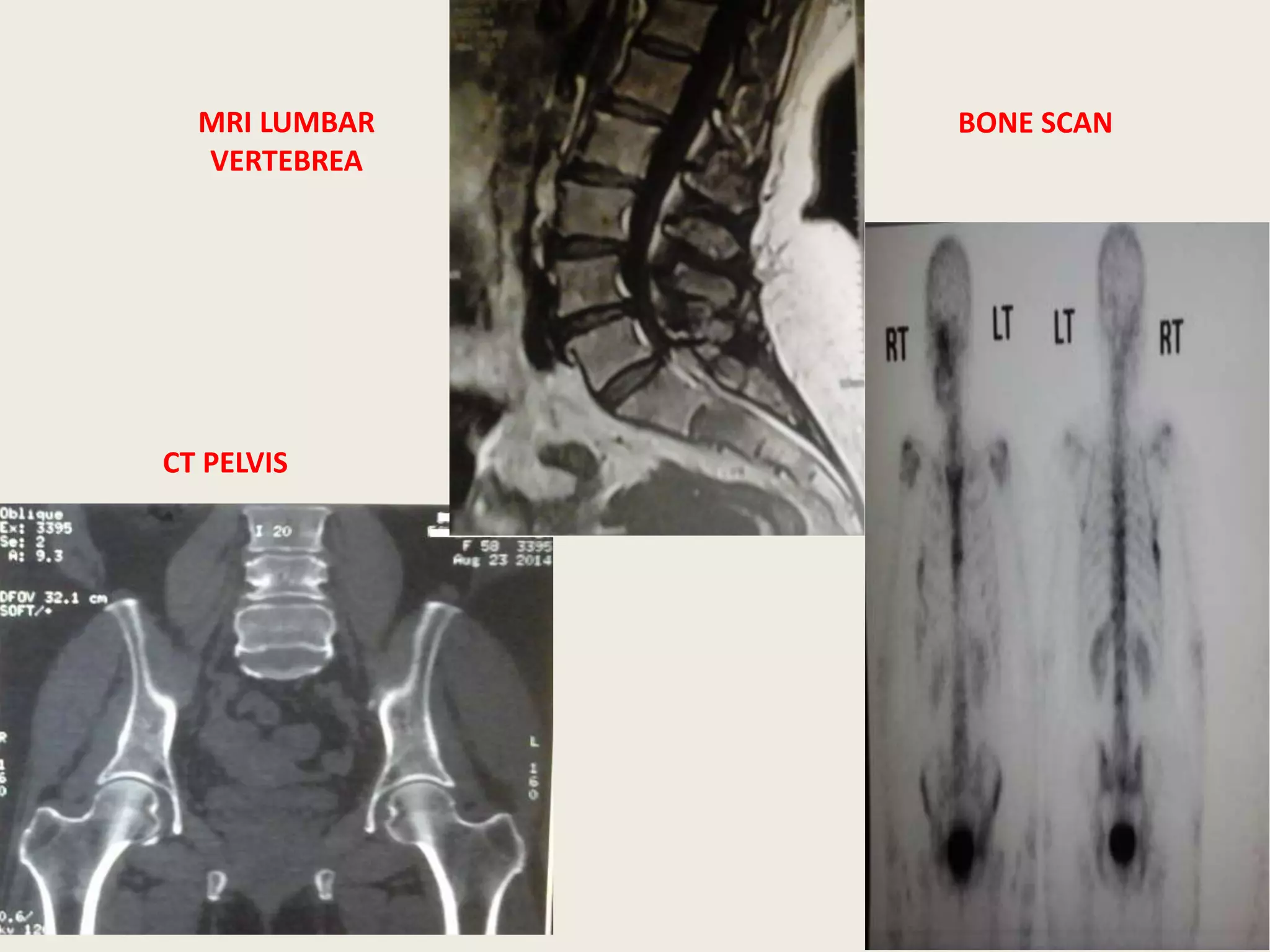
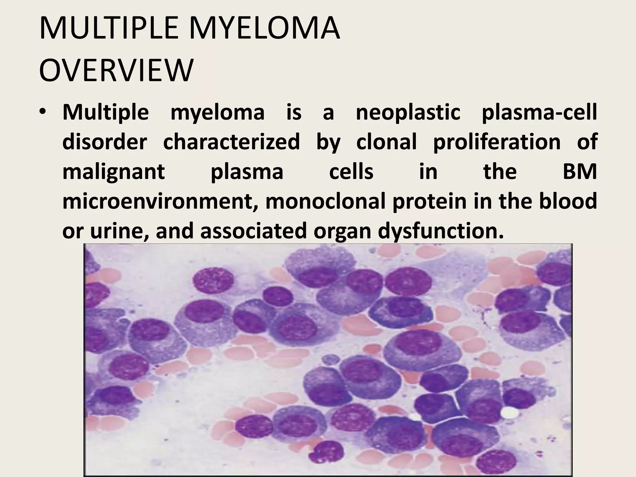
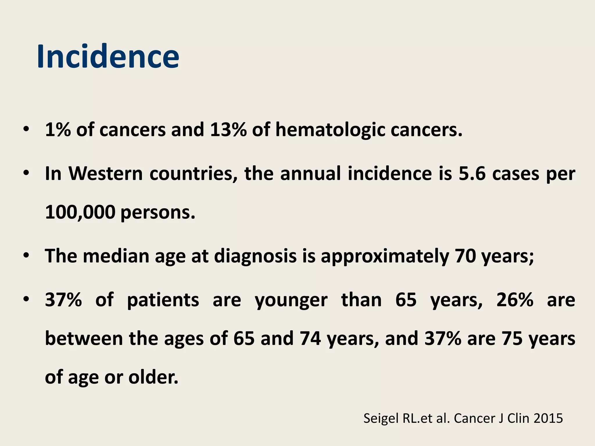
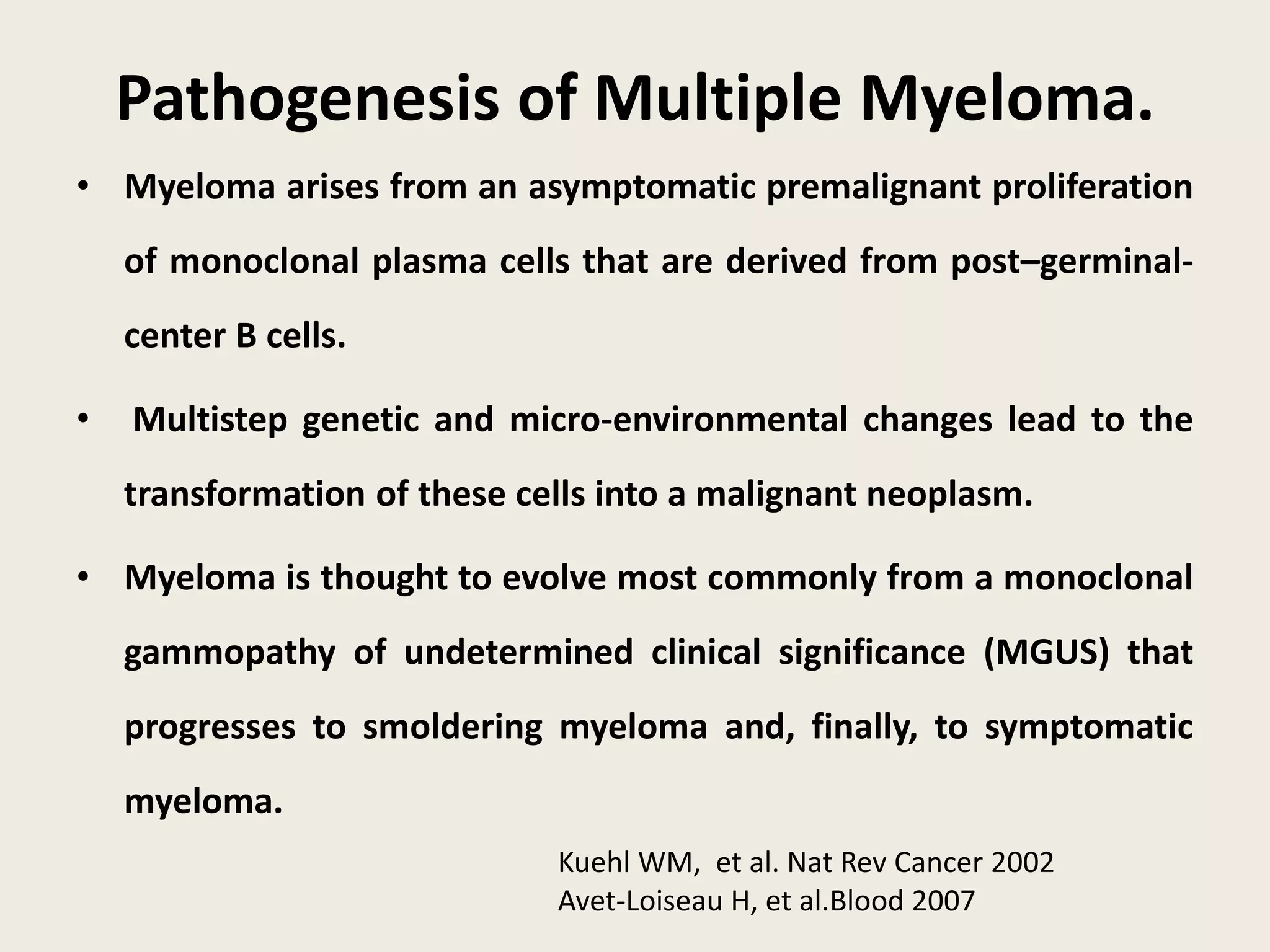
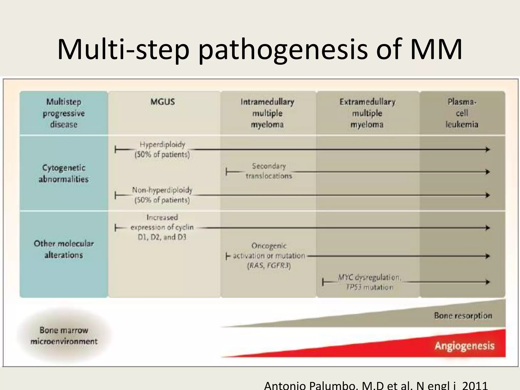
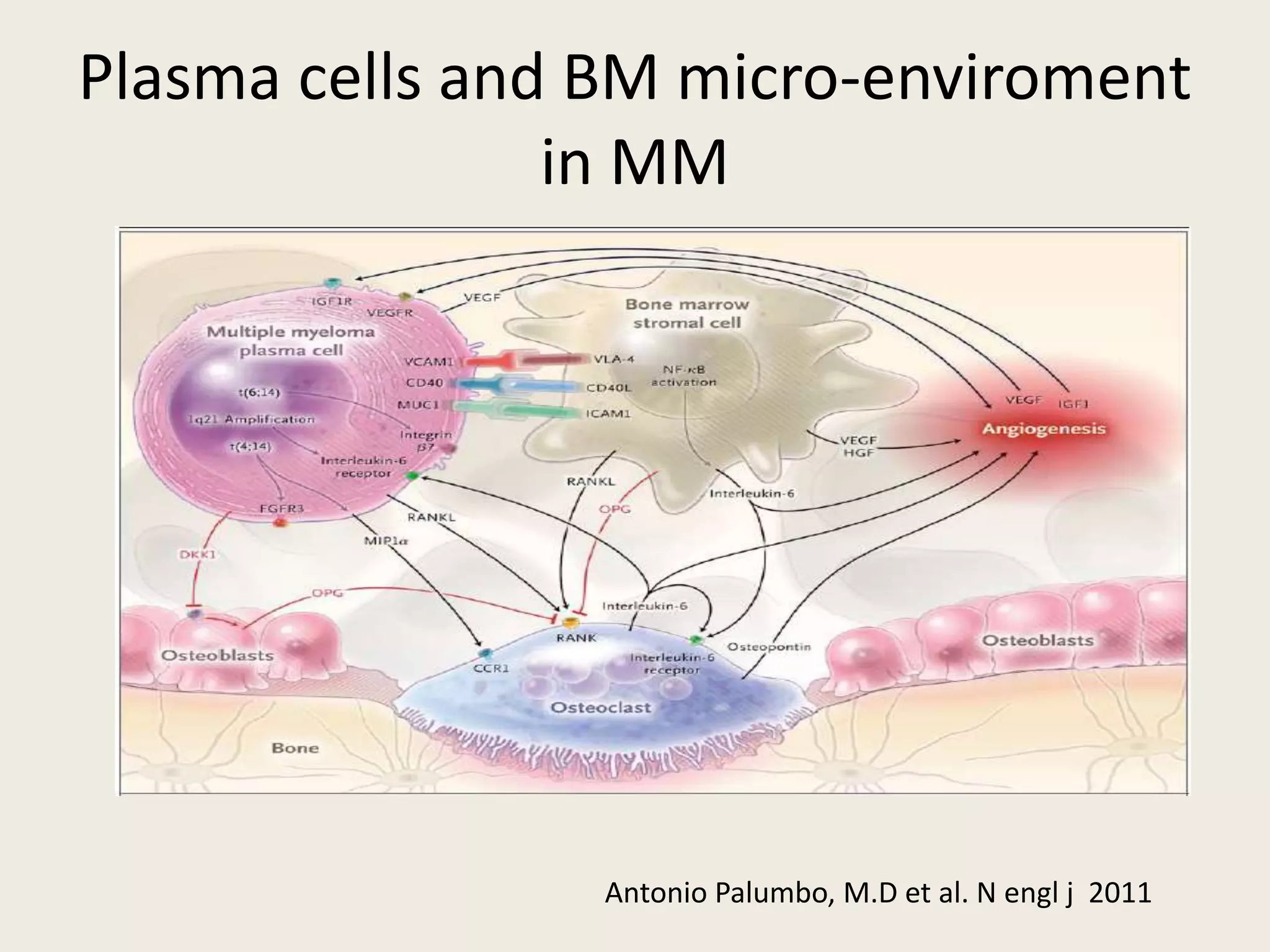
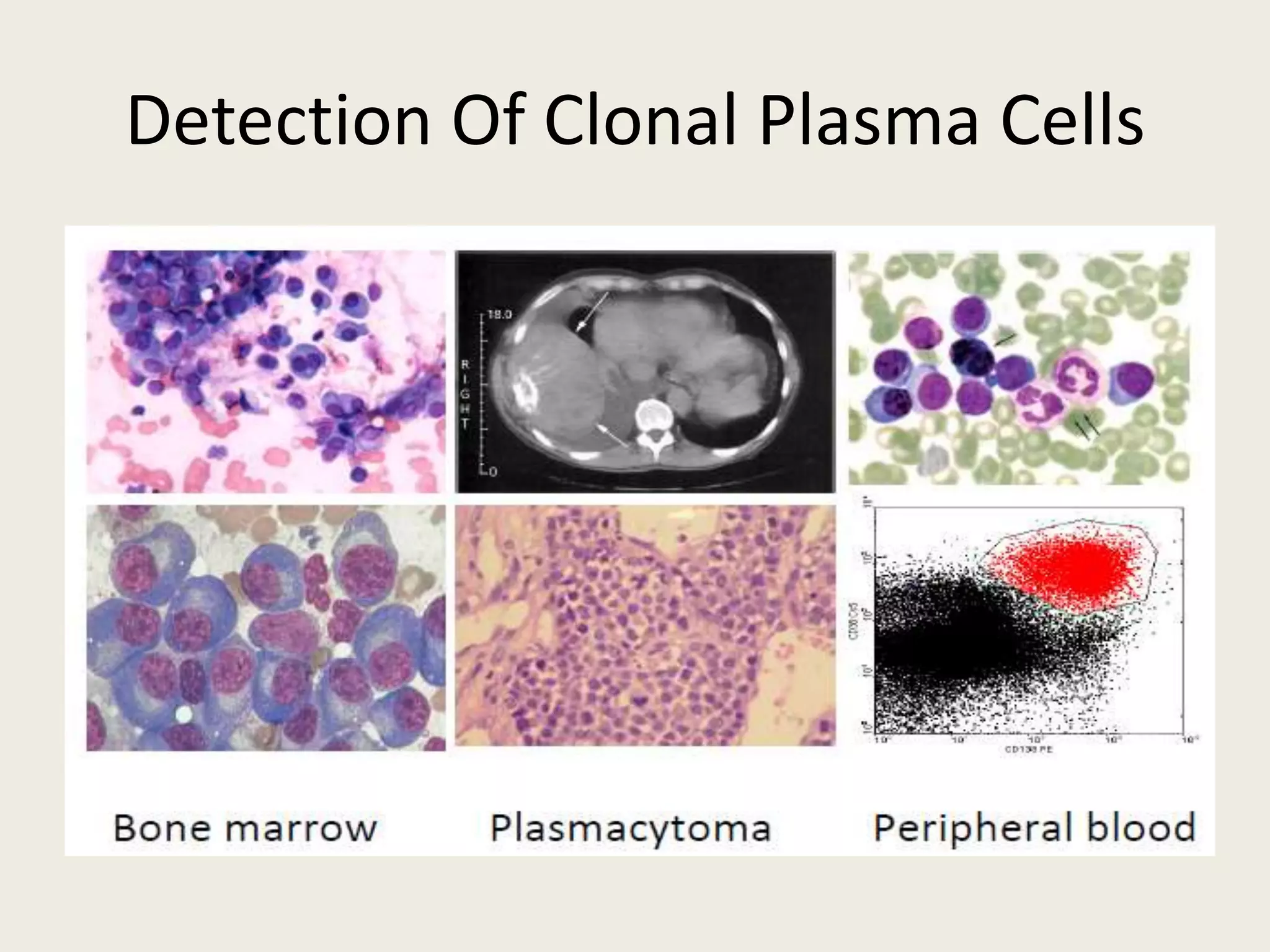
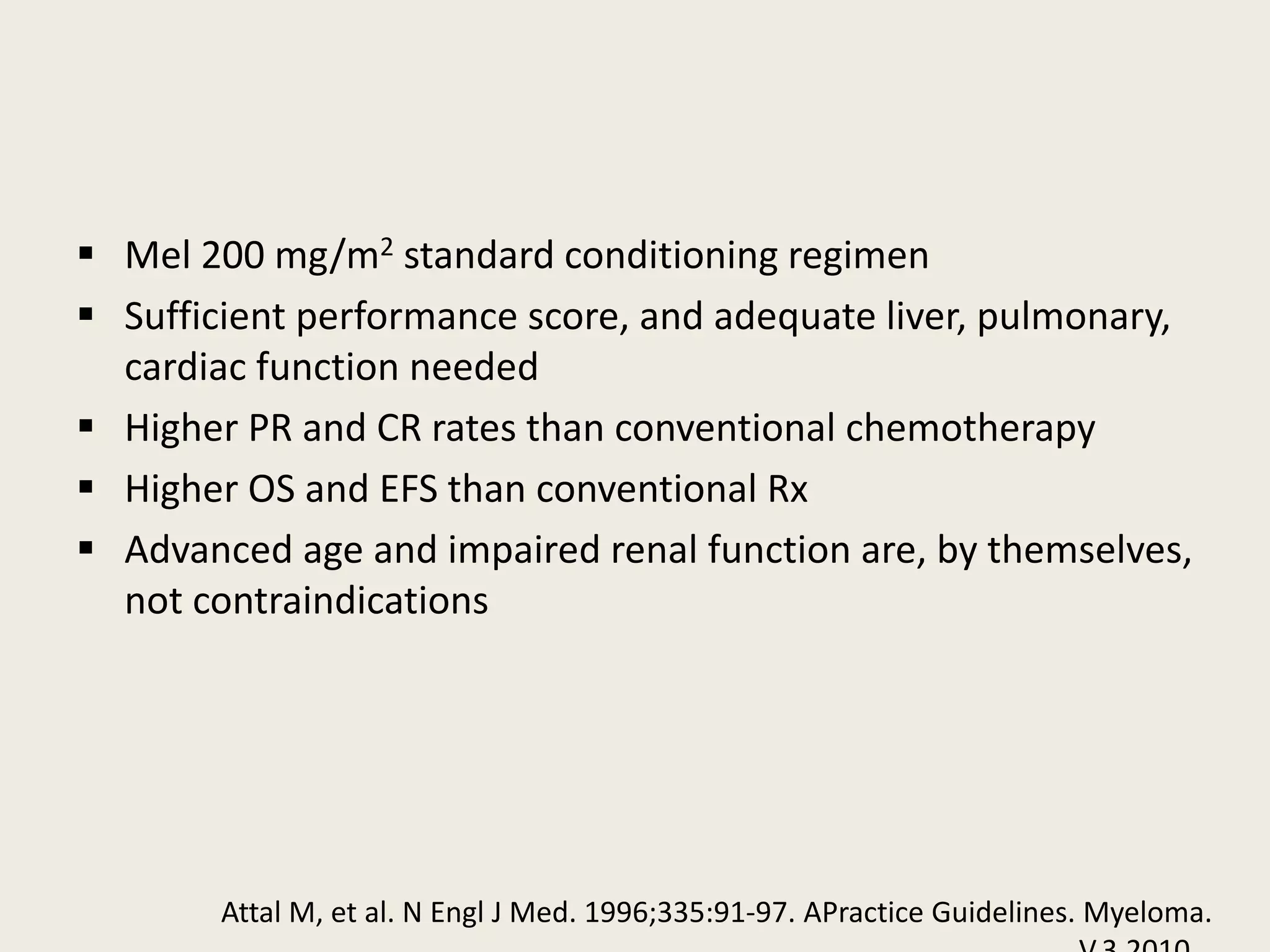
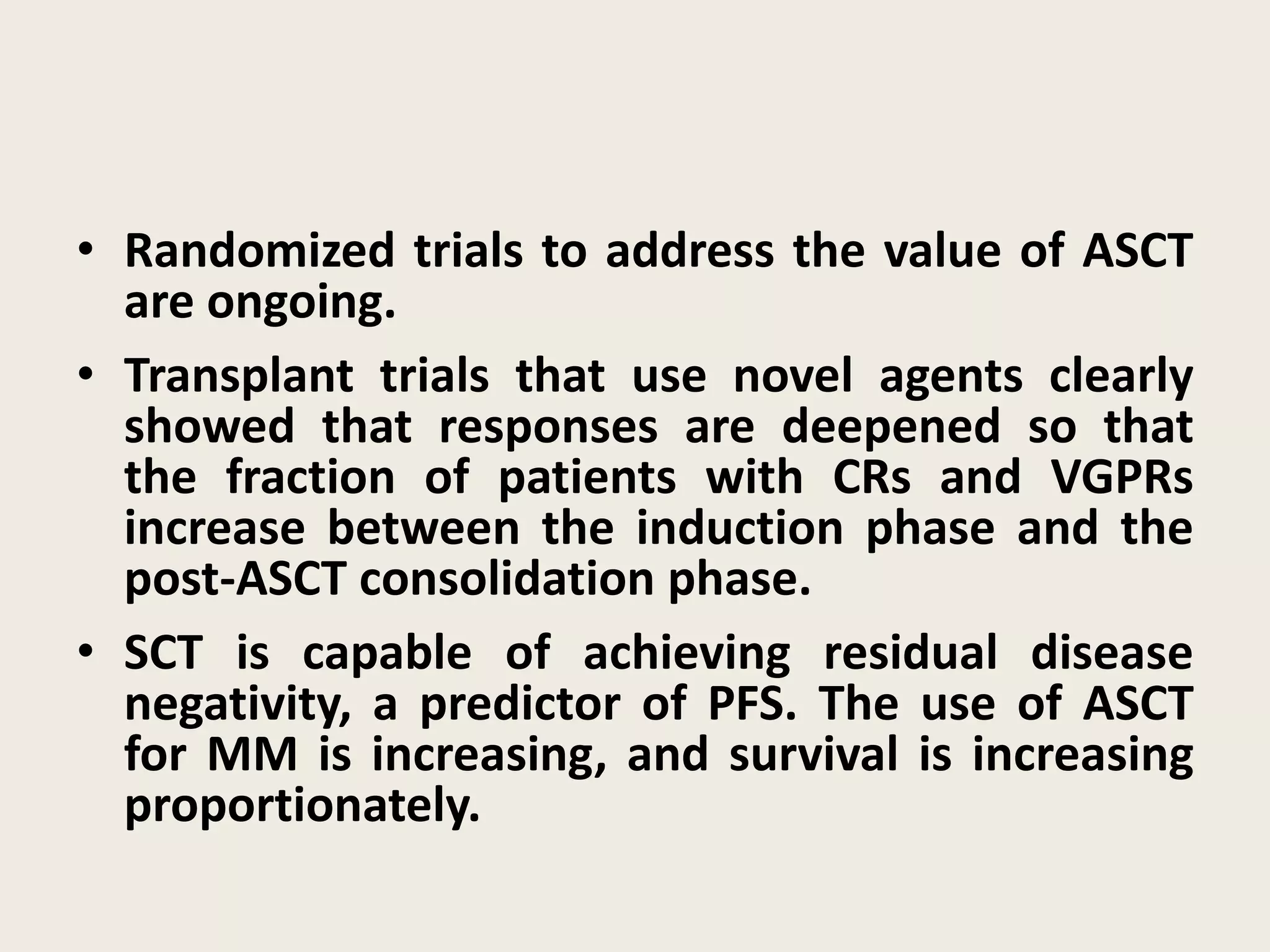
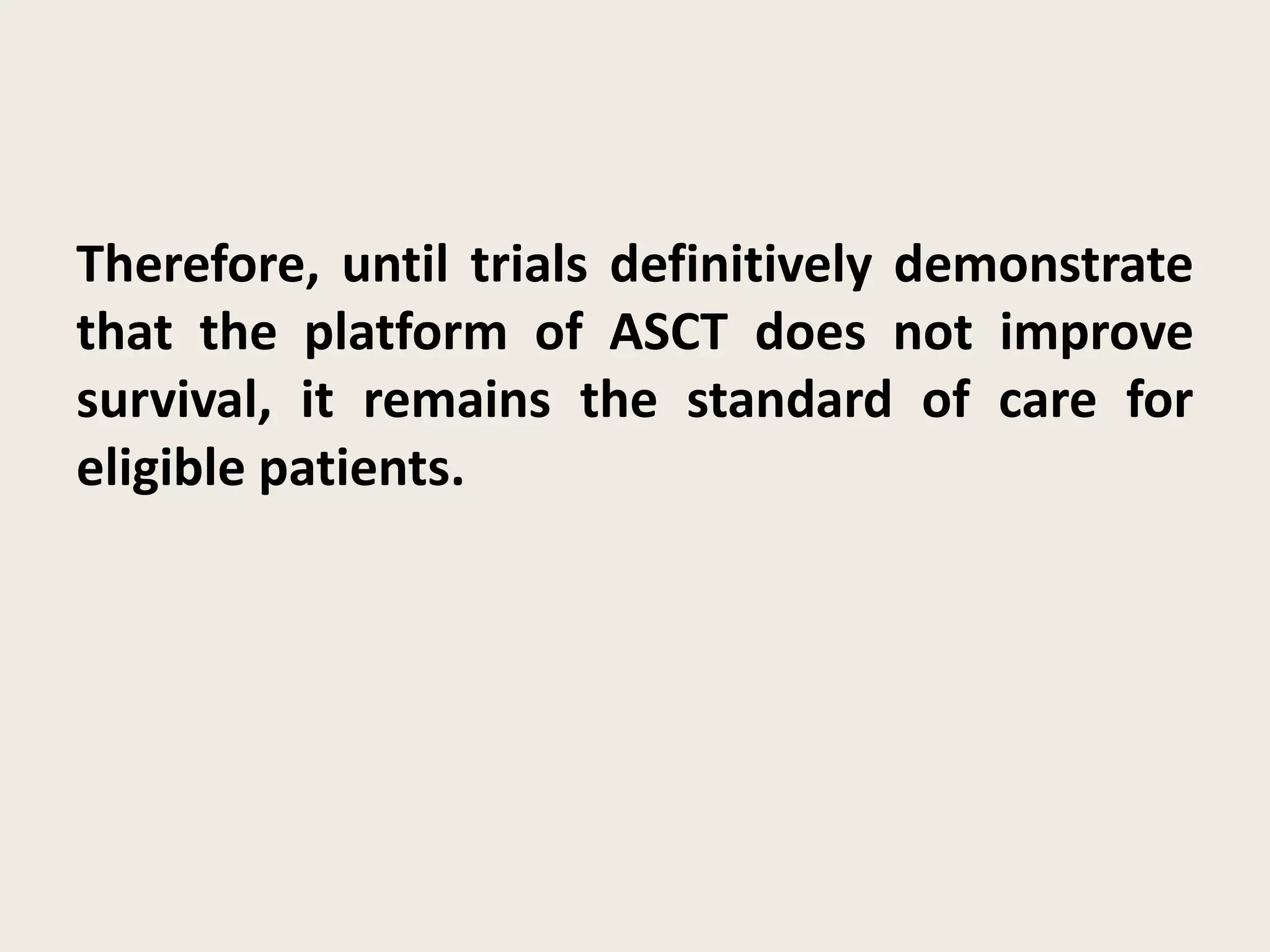
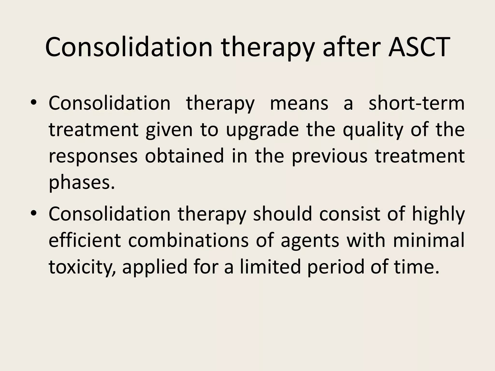
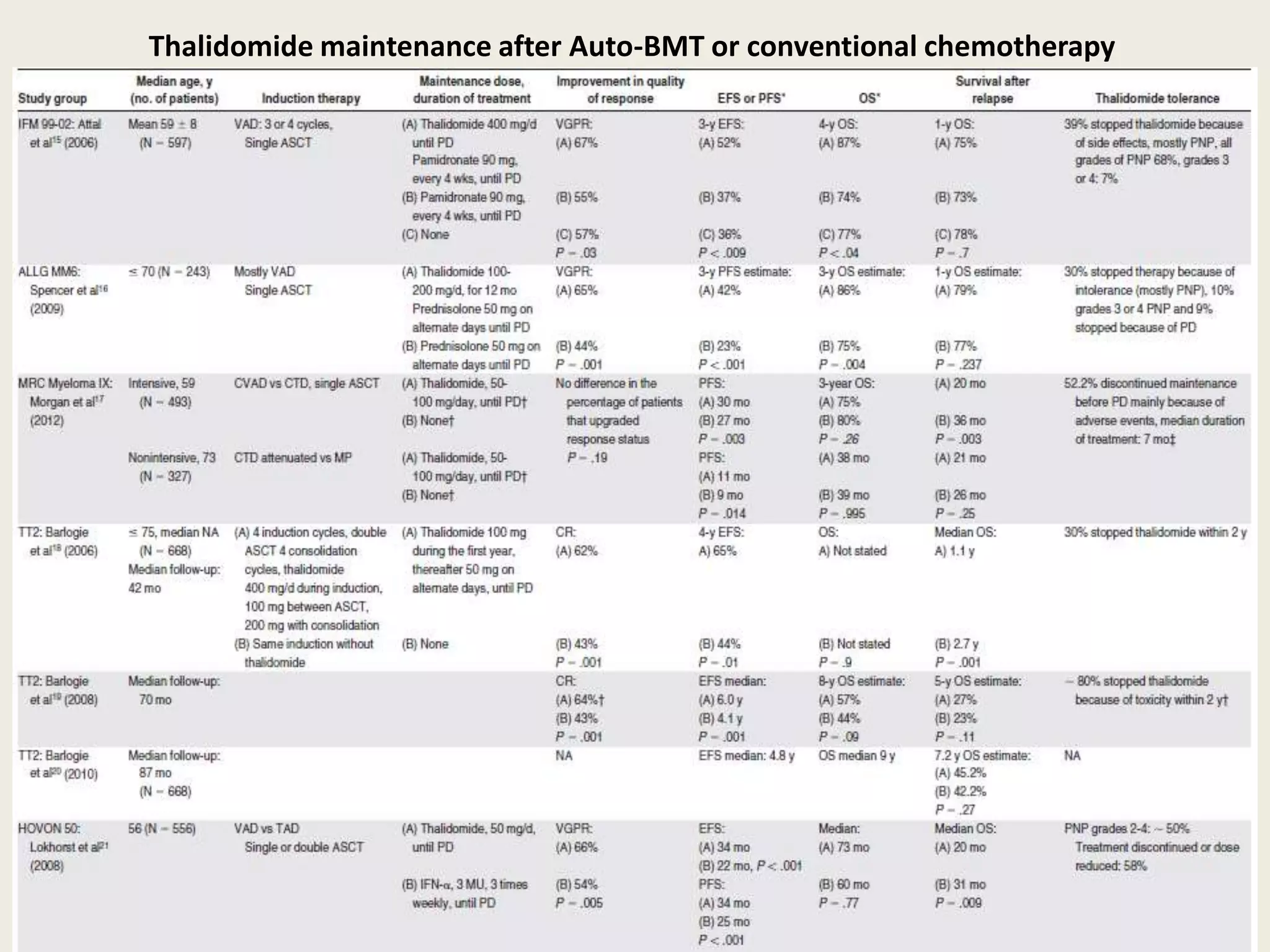
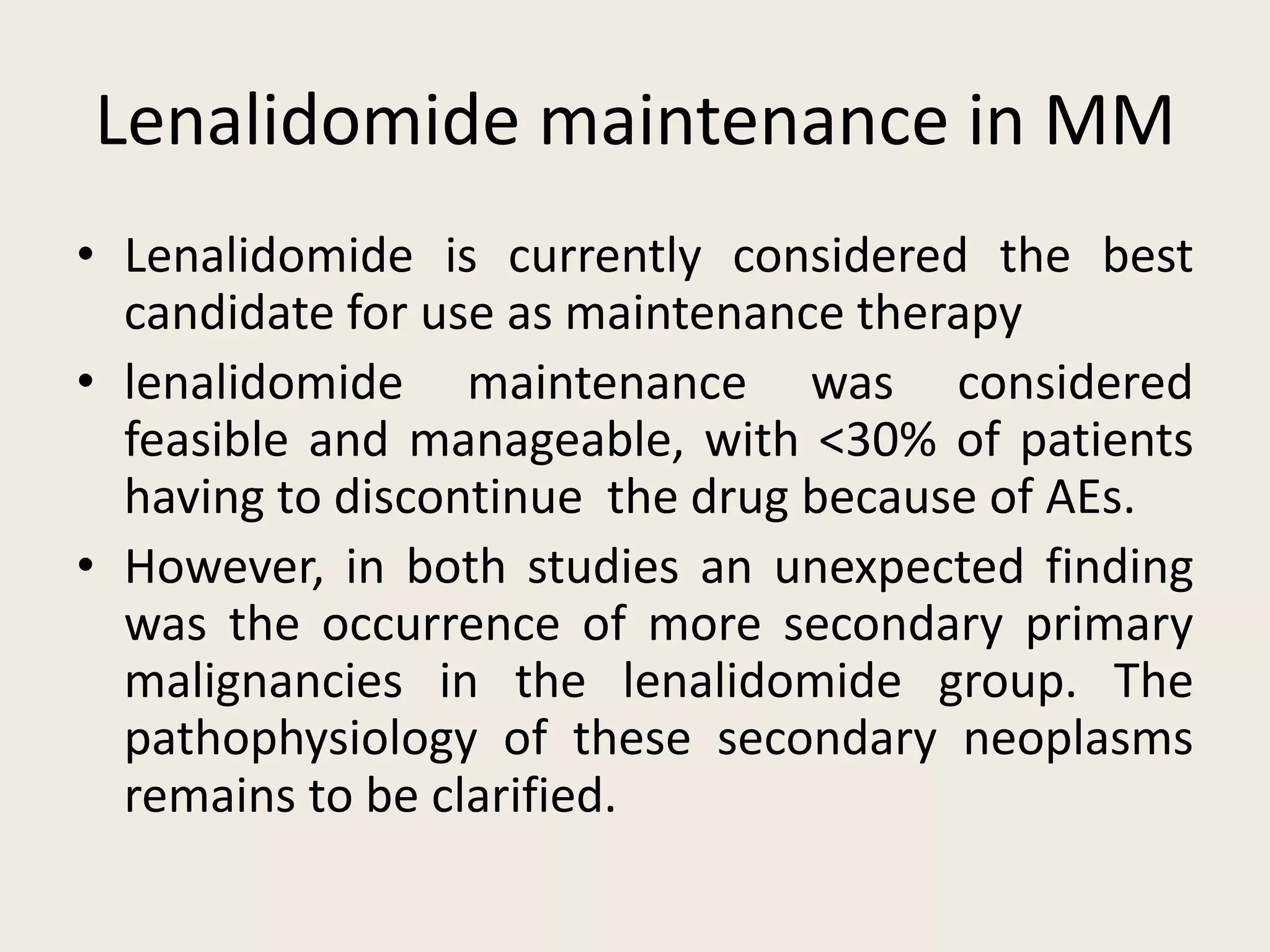
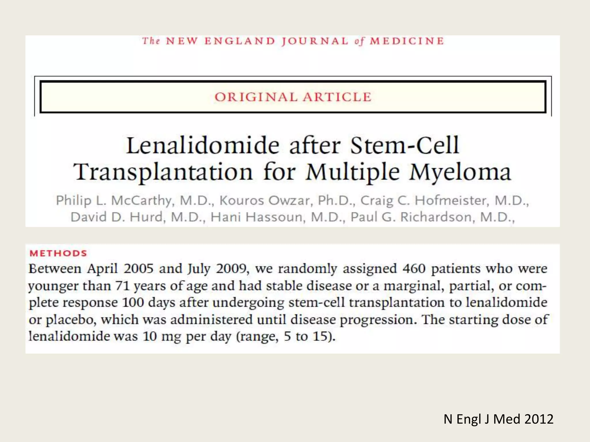
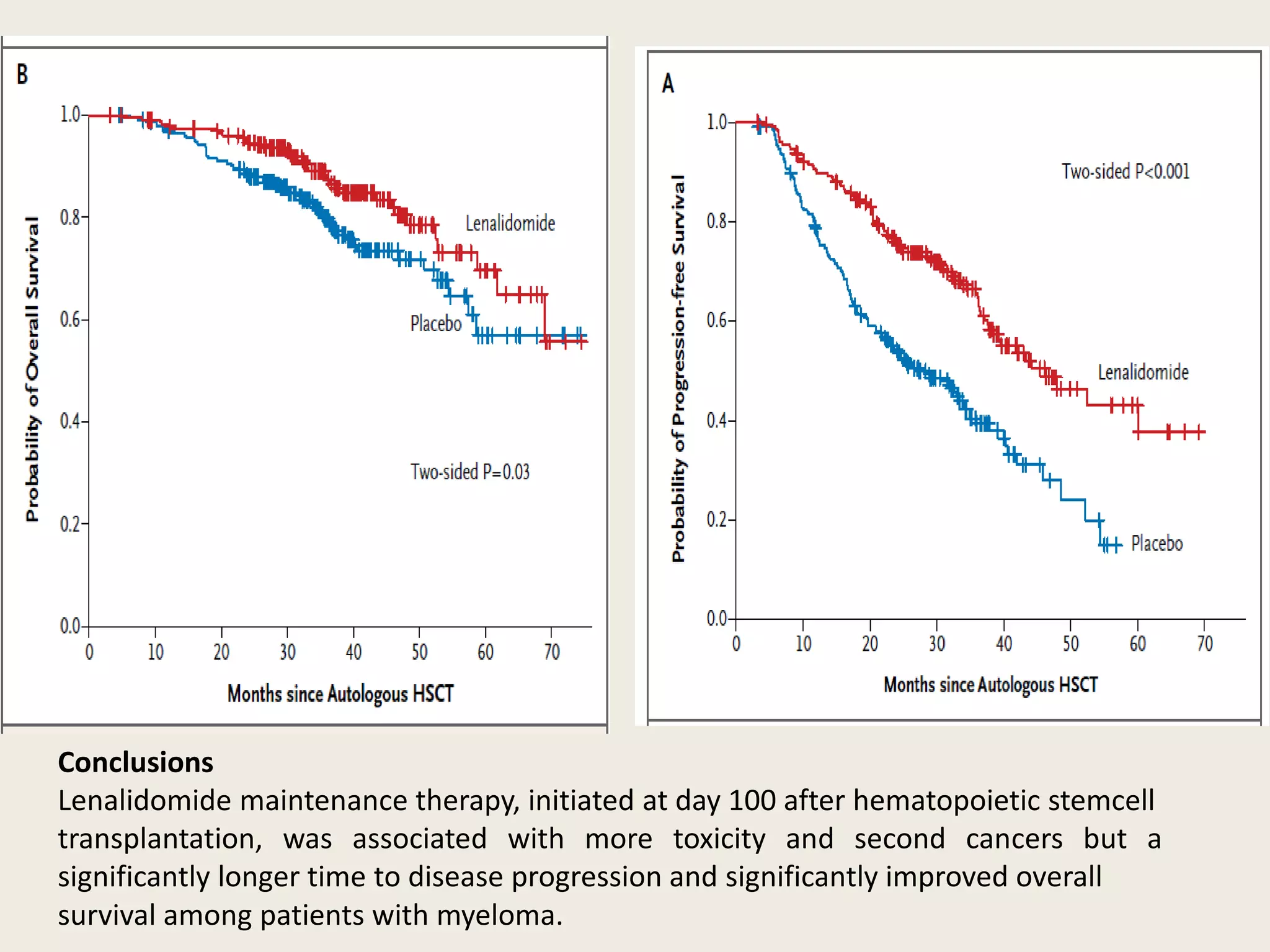
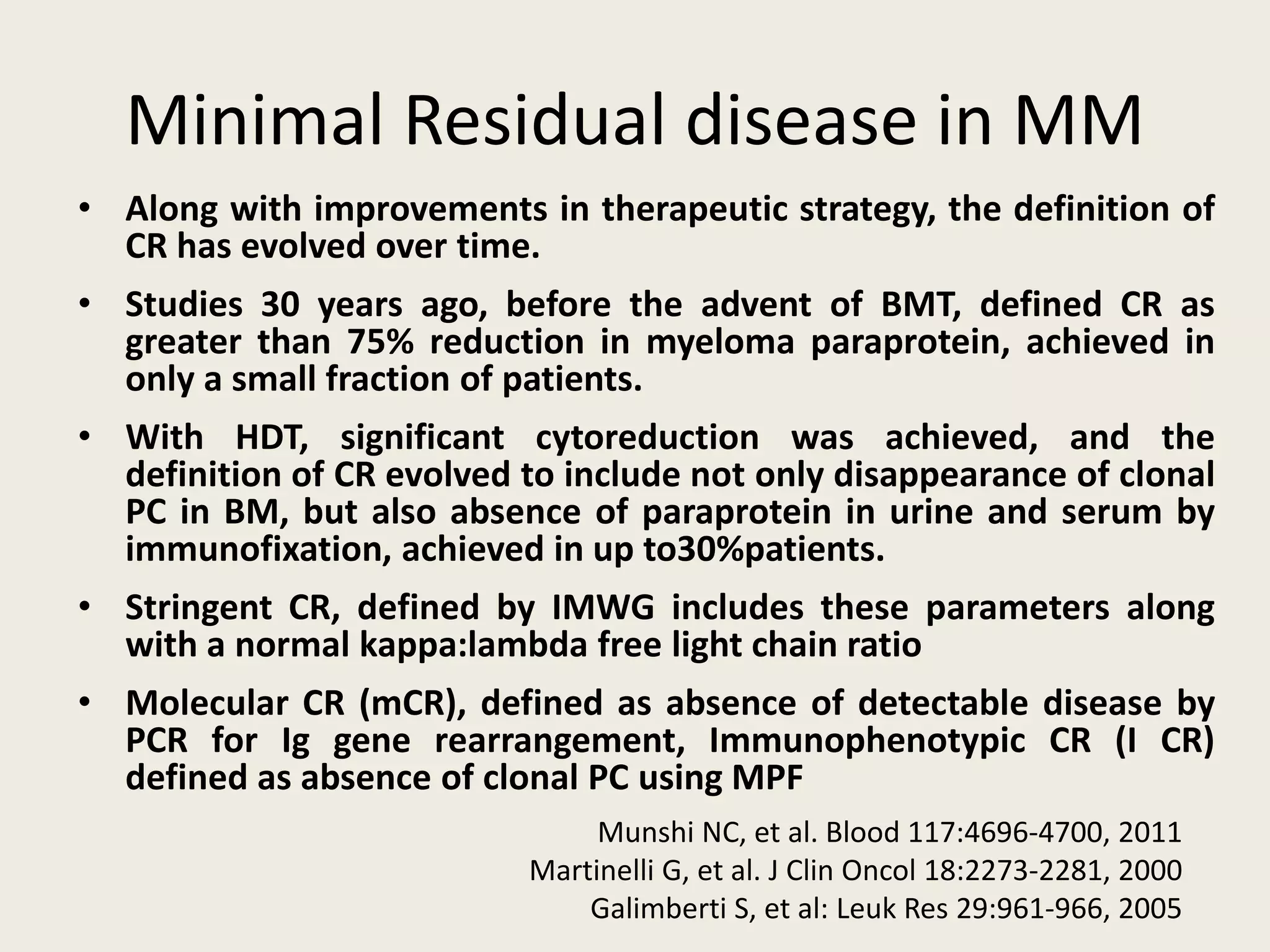
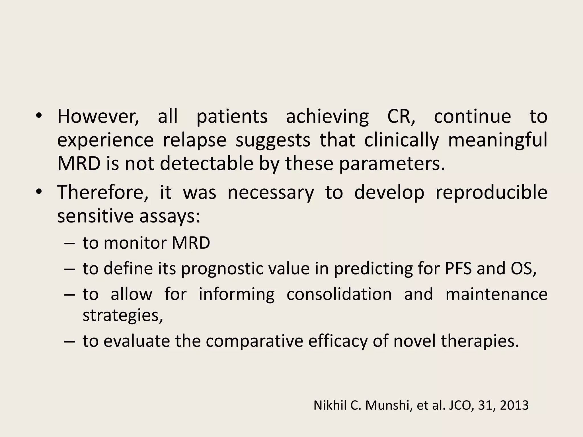
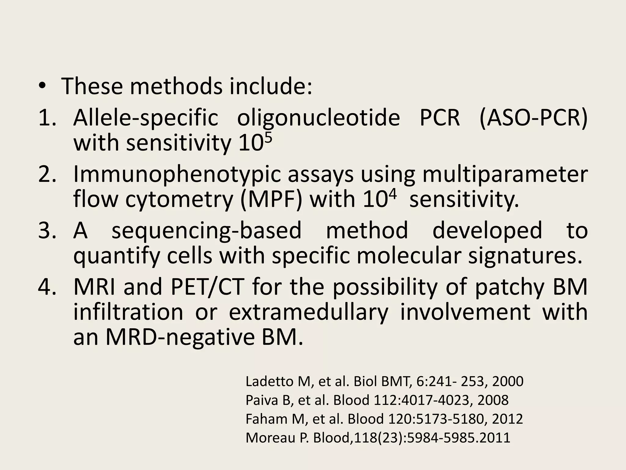
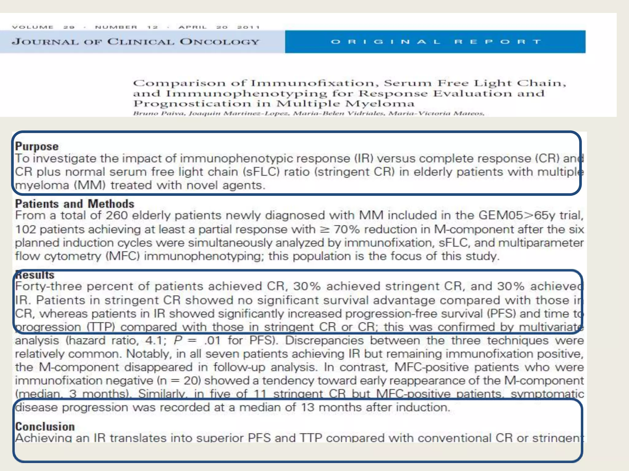
MULTIPLE MYELOMA
Case Presentation
DR MANAL BESSA
MD HEMATOLOGY,
ALEXANDRIA UNIVERSITY
Case:
• 59y old lady , was in good health till August 2014 when she
started to have low back pain that was severe ,more in the right
side and not relieved with analgesic.
• She did not report fever, changes in body weight, paraesthia or
weakness of LL, or bladder or bowel dysfunction
• Revision of systems was unremarkable.
• No past history of chronic disease or surgery..
• Unknown food or drug allergy.
• Family history irrelevant .
• She consulted orthopedic doctor. X ray dorsal and lumbosacral
study showed degenerative changes of lumbar and dorsal
vertebrae.
• With persistent pain , she did CT of pelvic bone that showed
osteolytic lesion involving the right aspect of SV2, SV1 with soft
tissue component encroaching upon the sacral canal
• Whole body bone scan with Tc 99 showed increase radioactivity
at 6th & 10th right rib, DV5, DV7, LV3 and right sacroiliac region
suggesting bone mets. MRI denied any cord infiltration or
compression.
• She was referred to hematologist for further management.
MRI LUMBAR
VERTEBREA
CT PELVIS
BONE SCAN
• On presentation:
– Average weight, conscious, oriented
– Moderate pain score 7/10
– Vital sign stable
– No pallor , no jaundice
– Chest and heart exam with no abnormal finding
– Abdomen was soft, lax with no detected organomegaly.
– Neurological assessment showed intact motor and
sensory system , intact reflex of both upper and lower
limb with tenderness upon palpation of lumbar and
sacral vertebrae.
Lab:
• CBC showed mild anemia, Hb 11 gm normochromic , normocytic, with
normal platelet and WBC count.
• Renal and liver function test are WNL
• Serum calcium was 9.9 mg/dL.
• High ESR (124 mm/hr) ,High CRP (4.8 mg/L)
• Normal serum albumin 4 gm/dL
• High LDH 561 U/L(up to 460) , β2 microglobulin 3.42mg/L(up to 3)
• PEP was positive for monoclonal band , Bence Jones protein was
undetected in urine.
• Serum IG assay showed high Ig G serum level 2.5gm/dL (N up to
1.6gm/dL)
• Serum immunofixation revealed monoclonal protein of 5.4 gm/dL with
serum free light chain showed Kappa restriction.
• Bone marrow biopsy showed hypercellular marrow
with infiltration of 20% plasma cells.
• The BM immunophenotyping revealed that the
plasma cells are positive for CD38, CD138, and
CD56, negative for CD19.
• BM cytogenetic showed normal female Karyotype
46, XX.
Management :
• The diagnosis was confirmed as symptomatic MM, Duri
Salmon IIIA, ISS II.
• The disease prognosis and plan of treatment was
discussed with the patient.
• She was started on Cy/Bor/D protocol with monthly
Zometa for bone disease.
• She was tolerating treatment with no reported toxicity.
• She completed 4 cycles last was on Dec 2014 and
evaluation showed achievement of CR (bone marrow
biopsy showed 2% plasma cells, negative PEP, and
normalization of κ/λ ratio).
• Subsequently, she underwent auto-BMT with
conditioning MEL 140 followed by thalidomide
maintenance dose of 50 mg/day
• Currently, she is doing well , her follow up lab
is acceptable with no evidence of MM and she
is tolerating thalidomide.
outline
• MM overview
• Management
• Role of consolidation
• Role of maintenance therapy
• Minimal residual disease in MM
MULTIPLE MYELOMA
OVERVIEW
• Multiple myeloma is a neoplastic plasma-cell
disorder characterized by clonal proliferation of
malignant plasma cells in the BM
microenvironment, monoclonal protein in the blood
or urine, and associated organ dysfunction.
Incidence
• 1% of cancers and 13% of hematologic cancers.
• In Western countries, the annual incidence is 5.6 cases per
100,000 persons.
• The median age at diagnosis is approximately 70 years;
• 37% of patients are younger than 65 years, 26% are
between the ages of 65 and 74 years, and 37% are 75 years
of age or older.
Seigel RL.et al. Cancer J Clin 2015
Pathogenesis of Multiple Myeloma.
• Myeloma arises from an asymptomatic premalignant proliferation
of monoclonal plasma cells that are derived from post–germinal-
center B cells.
• Multistep genetic and micro-environmental changes lead to the
transformation of these cells into a malignant neoplasm.
• Myeloma is thought to evolve most commonly from a monoclonal
gammopathy of undetermined clinical significance (MGUS) that
progresses to smoldering myeloma and, finally, to symptomatic
myeloma.
Kuehl WM, et al. Nat Rev Cancer 2002
Avet-Loiseau H, et al.Blood 2007
Multi-step pathogenesis of MM
Plasma cells and BM micro-enviroment
in MM
Antonio Palumbo, M.D et al. N engl j 2011
Multiple Myeloma
Clinical Presentation,
Clinical manifestations are related to malignant
behaviour of plasma cells and abnormalities produced
by M protein
• Plasma cell proliferation:
multiple osteolytic bone lesions
hypercalcemia
bone marrow suppression ( pancytopenia )
• Monoclonal protein
decreased level of normal immunoglobulins
hyperviscosity
Multiple Myeloma
Clinical Presentation,
• Anemia, in about 73% of patients at diagnosis.
• Bony lesions develop in almost 58% of patients
with newly diagnosed disease;
• Renal impairment occurs in 20 to 40% of
patients with newly diagnosed disease,
• Infection is increased with active disease but
decreases with response to therapy.
• Fatigue/generalized weakness - 32 percent
• Hypercalcemia
Kyle RA, et al. Mayo Clin Proc, 2003.
Multiple Myeloma
Diagnosis
All 3 criteria must be met:
1.Presence of a serum or urinary monoclonal
protein
2.Presence of clonal plasma cells in the bone
marrow or a plasmacytoma
3.Presence of end organ damage related to the
plasma cell dyscrasia, such as:
– Increased calcium concentration
– Lytic bone lesions
– Anemia
– Renal failure
Detection Of Clonal Plasma Cells
Detection Of Monoclonal protein
Serum free light chain assay
Differential Diagnosis of MM
• Monoclonal gammopathy of undetermined
significance (MGUS)
• Smoldering multiple myeloma (SMM)
• Waldenstrom macroglobulinemia
• Solitary plasmacytoma
• Primary amyloidosis (AL)
• POEMS syndrome
• Metastatic carcinoma
Kyle et al, NEJM, Volume 356:2582-2590, June 21, 2007
Multiple Myeloma
Staging System
International Staging System ISS
Griepp, P, R et al. J Clin Onco, 23;3412-3420, 2005
Cytogenetic Abnormalities
Chromsome 1 abnormaities
Multiple Myeloma
Management
Treatment History
1958 1962 1983-86 1996 20031999 2006 2012
Melphalan/
prednisone
HD dexamethasone
VAD
HD melphalan
Autologous BM
transplants
High-dose therapy with
autologous stem cell
support
Lenalidomide
Thalidomide
Bortezomib
Carfilzomib
Pomalidomide
Elotuzumab
Daratumumab
0
20
40
60
80
100
Dex VAD Thal+Dex CTD VD PAD Rd VTD RVD CRd
Patientswith≥VGPR(%)
S Kumar et al. Cancer Treatment Reviews 2010; Korde et al. ASH 2013.
Sarcolysine
MM: INITIAL THERAPY
• The paradigm of management of MM has
been changed recently with the availability of
novel drugs.
• The aim of treatment also is evolving from
disease control without cure to have complete
remission with possible molecular CR.
Response Criteria
• Assessing the response is the key determent
of myeloma treatment.
• IMWG response criteria is developed from
EBMT/IBMTR/ABMTR to help uniform
reporting.
• Update in response criteria definitions is
adding immunophenotypic CR and molecular
CR
Multiple Myeloma
Treatment Decisions :
• Indications for treatment : presence of any of CRAB ( bone
lesions can be diffuse osteopenia alone)
• Risk Stratification : High-risk factors at diagnosis
• Cytogenetics: del(13), t(4;14), t(14;16), del(17p), del(1p),
and 1q
• Renal failure, elevated LDH, plasma cell leukemia, GEP 70,
EMC-92
• Comorbidities limiting therapy
• International Scoring System stage II or III
Bergsagel PL, et al. Blood2013;
Munshi NC, et al. Blood. 2011
Clearly not transplantation
candidate based on age, performance
score, and comorbidity
MPT, MPV, Len/dex
or clinical trial*
Potential transplantation
candidate
Nonalkylator-based
induction x 4 cycles
Stem cell harvest
Initial Approach to Treatment of MM
• Is there any role for stem cell transplantation
in the novel-agent era?
• Before novel therapy era…
54
42
Attal M, et al. N Engl J Med. 1996;335:91-97. Child JA, et al. N Engl J Med. 2003;348:1875-1883.
15 30 45 60
25
50
75
100
OS(%)
0
0
High dose
Conventional dose
Mos
20 40 60 80
25
50
75
100
Survival(%)
0
0
Intensive therapy
Standard therapy
Mos
P = .03 by Wilcoxon test
P = .04 by log-rank test
Transplantation vs Conventional
Chemotherapy
In the 2 largest studies, autologous SCT improvement in OS compared with
non-transplant cohorts.
 Mel 200 mg/m2 standard conditioning regimen
 Sufficient performance score, and adequate liver, pulmonary,
cardiac function needed
 Higher PR and CR rates than conventional chemotherapy
 Higher OS and EFS than conventional Rx
 Advanced age and impaired renal function are, by themselves,
not contraindications
Attal M, et al. N Engl J Med. 1996;335:91-97. APractice Guidelines. Myeloma.
In the era of novel therapy……
• Randomized trials to address the value of ASCT
are ongoing.
• Transplant trials that use novel agents clearly
showed that responses are deepened so that
the fraction of patients with CRs and VGPRs
increase between the induction phase and the
post-ASCT consolidation phase.
• SCT is capable of achieving residual disease
negativity, a predictor of PFS. The use of ASCT
for MM is increasing, and survival is increasing
proportionately.
Therefore, until trials definitively demonstrate
that the platform of ASCT does not improve
survival, it remains the standard of care for
eligible patients.
Consolidation therapy after ASCT
• Consolidation therapy means a short-term
treatment given to upgrade the quality of the
responses obtained in the previous treatment
phases.
• Consolidation therapy should consist of highly
efficient combinations of agents with minimal
toxicity, applied for a limited period of time.
• Initial results of phase 2 and phase 3 trials
investigating novel agent–based consolidation
therapy show that this strategy may result in
the achievement of deep molecular–defined
or flow cytometry–defined CRs.
• Yet the impact of consolidation on OS, as well
as the optimal consolidation regimen is not
determined.
Phase 2 and 3 trials incorporating novel agent–based consolidation following ASCT
Ladetto M, et al. J Clin Oncol. 2010,Cavo M, et al. Blood. 2012.,Attal M, et al.N Engl J Med. 2012,
Mellqvist UH,et al. Blood. 2013; Leleu X, et al; Leukemia2013,Roussel M, et al. J Clin Oncol. 2014
Maintenance treatment in MM
• Maintaining the response of first-line therapy
is an important objective in MM.
• Maintenance therapy is applied for a
prolonged period of time with the goal of
preventing tumor progression prolonging
response duration, PFS, and, ultimately, OS,
while keeping toxicity minimal.
Requisite for maintenance in MM
• Patient must have:
– Disease that is in remission (undetectable or at a low
level)
– Recovered from all previous toxicities
• Maintenance agent must have:
– Minimal toxicity or at least not overlapping with the
toxicity of the induction regimen
– Convenient dosing
– Convenient route of administration
• The high efficacy of the novel agents,
observed in the front-line settings and
relapse has provided the rationale to also
test their capacity to maintain the benefits
of first-line therapy to prolong remission, and
importantly, to extend overall survival (OS).
Thalidomide in MM maintenance
• Thalidomide was the first novel agent examined in this
setting.
• Six randomized studies have been published, with all of
them showing a significant benefit for thalidomide in terms
of response and PFS, while OS was improved in 3 of them.
• The occurrence of peripheral neuropathy, which is
cumulative and was found to be the main cause of
treatment discontinuation, may hinder its use as long-term
maintenance therapy.
• There is a concern about possible emergence of tumor-
resistant clones in patients with prolonged exposure to
thalidomide and its lack of efficacy in patients with adverse
cytogenetic abnormalities.
Thalidomide maintenance after Auto-BMT or conventional chemotherapy
Lenalidomide maintenance in MM
• Lenalidomide is currently considered the best
candidate for use as maintenance therapy
• lenalidomide maintenance was considered
feasible and manageable, with <30% of patients
having to discontinue the drug because of AEs.
• However, in both studies an unexpected finding
was the occurrence of more secondary primary
malignancies in the lenalidomide group. The
pathophysiology of these secondary neoplasms
remains to be clarified.
N Engl J Med 2012
Conclusions
Lenalidomide maintenance therapy, initiated at day 100 after hematopoietic stemcell
transplantation, was associated with more toxicity and second cancers but a
significantly longer time to disease progression and significantly improved overall
survival among patients with myeloma.
CONCLUSIONS
Maintenance, as compared with no maintenance, significantly prolonged
progression-free survival. but 3-year overall survival was not significantly prolonged
(88.0% vs. 79.2%; hazard ratio for death, 0.64; 95% CI, 0.36 to 1.15; P = 0.14).
PAD + Bortezomib Maintenance vs VAD + Thalidomide
Maintenance: HOVON Trial
Sonneveld P, et al. ASH 2010. Abstract 40.
Efficacy Parameter PAD → bortezomib VAD → thalidomide P Value
3-yr PFS 48% 42% .005
3-yr OS 78% 71% .02
MM Stage II or III, Age 18–65
CAD + GCSF
3 x VAD
CAD + GCSF
3 x PAD
MEL 200 + PBSCT
In GMMG 2nd
MEL 200 + PBSCT
MEL 200 + PBSCT
In GMMG 2nd
MEL 200 + PBSCT
Thalidomide maintenance
50 mg/day for 2 yrs
Allogeneic
Tx
Bortezomib maintenance
1.3 mg/m2/2 weeks for 2 yrs
Bortezomib 1.3 mg/m2 IV
Doxorubicin 9 mg/m2
Dexameth 40 mg
Randomization
Bortezomib + Thalidomide vs Bortezomib + Prednisone as
Maintenance: GEM2005MAS65
Arm ORR CR Median PFS
VT maintenance 95% 46% 39 months
VP maintenance 97% 39% 32 months
Mateos MV, et al. Lancet Oncol. 2010;10:934-41.
No significant differences between VMP and VTP in ORR
(80% and 81%) and CR rate (20% and 27%)
Maintenance
Bort/Thal
(VT)
Bort/Pred
(VP)
Bort/Thal
(VT)
Bort/Pred
(VP)
Induction
(6 cycles)
Bort/Mel/Pred
(VMP)
Bort/Thal/Pred
(VTP)
vs
Series of 260 elderly untreated MM patients included in the GEM2005 Spanish trial
Bortezomib + Thalidomide Maintenance GIMEMA STUDY
Arm 3 y OS CR 3 Y PFS
VT maintenance 89% 38% 56%
No maintenance 87% 24% 41%
Mateos MV, et al. Lancet Oncol. 2010;10:934-41.
Maintenance
Bort/Thal
(VT)
No Maintenance
Induction
(9 cycles)
Bort/Mel/Pred/Thal
(VMPT)
Bort/Thal/Pred
(VMP)
vs
Series of 511 elderly untreated MM patients included in phase III trial
Conclusions About Maintenance Therapy
for Multiple Myeloma
• Maintenance therapy prolongs PFS.
• Low-dose oral agents preferable for maintenance therapy.
• Both bortezomib and lenalidomide are useful maintenance agents
and may need to be combined for patients with high-risk disease.
• Slight increase in incidence of secondary malignancy after
lenalidomide maintenance.
• Overall, everyone who meets prerequisites for maintenance therapy
should be considered candidates for treatment. The real question is
not if maintenance therapy should be given, but how long.
Minimal Residual disease in MM
• Along with improvements in therapeutic strategy, the definition of
CR has evolved over time.
• Studies 30 years ago, before the advent of BMT, defined CR as
greater than 75% reduction in myeloma paraprotein, achieved in
only a small fraction of patients.
• With HDT, significant cytoreduction was achieved, and the
definition of CR evolved to include not only disappearance of clonal
PC in BM, but also absence of paraprotein in urine and serum by
immunofixation, achieved in up to30%patients.
• Stringent CR, defined by IMWG includes these parameters along
with a normal kappa:lambda free light chain ratio
• Molecular CR (mCR), defined as absence of detectable disease by
PCR for Ig gene rearrangement, Immunophenotypic CR (I CR)
defined as absence of clonal PC using MPF
Munshi NC, et al. Blood 117:4696-4700, 2011
Martinelli G, et al. J Clin Oncol 18:2273-2281, 2000
Galimberti S, et al: Leuk Res 29:961-966, 2005
• However, all patients achieving CR, continue to
experience relapse suggests that clinically meaningful
MRD is not detectable by these parameters.
• Therefore, it was necessary to develop reproducible
sensitive assays:
– to monitor MRD
– to define its prognostic value in predicting for PFS and OS,
– to allow for informing consolidation and maintenance
strategies,
– to evaluate the comparative efficacy of novel therapies.
Nikhil C. Munshi, et al. JCO, 31, 2013
• These methods include:
1. Allele-specific oligonucleotide PCR (ASO-PCR)
with sensitivity 105
2. Immunophenotypic assays using multiparameter
flow cytometry (MPF) with 104 sensitivity.
3. A sequencing-based method developed to
quantify cells with specific molecular signatures.
4. MRI and PET/CT for the possibility of patchy BM
infiltration or extramedullary involvement with
an MRD-negative BM.
Ladetto M, et al. Biol BMT, 6:241- 253, 2000
Paiva B, et al. Blood 112:4017-4023, 2008
Faham M, et al. Blood 120:5173-5180, 2012
Moreau P. Blood,118(23):5984-5985.2011
A critical question????
• Does the ability to detect MRD by MPF or
ASO-PCR have clinical implications for PFS and
OS???
• May it inform therapy and allow us to tailor
therapeutic decisions????.
Number of studies published in PUBMED per MRD technique showing prognostic value for
progression-free survival (PFS) and overall survival (OS) specifically among patients in CR
after therapy.
Paiva B,et al. Blood. 2012. Martinez-LJ,et al. Blood. 2014. Bakkus MH, et al.Br J Haematol. 2004. Puig N, et
al. Leukemia. 2014. Corradini P, et al. Blood. 2003; Rawstron AC,et al. Blood. 2002. Paiva B, et al. Blood.
2008. Rawstron AC,et al JCO. 2013. Zamagni E,et al.Blood. 2011.
What is the clinical significance of MRD
monitoring in MM?
So far, no clinical trial has randomized MM
patients according to their MRD status and,
thereby, investigated the role of MRD for
individualized therapy. However, many studies
have shown the value of MRD diagnostics for
evaluation of the efficacy of specific treatment
stages and, therefore, potential treatment
decisions.
Bruno Paiva, et al blood 2015.
• Both the Spanish and the United Kingdom study groups
have shown that MRD kinetics before and after
HDT/ASCT allow the identification of chemosensitive
(MRD-negative cases at 2 time points), intermediate,
and chemoresistant patients (MRD-positive patients at
2 time points).
• For the latter, it could be hypothesized that
consolidation is needed to improve outcomes
• When such analysis is restricted to CR patients after
induction, those failing to eradicate MRD levels before
HDT/ASCT will show significantly superior PFS if MRD
negativity is achieved after HDT/ASCT,
Paiva B, et al. Blood. 2008;112(10): 4017-4023.
Rawstron AC,et al JCOl. 2013;31(20):2540-2547.
• Maintenance therapy represents another
illustrating example. In the Gruppo Italiano
Malattie EMatologiche dell’Adulto (GIMEMA)
reported PFS rates at median follow up of
100% vs 57% for patients in molecular-CR vs
MRD-positive cases, respectively.
Ladetto M,. J Clin Oncol. 2010;
Limitation
• Discordant results between MRD (by
immunophenotypic, molecular, and imaging
techniques) vs conventional response assessment
has questioned the sensitivity and specificity of
MRD monitoring over traditional paraprotein
measurement.
• False negative MRD because the patchy pattern
of BM infiltration lead to a degree of uncertainty
regarding MRD-negative results (ie, are clonal
PCs truly absent, or for non-representative BM
sampling?).
Zamagni E et al,Blood. 2011
Paiva B, et al.J Clin Oncol. 2011
Conclusion remark
• MRD clearance is achievable in the era of novel
and more effective treatment strategies and it is
predictive of superior outcomes.
• Persistence of MRD is always an adverse
prognostic feature, even among CR patients.
• Some of the limitations could be potentially
overcome by parallel usage of sensitive imaging
techniques.
• The importance of the 2 consecutive protein
response assessments before the institution of
any new therapy to confirm a response category.
Toward an optimal strategy of induction, HDT
plus ASCT,
consolidation, and maintenance
Roussel M, et al. J Clin Oncol. 2014; Usmani SZ, et al.Leukemia. 2013;
MULTIPLE MYELOMA UPDATE

MULTIPLE MYELOMA UPDATE

  • 1.
    MULTIPLE MYELOMA Case Presentation DRMANAL BESSA MD HEMATOLOGY, ALEXANDRIA UNIVERSITY
  • 2.
    Case: • 59y oldlady , was in good health till August 2014 when she started to have low back pain that was severe ,more in the right side and not relieved with analgesic. • She did not report fever, changes in body weight, paraesthia or weakness of LL, or bladder or bowel dysfunction • Revision of systems was unremarkable. • No past history of chronic disease or surgery.. • Unknown food or drug allergy. • Family history irrelevant .
  • 3.
    • She consultedorthopedic doctor. X ray dorsal and lumbosacral study showed degenerative changes of lumbar and dorsal vertebrae. • With persistent pain , she did CT of pelvic bone that showed osteolytic lesion involving the right aspect of SV2, SV1 with soft tissue component encroaching upon the sacral canal • Whole body bone scan with Tc 99 showed increase radioactivity at 6th & 10th right rib, DV5, DV7, LV3 and right sacroiliac region suggesting bone mets. MRI denied any cord infiltration or compression. • She was referred to hematologist for further management.
  • 4.
  • 5.
    • On presentation: –Average weight, conscious, oriented – Moderate pain score 7/10 – Vital sign stable – No pallor , no jaundice – Chest and heart exam with no abnormal finding – Abdomen was soft, lax with no detected organomegaly. – Neurological assessment showed intact motor and sensory system , intact reflex of both upper and lower limb with tenderness upon palpation of lumbar and sacral vertebrae.
  • 6.
    Lab: • CBC showedmild anemia, Hb 11 gm normochromic , normocytic, with normal platelet and WBC count. • Renal and liver function test are WNL • Serum calcium was 9.9 mg/dL. • High ESR (124 mm/hr) ,High CRP (4.8 mg/L) • Normal serum albumin 4 gm/dL • High LDH 561 U/L(up to 460) , β2 microglobulin 3.42mg/L(up to 3) • PEP was positive for monoclonal band , Bence Jones protein was undetected in urine. • Serum IG assay showed high Ig G serum level 2.5gm/dL (N up to 1.6gm/dL) • Serum immunofixation revealed monoclonal protein of 5.4 gm/dL with serum free light chain showed Kappa restriction.
  • 7.
    • Bone marrowbiopsy showed hypercellular marrow with infiltration of 20% plasma cells. • The BM immunophenotyping revealed that the plasma cells are positive for CD38, CD138, and CD56, negative for CD19. • BM cytogenetic showed normal female Karyotype 46, XX.
  • 8.
    Management : • Thediagnosis was confirmed as symptomatic MM, Duri Salmon IIIA, ISS II. • The disease prognosis and plan of treatment was discussed with the patient. • She was started on Cy/Bor/D protocol with monthly Zometa for bone disease. • She was tolerating treatment with no reported toxicity. • She completed 4 cycles last was on Dec 2014 and evaluation showed achievement of CR (bone marrow biopsy showed 2% plasma cells, negative PEP, and normalization of κ/λ ratio).
  • 9.
    • Subsequently, sheunderwent auto-BMT with conditioning MEL 140 followed by thalidomide maintenance dose of 50 mg/day • Currently, she is doing well , her follow up lab is acceptable with no evidence of MM and she is tolerating thalidomide.
  • 10.
    outline • MM overview •Management • Role of consolidation • Role of maintenance therapy • Minimal residual disease in MM
  • 11.
    MULTIPLE MYELOMA OVERVIEW • Multiplemyeloma is a neoplastic plasma-cell disorder characterized by clonal proliferation of malignant plasma cells in the BM microenvironment, monoclonal protein in the blood or urine, and associated organ dysfunction.
  • 12.
    Incidence • 1% ofcancers and 13% of hematologic cancers. • In Western countries, the annual incidence is 5.6 cases per 100,000 persons. • The median age at diagnosis is approximately 70 years; • 37% of patients are younger than 65 years, 26% are between the ages of 65 and 74 years, and 37% are 75 years of age or older. Seigel RL.et al. Cancer J Clin 2015
  • 13.
    Pathogenesis of MultipleMyeloma. • Myeloma arises from an asymptomatic premalignant proliferation of monoclonal plasma cells that are derived from post–germinal- center B cells. • Multistep genetic and micro-environmental changes lead to the transformation of these cells into a malignant neoplasm. • Myeloma is thought to evolve most commonly from a monoclonal gammopathy of undetermined clinical significance (MGUS) that progresses to smoldering myeloma and, finally, to symptomatic myeloma. Kuehl WM, et al. Nat Rev Cancer 2002 Avet-Loiseau H, et al.Blood 2007
  • 14.
  • 15.
    Plasma cells andBM micro-enviroment in MM Antonio Palumbo, M.D et al. N engl j 2011
  • 16.
    Multiple Myeloma Clinical Presentation, Clinicalmanifestations are related to malignant behaviour of plasma cells and abnormalities produced by M protein • Plasma cell proliferation: multiple osteolytic bone lesions hypercalcemia bone marrow suppression ( pancytopenia ) • Monoclonal protein decreased level of normal immunoglobulins hyperviscosity
  • 17.
    Multiple Myeloma Clinical Presentation, •Anemia, in about 73% of patients at diagnosis. • Bony lesions develop in almost 58% of patients with newly diagnosed disease; • Renal impairment occurs in 20 to 40% of patients with newly diagnosed disease, • Infection is increased with active disease but decreases with response to therapy. • Fatigue/generalized weakness - 32 percent • Hypercalcemia Kyle RA, et al. Mayo Clin Proc, 2003.
  • 19.
    Multiple Myeloma Diagnosis All 3criteria must be met: 1.Presence of a serum or urinary monoclonal protein 2.Presence of clonal plasma cells in the bone marrow or a plasmacytoma 3.Presence of end organ damage related to the plasma cell dyscrasia, such as: – Increased calcium concentration – Lytic bone lesions – Anemia – Renal failure
  • 20.
    Detection Of ClonalPlasma Cells
  • 21.
    Detection Of Monoclonalprotein Serum free light chain assay
  • 22.
    Differential Diagnosis ofMM • Monoclonal gammopathy of undetermined significance (MGUS) • Smoldering multiple myeloma (SMM) • Waldenstrom macroglobulinemia • Solitary plasmacytoma • Primary amyloidosis (AL) • POEMS syndrome • Metastatic carcinoma
  • 23.
    Kyle et al,NEJM, Volume 356:2582-2590, June 21, 2007
  • 24.
  • 25.
    International Staging SystemISS Griepp, P, R et al. J Clin Onco, 23;3412-3420, 2005
  • 26.
  • 27.
  • 28.
    Treatment History 1958 19621983-86 1996 20031999 2006 2012 Melphalan/ prednisone HD dexamethasone VAD HD melphalan Autologous BM transplants High-dose therapy with autologous stem cell support Lenalidomide Thalidomide Bortezomib Carfilzomib Pomalidomide Elotuzumab Daratumumab 0 20 40 60 80 100 Dex VAD Thal+Dex CTD VD PAD Rd VTD RVD CRd Patientswith≥VGPR(%) S Kumar et al. Cancer Treatment Reviews 2010; Korde et al. ASH 2013. Sarcolysine
  • 29.
    MM: INITIAL THERAPY •The paradigm of management of MM has been changed recently with the availability of novel drugs. • The aim of treatment also is evolving from disease control without cure to have complete remission with possible molecular CR.
  • 30.
    Response Criteria • Assessingthe response is the key determent of myeloma treatment. • IMWG response criteria is developed from EBMT/IBMTR/ABMTR to help uniform reporting. • Update in response criteria definitions is adding immunophenotypic CR and molecular CR
  • 32.
    Multiple Myeloma Treatment Decisions: • Indications for treatment : presence of any of CRAB ( bone lesions can be diffuse osteopenia alone) • Risk Stratification : High-risk factors at diagnosis • Cytogenetics: del(13), t(4;14), t(14;16), del(17p), del(1p), and 1q • Renal failure, elevated LDH, plasma cell leukemia, GEP 70, EMC-92 • Comorbidities limiting therapy • International Scoring System stage II or III Bergsagel PL, et al. Blood2013; Munshi NC, et al. Blood. 2011
  • 33.
    Clearly not transplantation candidatebased on age, performance score, and comorbidity MPT, MPV, Len/dex or clinical trial* Potential transplantation candidate Nonalkylator-based induction x 4 cycles Stem cell harvest Initial Approach to Treatment of MM
  • 35.
    • Is thereany role for stem cell transplantation in the novel-agent era?
  • 36.
    • Before noveltherapy era…
  • 37.
    54 42 Attal M, etal. N Engl J Med. 1996;335:91-97. Child JA, et al. N Engl J Med. 2003;348:1875-1883. 15 30 45 60 25 50 75 100 OS(%) 0 0 High dose Conventional dose Mos 20 40 60 80 25 50 75 100 Survival(%) 0 0 Intensive therapy Standard therapy Mos P = .03 by Wilcoxon test P = .04 by log-rank test Transplantation vs Conventional Chemotherapy In the 2 largest studies, autologous SCT improvement in OS compared with non-transplant cohorts.
  • 38.
     Mel 200mg/m2 standard conditioning regimen  Sufficient performance score, and adequate liver, pulmonary, cardiac function needed  Higher PR and CR rates than conventional chemotherapy  Higher OS and EFS than conventional Rx  Advanced age and impaired renal function are, by themselves, not contraindications Attal M, et al. N Engl J Med. 1996;335:91-97. APractice Guidelines. Myeloma.
  • 39.
    In the eraof novel therapy……
  • 40.
    • Randomized trialsto address the value of ASCT are ongoing. • Transplant trials that use novel agents clearly showed that responses are deepened so that the fraction of patients with CRs and VGPRs increase between the induction phase and the post-ASCT consolidation phase. • SCT is capable of achieving residual disease negativity, a predictor of PFS. The use of ASCT for MM is increasing, and survival is increasing proportionately.
  • 41.
    Therefore, until trialsdefinitively demonstrate that the platform of ASCT does not improve survival, it remains the standard of care for eligible patients.
  • 42.
    Consolidation therapy afterASCT • Consolidation therapy means a short-term treatment given to upgrade the quality of the responses obtained in the previous treatment phases. • Consolidation therapy should consist of highly efficient combinations of agents with minimal toxicity, applied for a limited period of time.
  • 43.
    • Initial resultsof phase 2 and phase 3 trials investigating novel agent–based consolidation therapy show that this strategy may result in the achievement of deep molecular–defined or flow cytometry–defined CRs. • Yet the impact of consolidation on OS, as well as the optimal consolidation regimen is not determined.
  • 44.
    Phase 2 and3 trials incorporating novel agent–based consolidation following ASCT Ladetto M, et al. J Clin Oncol. 2010,Cavo M, et al. Blood. 2012.,Attal M, et al.N Engl J Med. 2012, Mellqvist UH,et al. Blood. 2013; Leleu X, et al; Leukemia2013,Roussel M, et al. J Clin Oncol. 2014
  • 45.
    Maintenance treatment inMM • Maintaining the response of first-line therapy is an important objective in MM. • Maintenance therapy is applied for a prolonged period of time with the goal of preventing tumor progression prolonging response duration, PFS, and, ultimately, OS, while keeping toxicity minimal.
  • 46.
    Requisite for maintenancein MM • Patient must have: – Disease that is in remission (undetectable or at a low level) – Recovered from all previous toxicities • Maintenance agent must have: – Minimal toxicity or at least not overlapping with the toxicity of the induction regimen – Convenient dosing – Convenient route of administration
  • 47.
    • The highefficacy of the novel agents, observed in the front-line settings and relapse has provided the rationale to also test their capacity to maintain the benefits of first-line therapy to prolong remission, and importantly, to extend overall survival (OS).
  • 48.
    Thalidomide in MMmaintenance • Thalidomide was the first novel agent examined in this setting. • Six randomized studies have been published, with all of them showing a significant benefit for thalidomide in terms of response and PFS, while OS was improved in 3 of them. • The occurrence of peripheral neuropathy, which is cumulative and was found to be the main cause of treatment discontinuation, may hinder its use as long-term maintenance therapy. • There is a concern about possible emergence of tumor- resistant clones in patients with prolonged exposure to thalidomide and its lack of efficacy in patients with adverse cytogenetic abnormalities.
  • 49.
    Thalidomide maintenance afterAuto-BMT or conventional chemotherapy
  • 50.
    Lenalidomide maintenance inMM • Lenalidomide is currently considered the best candidate for use as maintenance therapy • lenalidomide maintenance was considered feasible and manageable, with <30% of patients having to discontinue the drug because of AEs. • However, in both studies an unexpected finding was the occurrence of more secondary primary malignancies in the lenalidomide group. The pathophysiology of these secondary neoplasms remains to be clarified.
  • 51.
    N Engl JMed 2012
  • 52.
    Conclusions Lenalidomide maintenance therapy,initiated at day 100 after hematopoietic stemcell transplantation, was associated with more toxicity and second cancers but a significantly longer time to disease progression and significantly improved overall survival among patients with myeloma.
  • 54.
    CONCLUSIONS Maintenance, as comparedwith no maintenance, significantly prolonged progression-free survival. but 3-year overall survival was not significantly prolonged (88.0% vs. 79.2%; hazard ratio for death, 0.64; 95% CI, 0.36 to 1.15; P = 0.14).
  • 55.
    PAD + BortezomibMaintenance vs VAD + Thalidomide Maintenance: HOVON Trial Sonneveld P, et al. ASH 2010. Abstract 40. Efficacy Parameter PAD → bortezomib VAD → thalidomide P Value 3-yr PFS 48% 42% .005 3-yr OS 78% 71% .02 MM Stage II or III, Age 18–65 CAD + GCSF 3 x VAD CAD + GCSF 3 x PAD MEL 200 + PBSCT In GMMG 2nd MEL 200 + PBSCT MEL 200 + PBSCT In GMMG 2nd MEL 200 + PBSCT Thalidomide maintenance 50 mg/day for 2 yrs Allogeneic Tx Bortezomib maintenance 1.3 mg/m2/2 weeks for 2 yrs Bortezomib 1.3 mg/m2 IV Doxorubicin 9 mg/m2 Dexameth 40 mg Randomization
  • 56.
    Bortezomib + Thalidomidevs Bortezomib + Prednisone as Maintenance: GEM2005MAS65 Arm ORR CR Median PFS VT maintenance 95% 46% 39 months VP maintenance 97% 39% 32 months Mateos MV, et al. Lancet Oncol. 2010;10:934-41. No significant differences between VMP and VTP in ORR (80% and 81%) and CR rate (20% and 27%) Maintenance Bort/Thal (VT) Bort/Pred (VP) Bort/Thal (VT) Bort/Pred (VP) Induction (6 cycles) Bort/Mel/Pred (VMP) Bort/Thal/Pred (VTP) vs Series of 260 elderly untreated MM patients included in the GEM2005 Spanish trial
  • 57.
    Bortezomib + ThalidomideMaintenance GIMEMA STUDY Arm 3 y OS CR 3 Y PFS VT maintenance 89% 38% 56% No maintenance 87% 24% 41% Mateos MV, et al. Lancet Oncol. 2010;10:934-41. Maintenance Bort/Thal (VT) No Maintenance Induction (9 cycles) Bort/Mel/Pred/Thal (VMPT) Bort/Thal/Pred (VMP) vs Series of 511 elderly untreated MM patients included in phase III trial
  • 58.
    Conclusions About MaintenanceTherapy for Multiple Myeloma • Maintenance therapy prolongs PFS. • Low-dose oral agents preferable for maintenance therapy. • Both bortezomib and lenalidomide are useful maintenance agents and may need to be combined for patients with high-risk disease. • Slight increase in incidence of secondary malignancy after lenalidomide maintenance. • Overall, everyone who meets prerequisites for maintenance therapy should be considered candidates for treatment. The real question is not if maintenance therapy should be given, but how long.
  • 59.
    Minimal Residual diseasein MM • Along with improvements in therapeutic strategy, the definition of CR has evolved over time. • Studies 30 years ago, before the advent of BMT, defined CR as greater than 75% reduction in myeloma paraprotein, achieved in only a small fraction of patients. • With HDT, significant cytoreduction was achieved, and the definition of CR evolved to include not only disappearance of clonal PC in BM, but also absence of paraprotein in urine and serum by immunofixation, achieved in up to30%patients. • Stringent CR, defined by IMWG includes these parameters along with a normal kappa:lambda free light chain ratio • Molecular CR (mCR), defined as absence of detectable disease by PCR for Ig gene rearrangement, Immunophenotypic CR (I CR) defined as absence of clonal PC using MPF Munshi NC, et al. Blood 117:4696-4700, 2011 Martinelli G, et al. J Clin Oncol 18:2273-2281, 2000 Galimberti S, et al: Leuk Res 29:961-966, 2005
  • 60.
    • However, allpatients achieving CR, continue to experience relapse suggests that clinically meaningful MRD is not detectable by these parameters. • Therefore, it was necessary to develop reproducible sensitive assays: – to monitor MRD – to define its prognostic value in predicting for PFS and OS, – to allow for informing consolidation and maintenance strategies, – to evaluate the comparative efficacy of novel therapies. Nikhil C. Munshi, et al. JCO, 31, 2013
  • 61.
    • These methodsinclude: 1. Allele-specific oligonucleotide PCR (ASO-PCR) with sensitivity 105 2. Immunophenotypic assays using multiparameter flow cytometry (MPF) with 104 sensitivity. 3. A sequencing-based method developed to quantify cells with specific molecular signatures. 4. MRI and PET/CT for the possibility of patchy BM infiltration or extramedullary involvement with an MRD-negative BM. Ladetto M, et al. Biol BMT, 6:241- 253, 2000 Paiva B, et al. Blood 112:4017-4023, 2008 Faham M, et al. Blood 120:5173-5180, 2012 Moreau P. Blood,118(23):5984-5985.2011
  • 62.
    A critical question???? •Does the ability to detect MRD by MPF or ASO-PCR have clinical implications for PFS and OS??? • May it inform therapy and allow us to tailor therapeutic decisions????.
  • 64.
    Number of studiespublished in PUBMED per MRD technique showing prognostic value for progression-free survival (PFS) and overall survival (OS) specifically among patients in CR after therapy. Paiva B,et al. Blood. 2012. Martinez-LJ,et al. Blood. 2014. Bakkus MH, et al.Br J Haematol. 2004. Puig N, et al. Leukemia. 2014. Corradini P, et al. Blood. 2003; Rawstron AC,et al. Blood. 2002. Paiva B, et al. Blood. 2008. Rawstron AC,et al JCO. 2013. Zamagni E,et al.Blood. 2011.
  • 65.
    What is theclinical significance of MRD monitoring in MM? So far, no clinical trial has randomized MM patients according to their MRD status and, thereby, investigated the role of MRD for individualized therapy. However, many studies have shown the value of MRD diagnostics for evaluation of the efficacy of specific treatment stages and, therefore, potential treatment decisions. Bruno Paiva, et al blood 2015.
  • 66.
    • Both theSpanish and the United Kingdom study groups have shown that MRD kinetics before and after HDT/ASCT allow the identification of chemosensitive (MRD-negative cases at 2 time points), intermediate, and chemoresistant patients (MRD-positive patients at 2 time points). • For the latter, it could be hypothesized that consolidation is needed to improve outcomes • When such analysis is restricted to CR patients after induction, those failing to eradicate MRD levels before HDT/ASCT will show significantly superior PFS if MRD negativity is achieved after HDT/ASCT, Paiva B, et al. Blood. 2008;112(10): 4017-4023. Rawstron AC,et al JCOl. 2013;31(20):2540-2547.
  • 67.
    • Maintenance therapyrepresents another illustrating example. In the Gruppo Italiano Malattie EMatologiche dell’Adulto (GIMEMA) reported PFS rates at median follow up of 100% vs 57% for patients in molecular-CR vs MRD-positive cases, respectively. Ladetto M,. J Clin Oncol. 2010;
  • 68.
    Limitation • Discordant resultsbetween MRD (by immunophenotypic, molecular, and imaging techniques) vs conventional response assessment has questioned the sensitivity and specificity of MRD monitoring over traditional paraprotein measurement. • False negative MRD because the patchy pattern of BM infiltration lead to a degree of uncertainty regarding MRD-negative results (ie, are clonal PCs truly absent, or for non-representative BM sampling?). Zamagni E et al,Blood. 2011 Paiva B, et al.J Clin Oncol. 2011
  • 69.
    Conclusion remark • MRDclearance is achievable in the era of novel and more effective treatment strategies and it is predictive of superior outcomes. • Persistence of MRD is always an adverse prognostic feature, even among CR patients. • Some of the limitations could be potentially overcome by parallel usage of sensitive imaging techniques. • The importance of the 2 consecutive protein response assessments before the institution of any new therapy to confirm a response category.
  • 70.
    Toward an optimalstrategy of induction, HDT plus ASCT, consolidation, and maintenance Roussel M, et al. J Clin Oncol. 2014; Usmani SZ, et al.Leukemia. 2013;

Editor's Notes

  • #34 Len/dex, lenalidomide/dexamethasone; MM, multiple myeloma; MPT, melphalan/prednisone/thalidomide; MPV, melphalan/prednisone/bortezomib.   Patients considered eligible for transplantation typically have a good performance status; no significant comorbidities, such as severe cardiac disease or severe pulmonary disease; and are usually younger than 75 years of age. Stem cell transplantation is available through 3 centers in the VA Health System, located in Seattle, Washington; Nashville, Tennessee; and San Antonio, Texas.   Transplant candidates should not receive alkylator‑based therapy with agents such as melphalan hydrochloride, because these agents adversely affect stem cells as well as myeloma cells. Instead, non–alkylator‑based therapies are required for patients who are potential transplantation candidates. Once a patient is determined to be a transplantation candidate, he or she should undergo stem cell harvest.
  • #38 OS, overall survival.   Stem cell transplantation was shown to be better than conventional therapy in a trial reported by Attal and colleagues in 1996. Patients undergoing stem cell transplantation had a markedly improved median survival compared with patients receiving conventional therapy, with a median survival of approximately 60 months compared with 30 months, respectively. This translated into an overall survival benefit with stem cell transplantation in these patients.
  • #56 Next, we move on to the question of maintenance bortezomib. The HOVON trial randomized myeloma patients to VAD (a vincristine-based chemotherapy regimen) or PAD induction therapy, which replaces vincristine with bortezomib. All patients then received transplantation, which was followed by thalidomide maintenance (for VAD patients) or bortezomib maintenance (for PAD patients). The bortezomib arm showed a significant improvement in both PFS and OS. However, the study design prohibits any conclusions from being drawn about maintenance bortezomib, since it was not rigorously controlled. Rather, the most important point of this study is the conclusion that bortezomib can be tolerated as maintenance therapy, at least when using this regimen of 1.3 mg/m2 every other week.
  • #57 This next study asks whether 2 maintenance therapies – thalidomide and bortezomib – can be safely and effectively combined. This Spanish trial of 260 elderly patients with untreated myeloma randomized patients first to the bortezomib-containing VMP induction regimen or to VTP, which contains both bortezomib and thalidomide. After this stage, patients were again randomized to receive either bortezomib/prednisone maintenance or bortezomib/thalidomide maintenance. Bortezomib maintenance was administered on days 1, 4, 8, and 11 every 3 months. No difference in either ORR or CR was observed between the VMP and VTP induction regimens, but it is interesting to note that either maintenance therapy regimen produced an increase in complete responses. No significant PFS difference was observed between the 2 maintenance therapy regimens, with the bortezomib/thalidomide arm showing a 39-month PFS and the bortezomib/prednisone arm showing a 32-month PFS. The bortezomib/thalidomide arm also showed a slight increase in toxicity (12% vs 8%), which would be expected based on the toxicity profiles of the 2 agents.
  • #59 In conclusion, the data presented demonstrate that maintenance therapy prolongs PFS and supports the notion that low-dose oral agents are preferable for maintenance therapy. Both bortezomib and lenalidomide are useful maintenance agents, and these 2 agents may need to be combined for patients with high-risk disease. There is a slight increase in the incidence of secondary malignancy after lenalidomide maintenance. So, should everyone be treated with maintenance therapy? Overall, everyone who meets the prerequisites for maintenance therapy – achieving at least a PR and having resolution of prior toxicities -- should be considered candidates for maintenance treatment. The real question is not if maintenance therapy should be given, but how long.
  • #71 The implementation of an “optimal strategy,” consisting of novel agent–based induction, HDT, and the use of novel agents in consolidation and maintenance, may result in a 5-year survival rate of 80%,