Download as PDF, PPTX
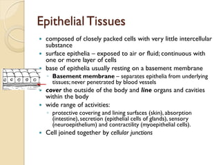
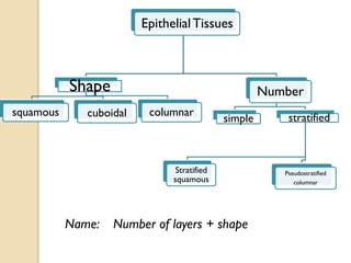
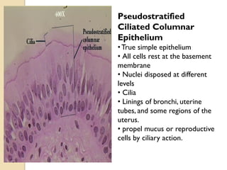
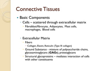
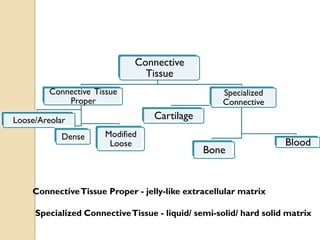

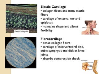
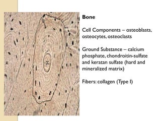
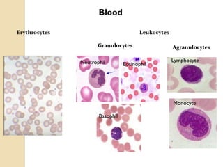
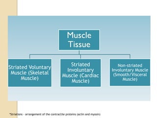
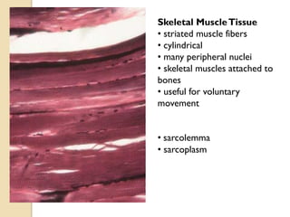
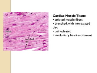
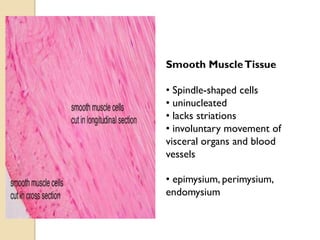
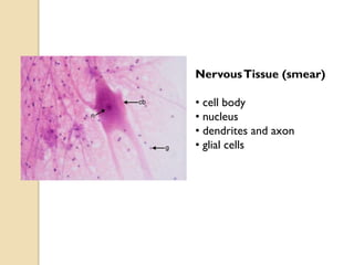
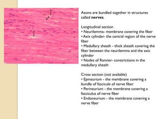


Tissues are groups of cells that work together to perform a specific function. There are four main types of tissues: epithelial, connective, muscular, and nervous. Epithelial tissues cover surfaces and line organs. Connective tissues connect and support other tissues. Muscular tissues allow for movement. Nervous tissues detect stimuli and transmit signals. Each tissue contains different cell types organized to carry out their role.


![animal_tissues_by_Deepak_b[presentation].pptx](https://cdn.slidesharecdn.com/ss_thumbnails/animaltissuesbydeepakb1-250228175730-fe92da00-thumbnail.jpg?width=640&height=640&fit=bounds)


