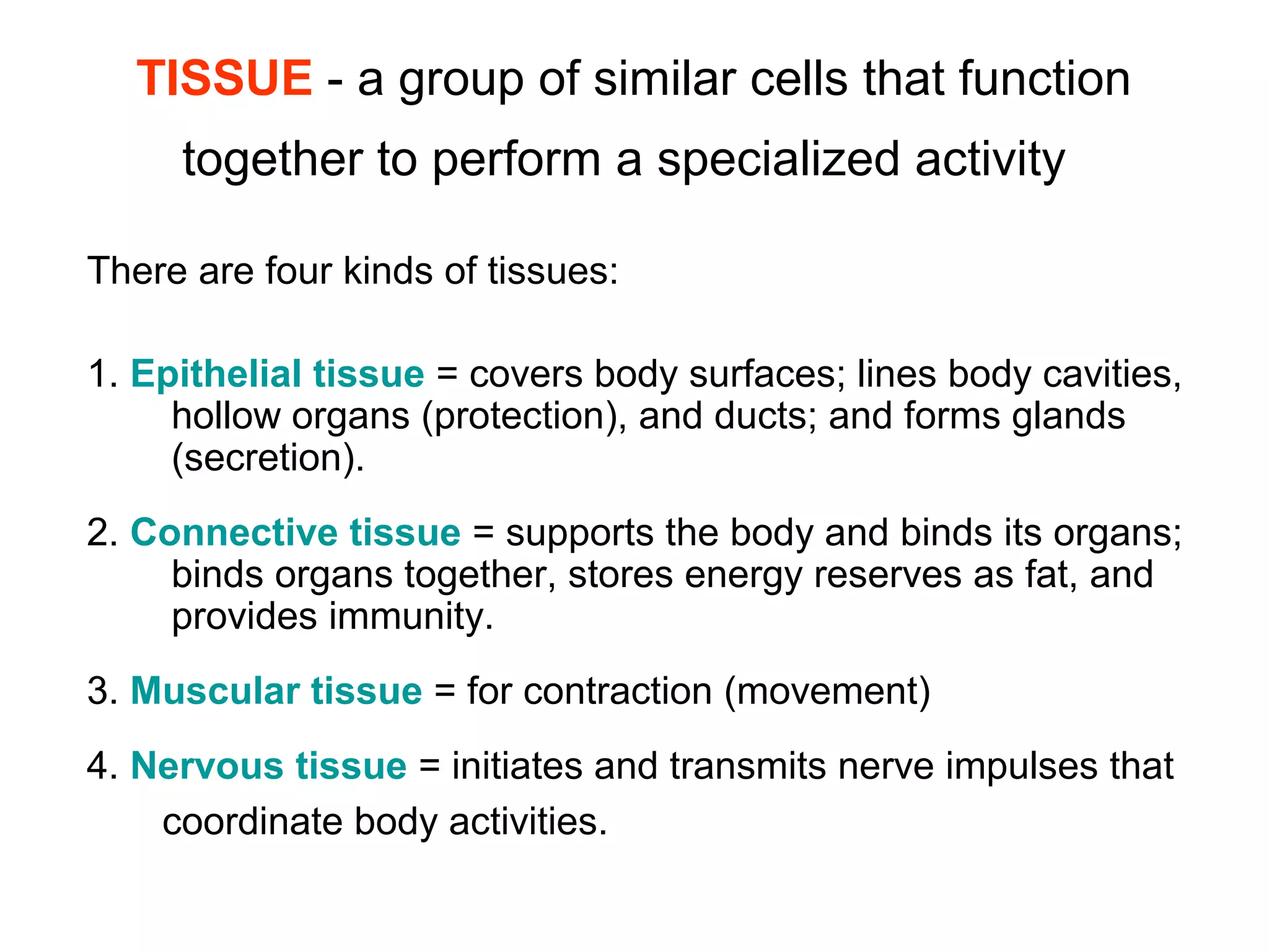
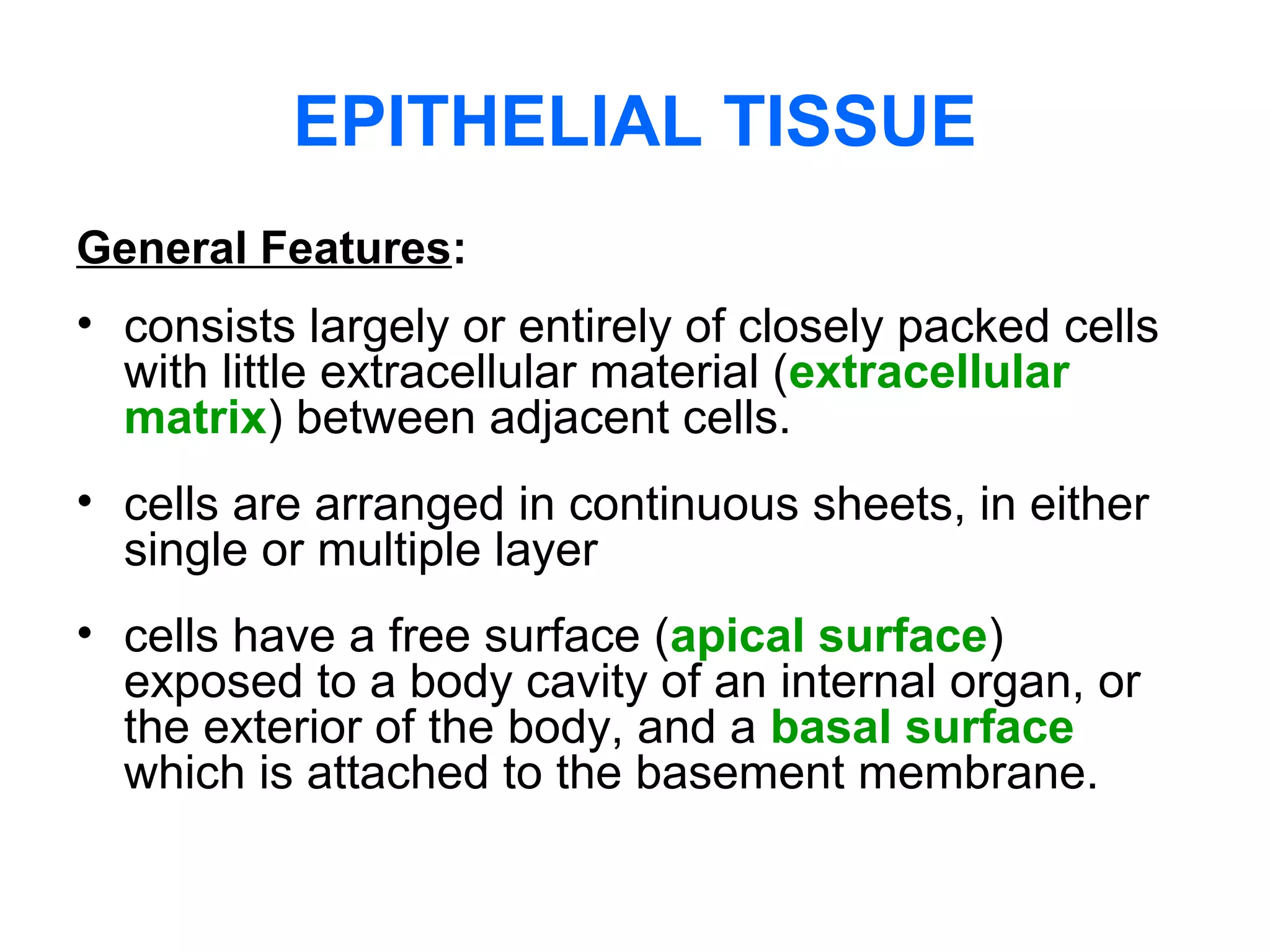
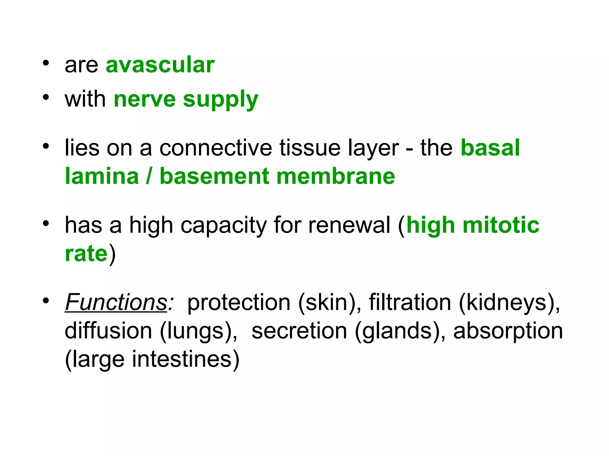
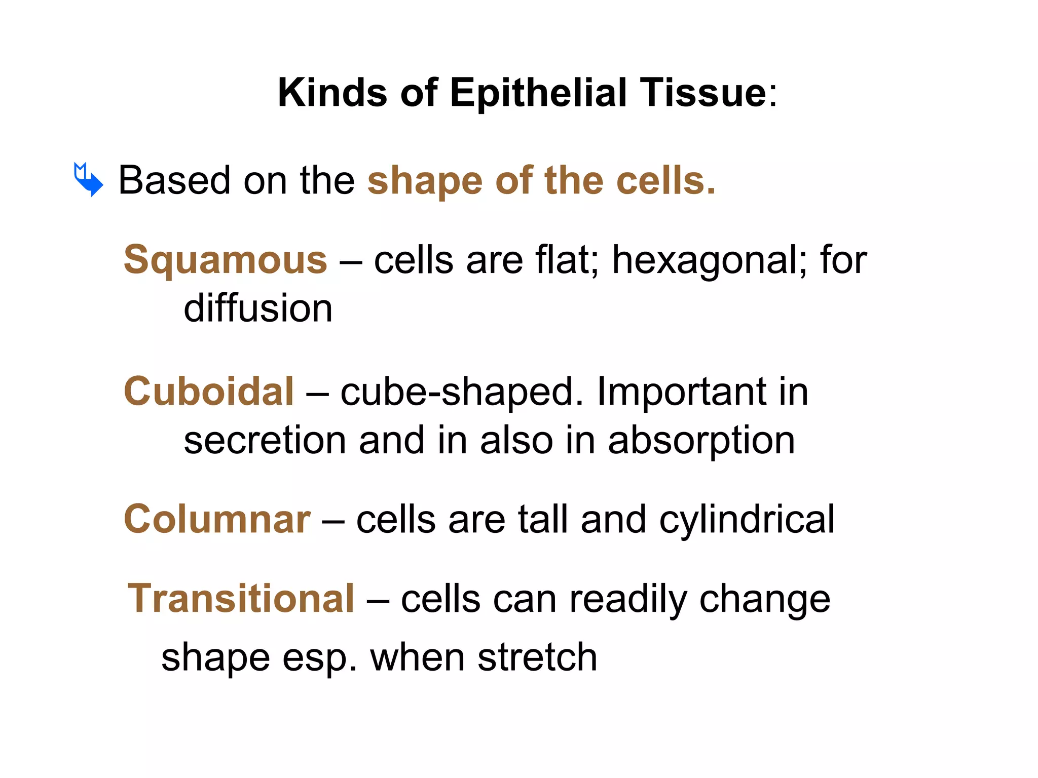
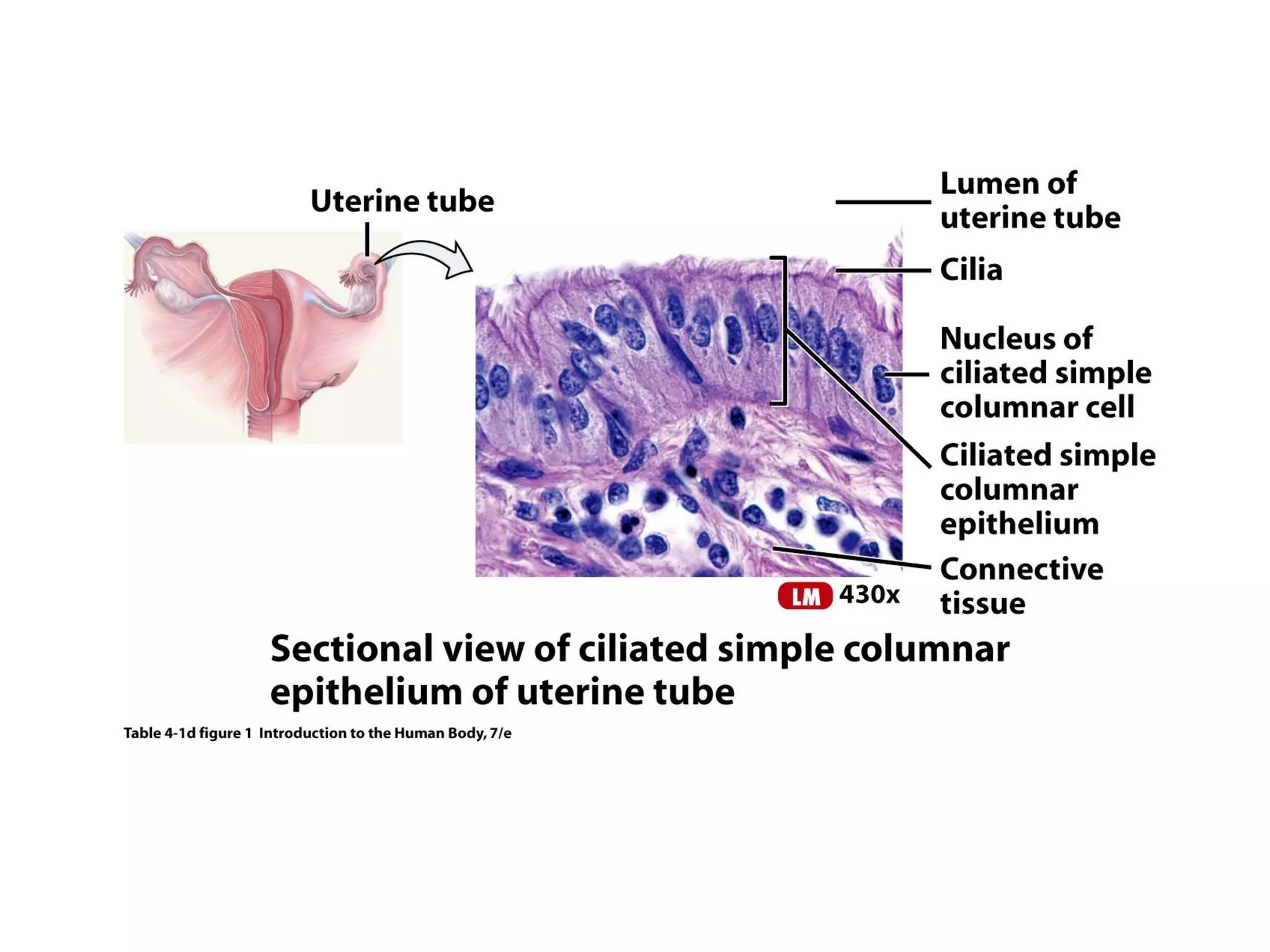
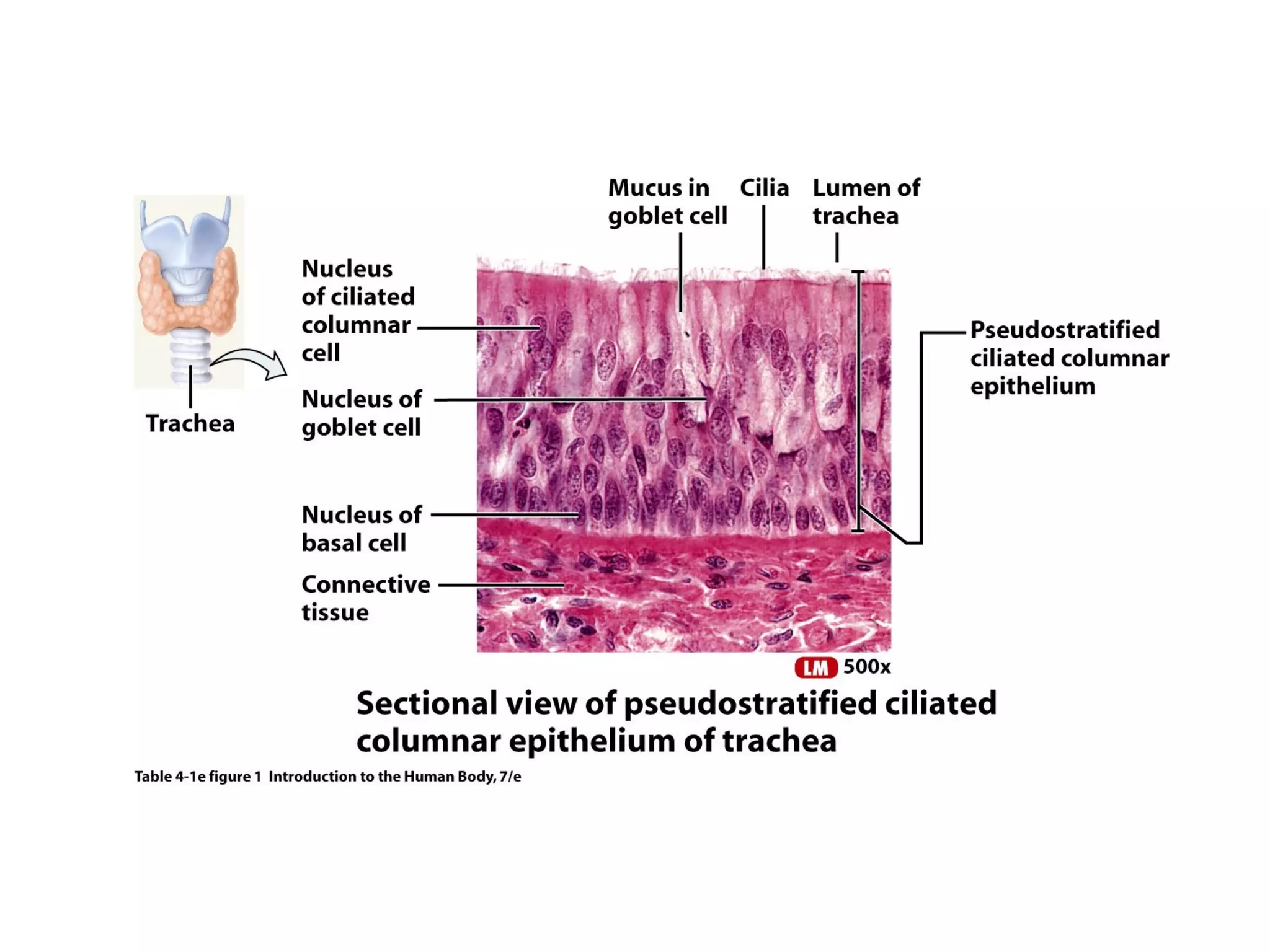
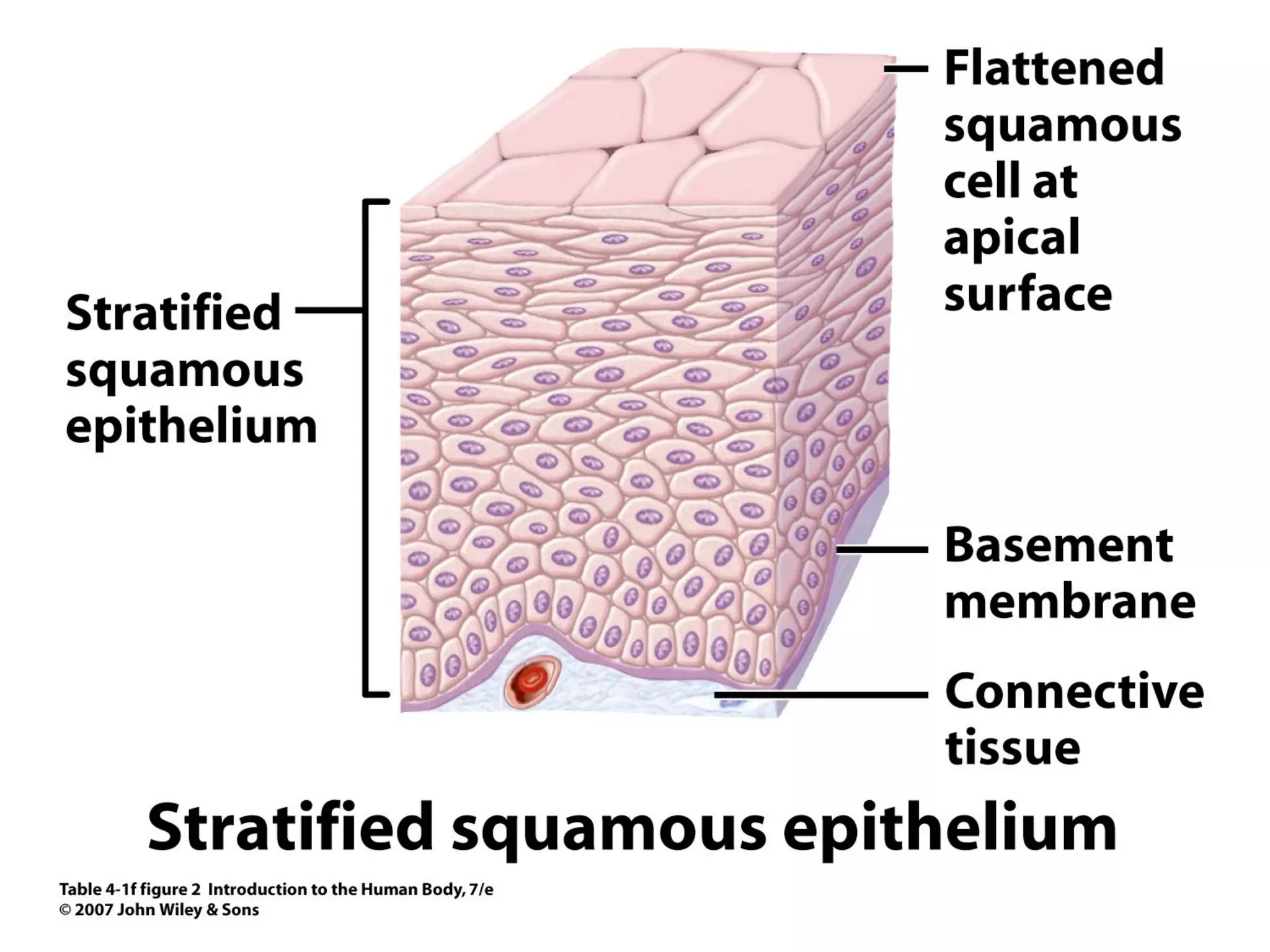
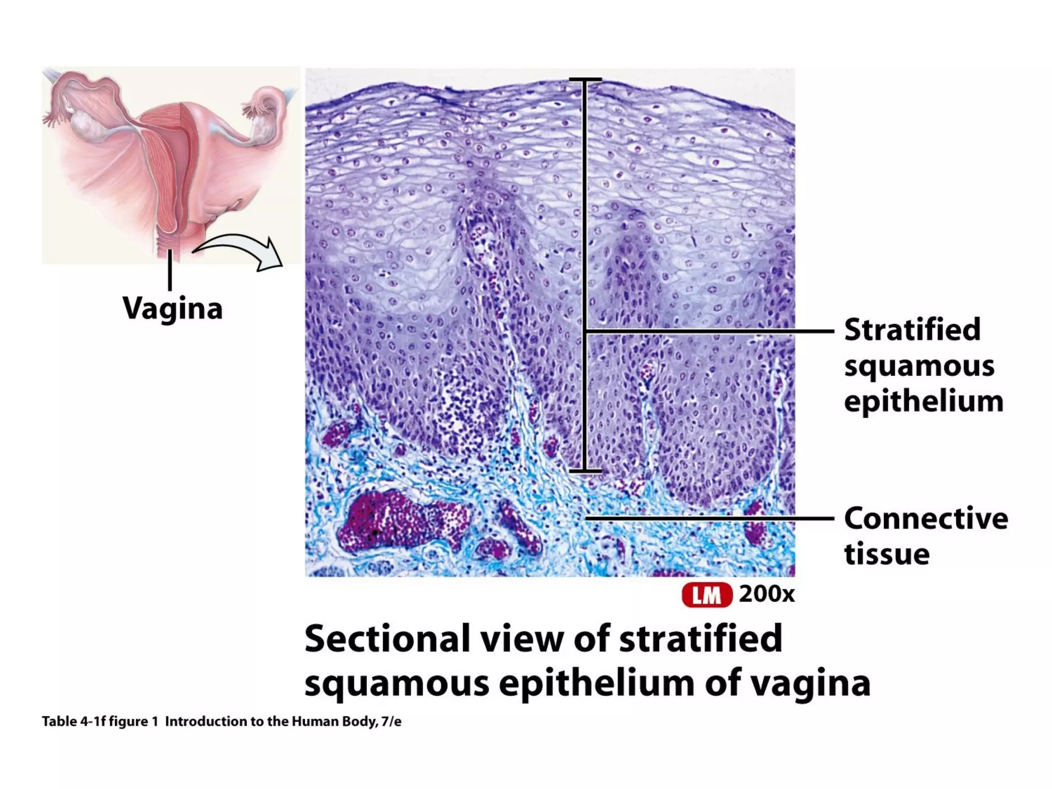
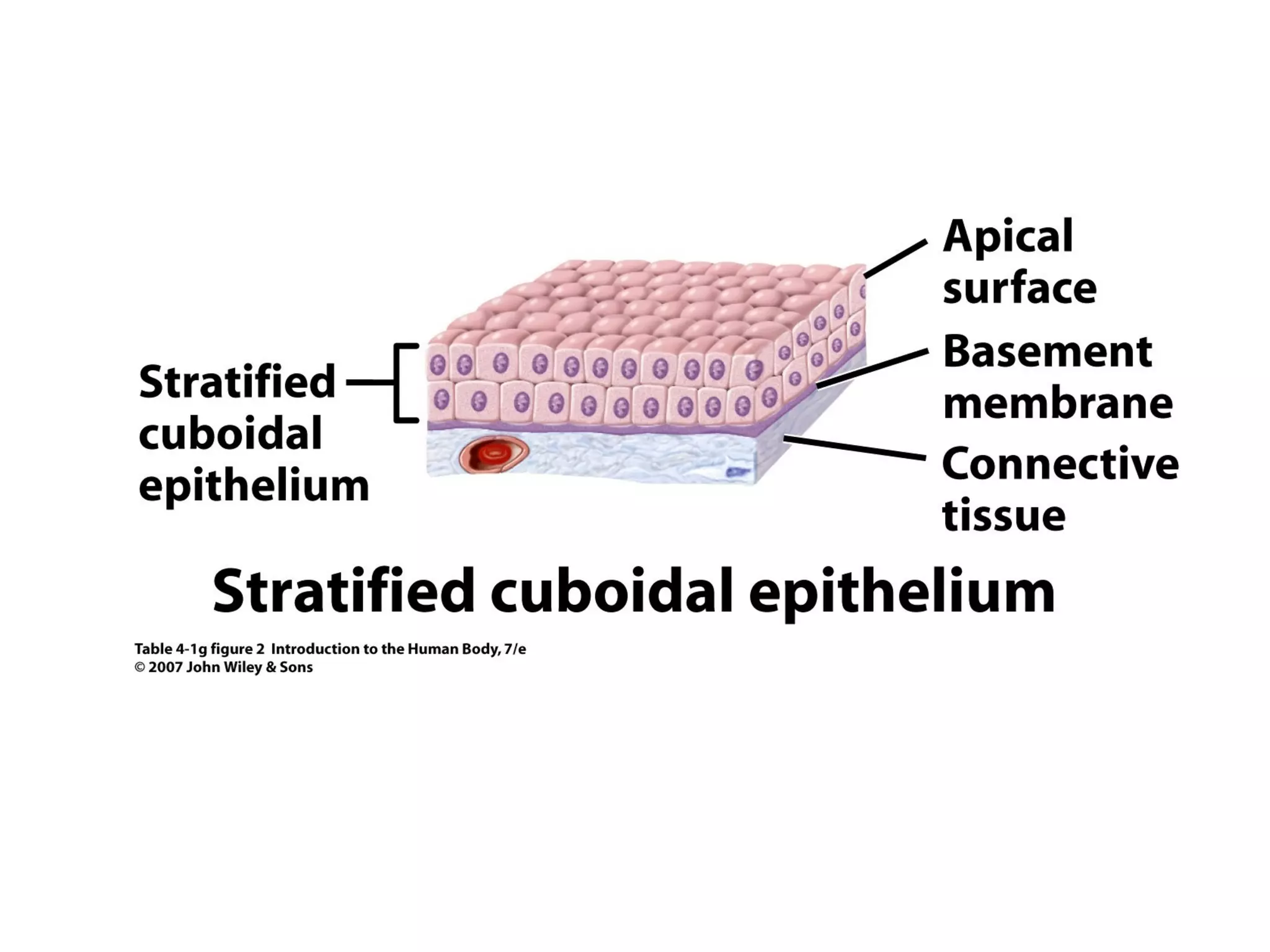
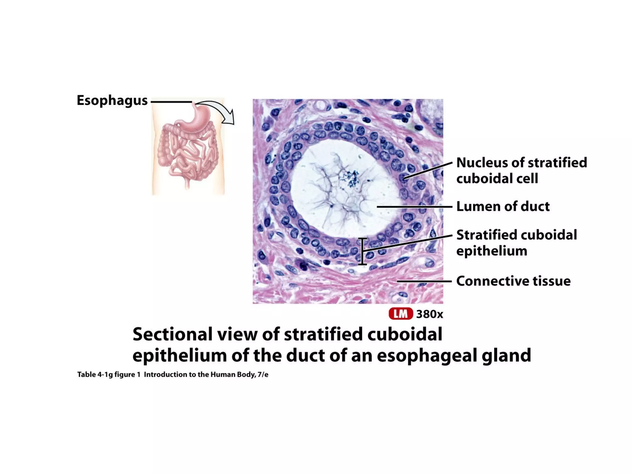
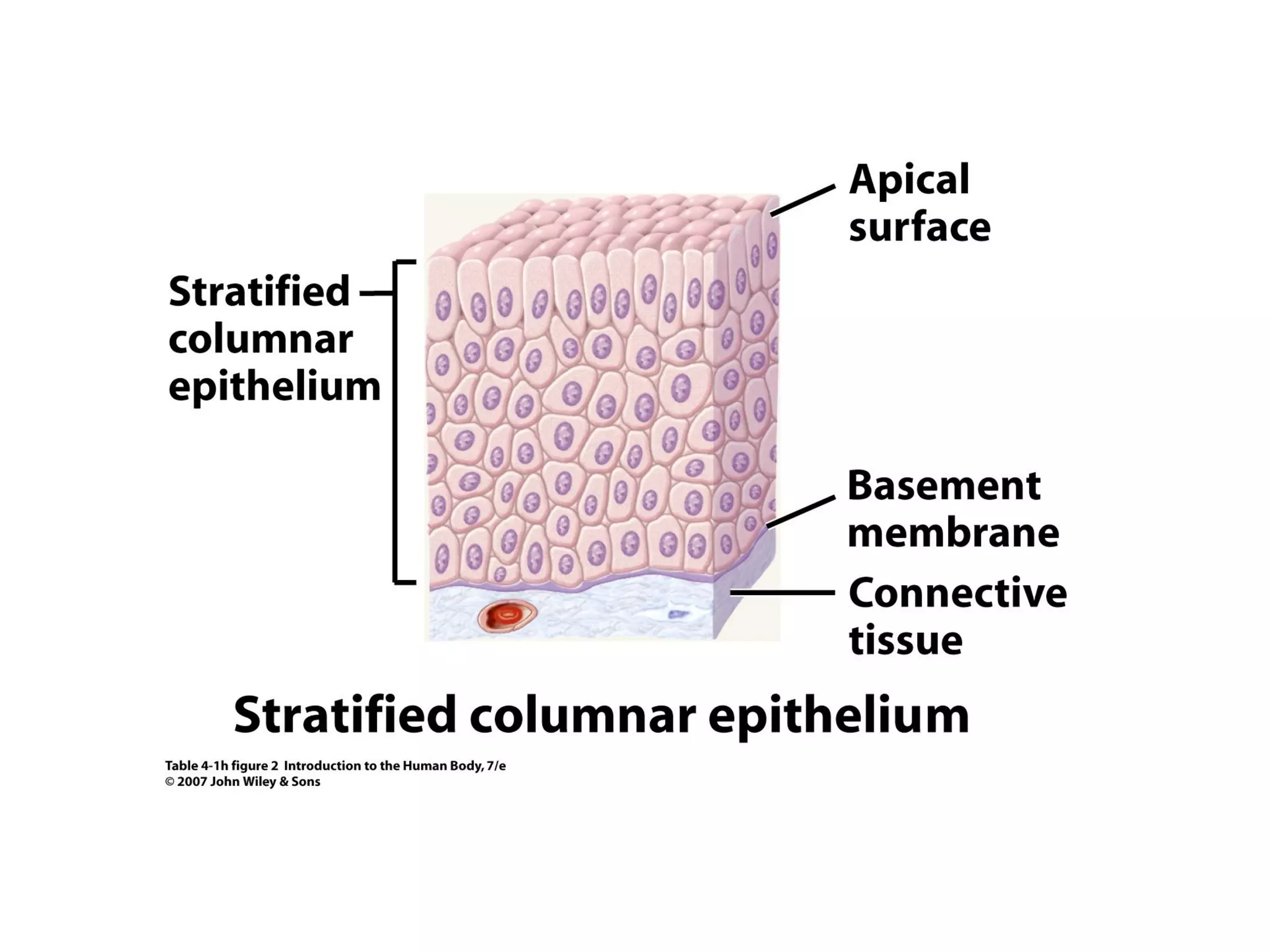
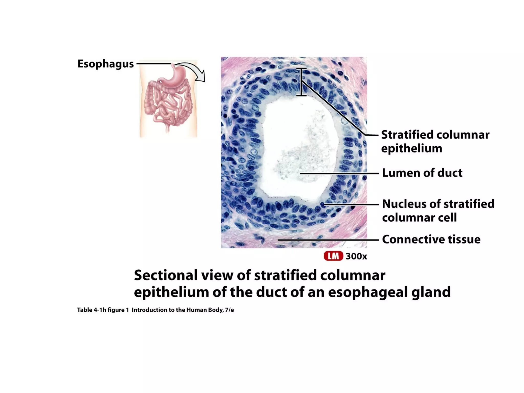
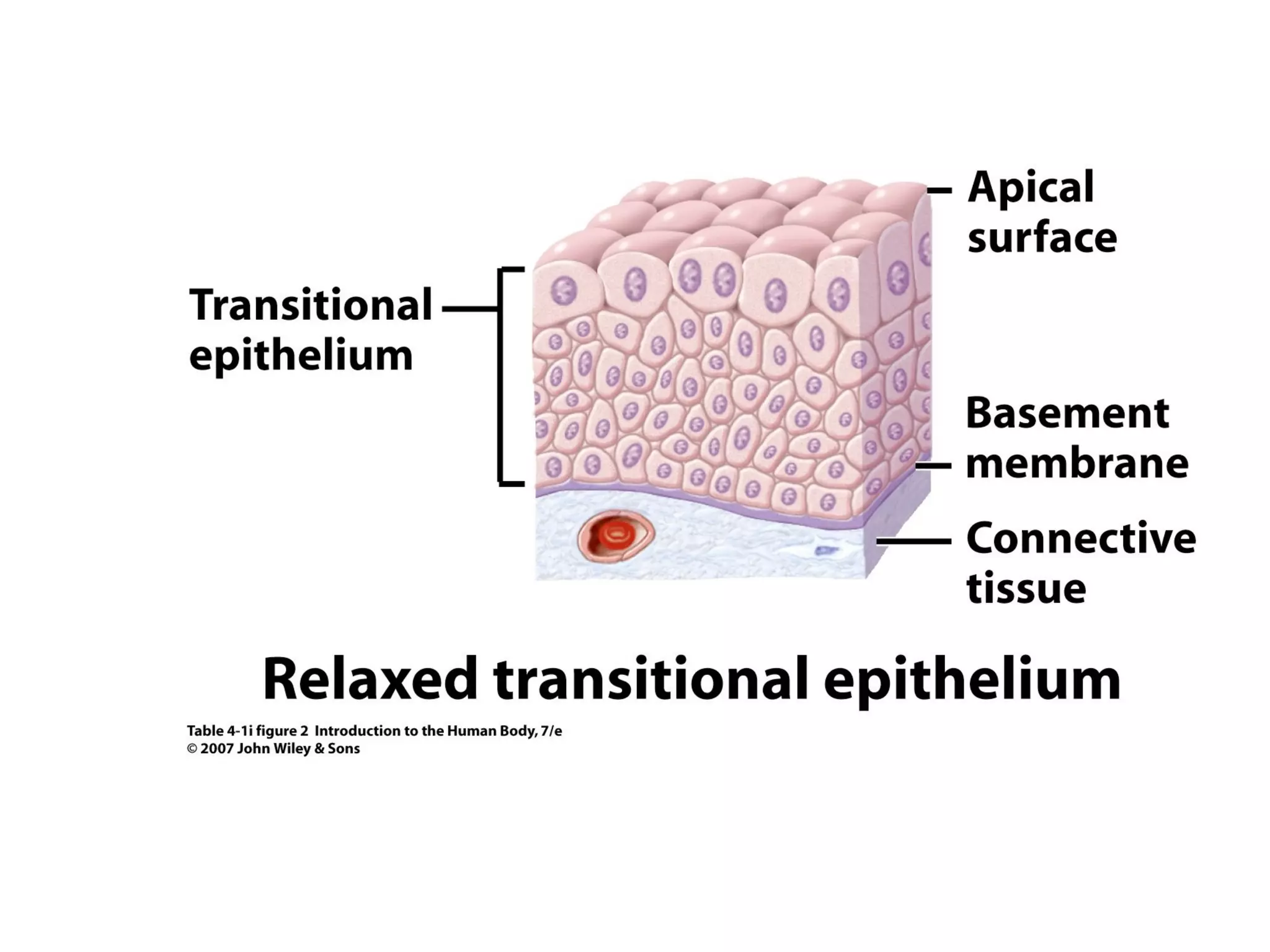
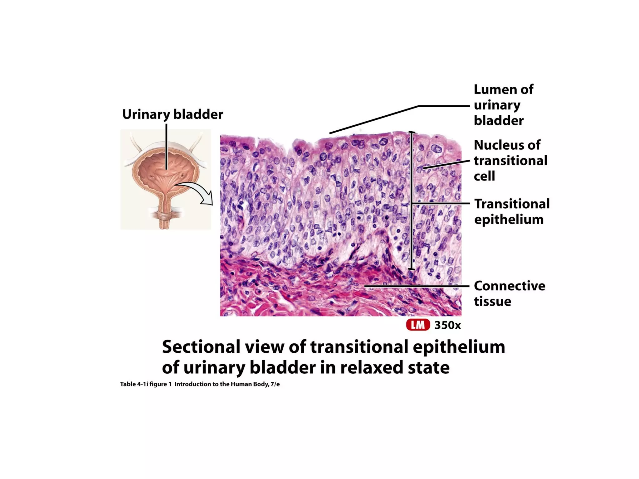
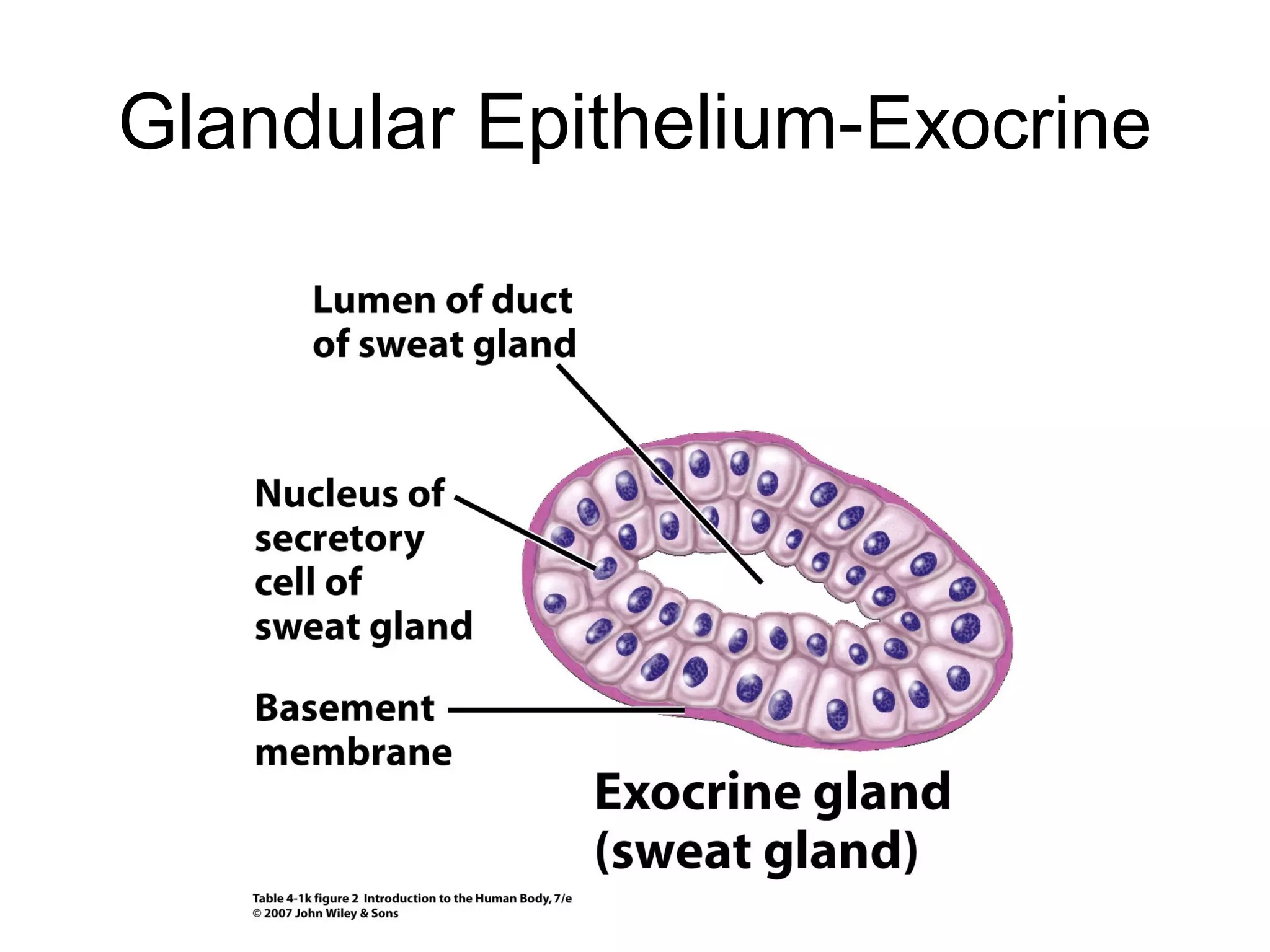
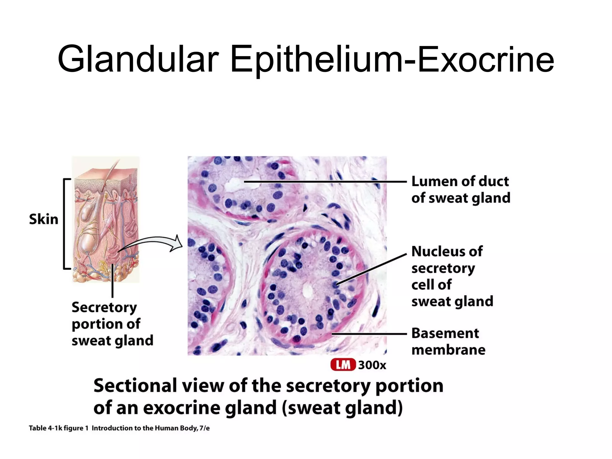
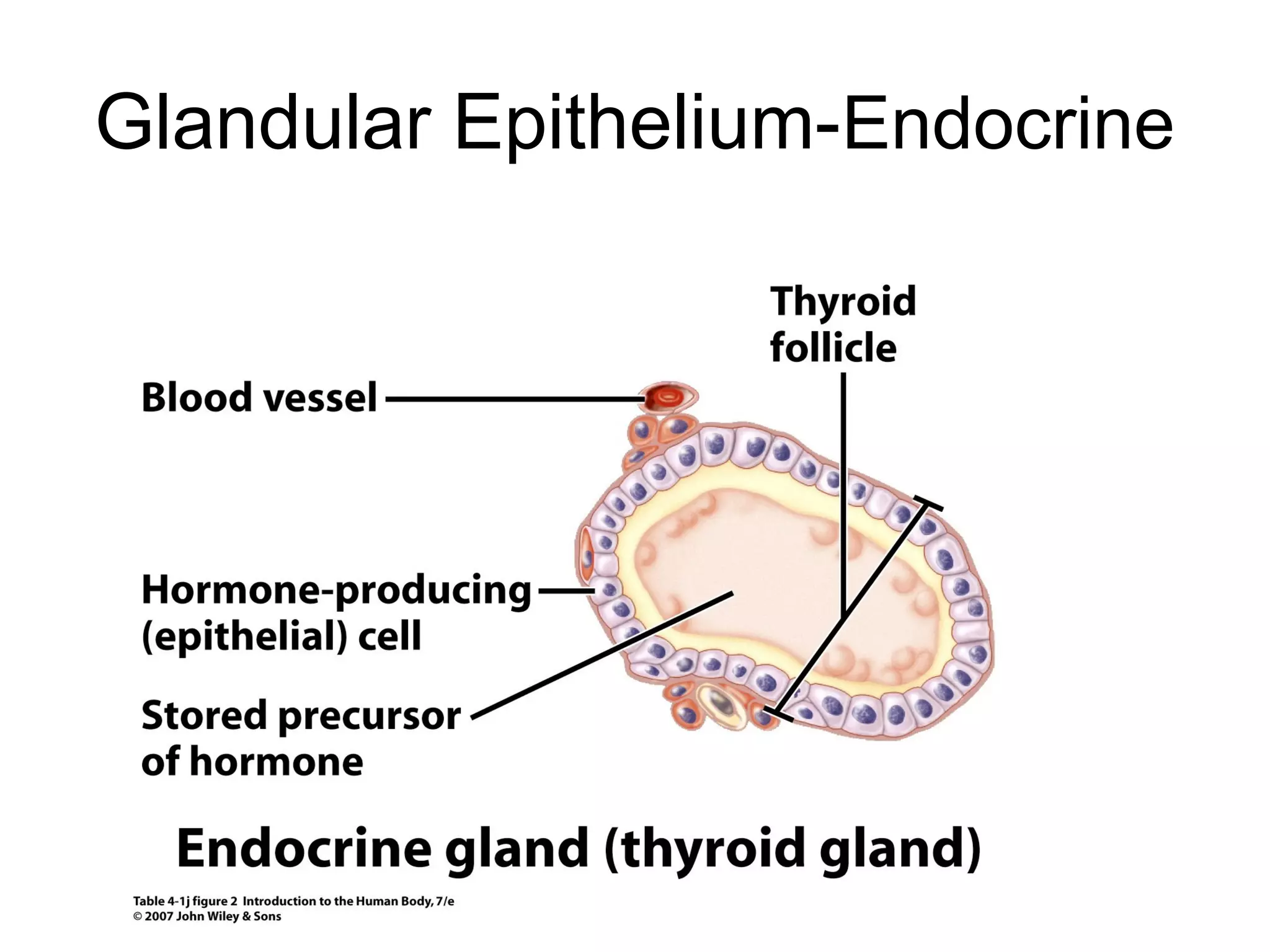
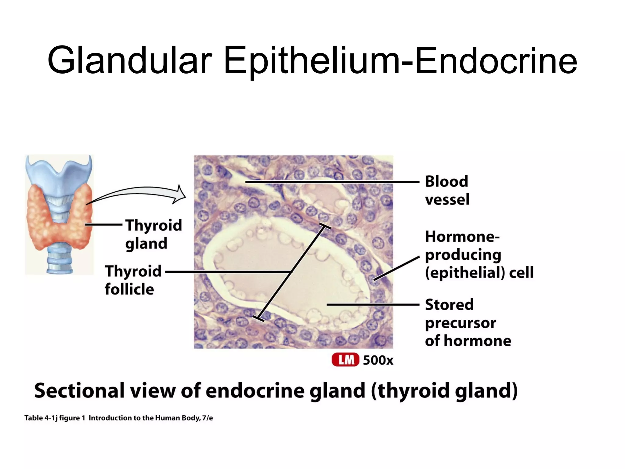
TISSUE - a group of similar cells that function
together to perform a specialized activity
There are four kinds of tissues:
1. Epithelial tissue = covers body surfaces; lines body cavities,
hollow organs (protection), and ducts; and forms glands
(secretion).
2. Connective tissue = supports the body and binds its organs;
binds organs together, stores energy reserves as fat, and
provides immunity.
3. Muscular tissue = for contraction (movement)
4. Nervous tissue = initiates and transmits nerve impulses that
coordinate body activities.
EPITHELIAL TISSUE
General Features:
• consists largely or entirely of closely packed cells
with little extracellular material (extracellular
matrix) between adjacent cells.
• cells are arranged in continuous sheets, in either
single or multiple layer
• cells have a free surface (apical surface)
exposed to a body cavity of an internal organ, or
the exterior of the body, and a basal surface
which is attached to the basement membrane.
• are avascular
• with nerve supply
• lies on a connective tissue layer - the basal
lamina / basement membrane
• has a high capacity for renewal (high mitotic
rate)
• Functions: protection (skin), filtration (kidneys),
diffusion (lungs), secretion (glands), absorption
(large intestines)
Kinds of Epithelial Tissue:
 Based on the shape of the cells.
Squamous – cells are flat; hexagonal; for
diffusion
Cuboidal – cube-shaped. Important in
secretion and in also in absorption
Columnar – cells are tall and cylindrical
Transitional – cells can readily change
shape esp. when stretch
 Based on the number of layers of cells.
Simple epithelium – single layer of cells;
found in areas where diffusion, osmosis,
filtration, secretion and absorption occur.
Stratified epithelium – contains two or more
layers of cells; protects underlying tissues;
found where there is wear and tear.
Pseudostratified epithelium – contains a single
layer of a mixture of cell types. Has a stratified
appearance, but is a single layer.
These cells are large, but quite thin, and have a prominent, protruding
nucleus. Found in linings of the peritoneal, pleural and pericardial
cavities. As well as in the glomerulus of the kidney, the walls of
capillaries, and the alveoli of the lungs.
SIMPLE SQUAMOUS EPITHELIUM
These are collecting ducts in the medulla of a mammalian kidney.
Each duct is lined by simple cuboidal epithelium, whose nuclei are
quite prominent. This type of epithelium is thicker than simple
squamous epithelium
SIMPLE CUBOIDAL EPITHELIUM
A cross section of the small intestine. The tall, vertical cells seen at
the top of the tissue are columnar cells (one is shown - red arrow),
which make up simple columnar epithelium (the cells at the bottom of
the image are connective tissue). Since columnar cells are quite thick,
they do not readily allow passive diffusion. They have numerous
microvilli (seen as a darker pink staining border at their lumenal
edge). The black arrow is indicating a goblet cell.
SIMPLE COLUMNAR EPITHELIUM
Glandular Epithelium-Exocrine
Glandular Epithelium-Endocrine
CONNECTIVE TISSUE
General Features:
• do not usually occur on free surfaces
• has a nerve supply (except for cartilage)
• usually is highly vascular (except cartilage & tendons)
• has three basic elements:
– cells
– ground substance
– fiber
The ground substance and the fibers, which are
outside the cells, make up the extracellular
matrix.
The matrix may be solid (as in bone), soft (as in
loose connective tissue), or liquid (as in blood)
• the most widespread and abundant type of tissue in
the human body
• Function: support, anchor and bind various parts of
the body
• also serves for: protection, blood formation, fat
storage, filling space
Kinds of Connective Tissue
1) Loose connective tissue
Has fibers which are far apart from each other, and
there are many cells.
• Areolar Connective Tissue
found in the lower layers of the skin, mucous
membranes, blood vessels, nerves, and around
body organs. Its function is to provide strength,
• Adipose Tissue
designed to store fat (triglycerides). The cells are
called adipocytes.
• Reticular Connective Tissue
contains a network of interlacing reticular fibers.
Its purpose is to form the framework of many
organs and bind together smooth muscle tissue
cells such as the lymph nodes, spleen and bone
marrow
2) Dense Connective Tissue
Also called dense fibrous tissue
Has collagen fibers as its main matrix element
They form strong, rope-like structures such as the
tendons – attach skeletal muscles to bones
ligaments – connect bones to bones at joints
also found in the dermis of the skin
3) Specialized CT:
A. Bone (osseus) tissue
Composed of bone cells (osteocytes) embedded in
small spaces called lacunae
Surrounded by concentric rings that consist of mineral
salts (calcium)
Small canals (canaliculi) project out of the lacunae
and provide a pathway for nutrients and wastes
Has rock-like hardness
B. Cartilage
Composed of cartilage cells chondrocytes
found in spaces called lacunae.
Surrounded by a membrane (perichondrium)
No blood vessels or nerves
Cartilage repairs very slowly after it has been
injured
Types of cartilage
1) Hyaline cartilage has abundant collagen fibers hidden
by a rubbery matrix with a glassy blue-white appearance
ex: ends of long bones, nose, anterior portion of
the ribs, and parts of the larynx, trachea,
bronchi, fetal skeleton
2) Fibrocartilage
highly compressible
serves as cushion
strongest type
ex: pubic symphysis,
intervertebral discs, and meniscus of the knee.
3) Elastic cartilage
highly elastic
ex: epiglottis of the larynx, tip of nose, external ear, and
auditory (Eustachian) tubes.
C. Vascular Tissue (Blood)
Plasma
liquid portion of the blood
consists mostly of water along with suspended
proteins, and dissolved substances, ions, food
molecules, and hormones
Formed Elements
solid portions of the blood
of three kinds:
1) erythrocytes - oxygen transporter
2) leukocytes – soldiers of the body, has
2 kinds
a. Granulocytes
neutrophils - most abundant of the
WBCs; squeeze through the capillary
walls and into infected tissue where they
kill the invaders and then engulf the
remnants by phagocytosis
eosinophils - numbers increase
sharply in certain diseases, especially
infections by parasitic worms; are
cytotoxic, releasing the contents of their
granules on the invader
basophils - increases during infection; play an
important part in some allergic responses
b. Agranulocytes
lymphocytes – 2 kinds: B and T lymphocytes
monocytes - functions in the ingestion of
bacteria (macrophage)
3) thrombocytes – important in blood clotting
Animal tissues epithelial & connective

Animal tissues epithelial & connective

  • 2.
    TISSUE - agroup of similar cells that function together to perform a specialized activity There are four kinds of tissues: 1. Epithelial tissue = covers body surfaces; lines body cavities, hollow organs (protection), and ducts; and forms glands (secretion). 2. Connective tissue = supports the body and binds its organs; binds organs together, stores energy reserves as fat, and provides immunity. 3. Muscular tissue = for contraction (movement) 4. Nervous tissue = initiates and transmits nerve impulses that coordinate body activities.
  • 3.
    EPITHELIAL TISSUE General Features: •consists largely or entirely of closely packed cells with little extracellular material (extracellular matrix) between adjacent cells. • cells are arranged in continuous sheets, in either single or multiple layer • cells have a free surface (apical surface) exposed to a body cavity of an internal organ, or the exterior of the body, and a basal surface which is attached to the basement membrane.
  • 4.
    • are avascular •with nerve supply • lies on a connective tissue layer - the basal lamina / basement membrane • has a high capacity for renewal (high mitotic rate) • Functions: protection (skin), filtration (kidneys), diffusion (lungs), secretion (glands), absorption (large intestines)
  • 5.
    Kinds of EpithelialTissue:  Based on the shape of the cells. Squamous – cells are flat; hexagonal; for diffusion Cuboidal – cube-shaped. Important in secretion and in also in absorption Columnar – cells are tall and cylindrical Transitional – cells can readily change shape esp. when stretch
  • 6.
     Based onthe number of layers of cells. Simple epithelium – single layer of cells; found in areas where diffusion, osmosis, filtration, secretion and absorption occur. Stratified epithelium – contains two or more layers of cells; protects underlying tissues; found where there is wear and tear. Pseudostratified epithelium – contains a single layer of a mixture of cell types. Has a stratified appearance, but is a single layer.
  • 8.
    These cells arelarge, but quite thin, and have a prominent, protruding nucleus. Found in linings of the peritoneal, pleural and pericardial cavities. As well as in the glomerulus of the kidney, the walls of capillaries, and the alveoli of the lungs. SIMPLE SQUAMOUS EPITHELIUM
  • 10.
    These are collectingducts in the medulla of a mammalian kidney. Each duct is lined by simple cuboidal epithelium, whose nuclei are quite prominent. This type of epithelium is thicker than simple squamous epithelium SIMPLE CUBOIDAL EPITHELIUM
  • 12.
    A cross sectionof the small intestine. The tall, vertical cells seen at the top of the tissue are columnar cells (one is shown - red arrow), which make up simple columnar epithelium (the cells at the bottom of the image are connective tissue). Since columnar cells are quite thick, they do not readily allow passive diffusion. They have numerous microvilli (seen as a darker pink staining border at their lumenal edge). The black arrow is indicating a goblet cell. SIMPLE COLUMNAR EPITHELIUM
  • 24.
  • 25.
  • 26.
  • 27.
  • 28.
    CONNECTIVE TISSUE General Features: •do not usually occur on free surfaces • has a nerve supply (except for cartilage) • usually is highly vascular (except cartilage & tendons) • has three basic elements: – cells – ground substance – fiber The ground substance and the fibers, which are outside the cells, make up the extracellular matrix.
  • 29.
    The matrix maybe solid (as in bone), soft (as in loose connective tissue), or liquid (as in blood) • the most widespread and abundant type of tissue in the human body • Function: support, anchor and bind various parts of the body • also serves for: protection, blood formation, fat storage, filling space
  • 30.
    Kinds of ConnectiveTissue 1) Loose connective tissue Has fibers which are far apart from each other, and there are many cells. • Areolar Connective Tissue found in the lower layers of the skin, mucous membranes, blood vessels, nerves, and around body organs. Its function is to provide strength, • Adipose Tissue designed to store fat (triglycerides). The cells are called adipocytes.
  • 31.
    • Reticular ConnectiveTissue contains a network of interlacing reticular fibers. Its purpose is to form the framework of many organs and bind together smooth muscle tissue cells such as the lymph nodes, spleen and bone marrow 2) Dense Connective Tissue Also called dense fibrous tissue Has collagen fibers as its main matrix element They form strong, rope-like structures such as the tendons – attach skeletal muscles to bones ligaments – connect bones to bones at joints also found in the dermis of the skin
  • 32.
    3) Specialized CT: A.Bone (osseus) tissue Composed of bone cells (osteocytes) embedded in small spaces called lacunae Surrounded by concentric rings that consist of mineral salts (calcium) Small canals (canaliculi) project out of the lacunae and provide a pathway for nutrients and wastes Has rock-like hardness
  • 34.
    B. Cartilage Composed ofcartilage cells chondrocytes found in spaces called lacunae. Surrounded by a membrane (perichondrium) No blood vessels or nerves Cartilage repairs very slowly after it has been injured Types of cartilage 1) Hyaline cartilage has abundant collagen fibers hidden by a rubbery matrix with a glassy blue-white appearance ex: ends of long bones, nose, anterior portion of the ribs, and parts of the larynx, trachea, bronchi, fetal skeleton
  • 35.
    2) Fibrocartilage highly compressible servesas cushion strongest type ex: pubic symphysis, intervertebral discs, and meniscus of the knee. 3) Elastic cartilage highly elastic ex: epiglottis of the larynx, tip of nose, external ear, and auditory (Eustachian) tubes.
  • 37.
    C. Vascular Tissue(Blood) Plasma liquid portion of the blood consists mostly of water along with suspended proteins, and dissolved substances, ions, food molecules, and hormones Formed Elements solid portions of the blood of three kinds:
  • 38.
    1) erythrocytes -oxygen transporter 2) leukocytes – soldiers of the body, has 2 kinds a. Granulocytes neutrophils - most abundant of the WBCs; squeeze through the capillary walls and into infected tissue where they kill the invaders and then engulf the remnants by phagocytosis eosinophils - numbers increase sharply in certain diseases, especially infections by parasitic worms; are cytotoxic, releasing the contents of their granules on the invader
  • 39.
    basophils - increasesduring infection; play an important part in some allergic responses b. Agranulocytes lymphocytes – 2 kinds: B and T lymphocytes monocytes - functions in the ingestion of bacteria (macrophage) 3) thrombocytes – important in blood clotting