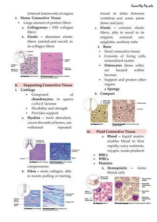
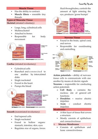
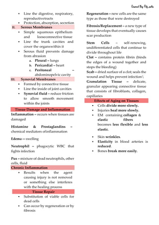
1. TISSUES
A group of cells with similar structure and
function that have similar extracellular
substances located between them.
Histology
The microscopic study of tissue
structure
Epithelial Tissue
▪ Covers external and internal
surfaces;
▪ Has a basement membrane,
▪ Little EM (extracellular matrix),
▪ No blood vessels (avascular).
▪ Regenerate easily if well-
nourished.
Functions of Epithelial Tissues (PAPSA)
1. Protecting underlying structures.
2. Acting as a barrier.
3. Permitting the passage of
substances.
4. Secreting substances.
5. Absorbing substances.
Classification of Epithelium
Accdg. To Layers
1. Simple E. – single layered
2. Stratified E. – multi--‐‐layered
3. Pseudostratified Columnar E –
single layeredbut appears to be
stratified
4. Transitional E – stratified x can
be greatlystretched
Accdg. To Shape
1. Squamous – flat; like fish scales
a. Non--‐‐keratinized --‐‐ in the
deepest and outermost layers,
moist
b. Keratinized --‐‐ composed of dead
cells containing the protein
keratin; durable, moisture--‐
‐resistant, dry character.
2. Cuboidal – cube--‐‐like
3. Columnar – tall and thin;
shaped like columns
Functional Characteristics
Simple Epithelia
▪ Functions in absorption,
secretion, and filtration
▪ Very thin (so not suited for
protection)
Simple Squamous E.
▪ diffusion, filtration, secretion, or
absorption; found in organs: to
move materials.
TISSUES
Owned By: Pdf_sells
2. ▪ Lines air sacs of the lungs
▪ Forms walls of capillaries
▪ Forms serous membranes
(serosae) that line and cover
organs in ventral cavity
Simple cuboidal E.
▪ Functions in secretion and
absorption; ciliated types propel
mucus or reproductive cells
▪ Common in glands and their ducts
▪ Forms walls of kidney tubules
▪ Covers the surface of ovaries
Simple Columnar E.
▪ Functions in secretion and
absorption; ciliated types propel
mucus or reproductive cells
Pseudostratified columnar E.
▪ All cells rest on a basement
membrane
▪ Single layer, but some cells are
shorter than others
▪ giving a false (pseudo) impression
of stratification
▪ Location: respiratory tract, where
it is ciliated and known as
pseudostratified ciliated columnar
epithelium
▪ Functions in absorption or
secretion
Stratified Epithelia
▪ Consist of two or more cell layers
▪ Function primarily in protection
Stratified Squamous E.
▪ Most common stratified
epithelium
▪ Named for cells present at the free
(apical) surface,
▪ which are squamous
▪ Functions as a protective covering
where friction is common
Stratified cuboidal E.
▪ two layers of cuboidal cells;
functions in protection
Stratified columnar E.
▪ surface cells are columnar, and
cells underneath vary in size and
shape; functions in protection
Owned By: Pdf_sells
3. Stratified cuboidal and columnar E.
▪ Rare in human body
▪ Found mainly in ducts of large
glands
Transitional E.
▪ Composed of modified stratified
squamous epithelium
▪ Shape of cells depends upon the
amount of stretching
▪ Functions in stretching and the
ability to return to
▪ normal shape
▪ Location: lining of urinary system
organs
Glandular Epithelia
▪ One or more cells responsible for
secreting a particular product
▪ Secretions contain protein
molecules in an aqueous (water-
based) fluid
▪ Secretion is an active process
Free Surfaces
▪ Smooth free surface – reduces
friction
▪ Microvilli – increase the free
surface area
▪ Cilia – propel materials
▪ Goblet Cells – specialized
mucus--‐‐producing cells
Cell Connections
▪ Tight Junctions --‐‐bind adjacent
cells together and form
permeability barriers; prevent
the passage of materials
between epithelial cells
▪ Desmosomes – mechanical
links that bind cells together
➢ Hemidesmosomes --‐‐ anchor
cells to the basement
membrane
▪ Gap Junctions – allow small
molecules and ions to pass
Glands
Glands – secrete substances onto a
surface
a. Exocrine – secrete through
ducts
➢ Unicellular
➢ Multicellular
❖ Simple – non
branched
❖ Compound- branched
• Tubular
o Straight
o Coiled
• Acinus(grapeli
ke) or Alveolus
(small cavity)
Mode of Secretion
➢ Merocrine – products are
released but no actual
cellular material is lost;
pancreas
➢ Apocrine – secretory
products are released as
fragments; mammary
glands
➢ Holocrine --‐‐ shedding of
entire cells; sebaceous
glands
b. Endocrine --‐‐ release hormones
that are absorbed directly into
the blood
Owned By: Pdf_sells
4. Connective Tissue
▪ Large amounts of EM
Functions of Connective Tissue (EC2S2PT)
1. Enclosing and separating other
tissues.
2. Connecting tissues to one
another.
3. Supporting and moving parts
of the body.
4. Storing compounds.
5. Cushioning and insulating.
6. Transporting.
7. Protecting.
Cells of Connective Tissue
▪ Blast (germ) – froms the matrix
▪ Cyte (cells) – maintains
▪ Clast (break) – breaks down
▪ Adipocytes – contain large
amount of lipids
▪ Macrophages – WBCs that
move about andingest foreign
substances
▪ Mast Cells – play an
important role ininflammation
▪ Mesenchymal Cells – have the
potential to differentiate to
form adult cell types
Extracellular Matrix
▪ Protein Fibers
➢ Collagen Fibers –
microscopic ropes;
flexible but resist
stretching
➢ Reticular Fibers – fine,
short that branch;
support network
➢ Elastic Fibers – coiled;
can recoil back toshape
▪ Ground Substance – shapeless
background where cells and
collagen fibers can be seen;
highly structured molecules
➢ Proteoglycans – pine
trees: branches =
proteins, pine
needles =
polysaccharides; trap
large quantities of water
▪ Fluid
Classification of Connective Tissue
Adult Connective Tissue
I. Connective Tissue Proper
1. Loose Connective Tissue
▪ Few protein fibers, numerous
spaces
a. Areolar – EM: collagen fibers
and few elastic fibers
b. Adipose – consists of
adipocytes (fat cells): contain
large amounts of lipid (for
energy storage); EM: loose
arranged collagen and
reticular fibers, scattered
elastic fibers
c. Reticular - Delicate network
of interwoven fibers with
reticular cells (like
fibroblasts); Forms stroma
Owned By: Pdf_sells
5. (internal framework) of organs
2. Dense Connective Tissue
▪ Large amount of protein fibers
a. Collagenous – EM: collagen
fibers
b. Elastic – abundant elastic
fibers (stretch and recoil) in
its collagen fibers
II. Supporting Connective Tissue
1. Cartilage
▪ Composed of
chondrocytes, in spaces
called lacunae
▪ Flexibility and strength
▪ Provides support
a. Hyaline – most abundant,
covers the ends of bones, can
withstand repeated
compressions
b. Fibro – more collagen, able
to resists pulling or tearing,
found in disks between
vertebrae and some joints
(knee and jaw)
c. Elastic – contains elastic
fibers, able to recoil to its
original, external ear,
epiglottis, auditory tube
2. Bone
▪ Hard connective tissue
▪ Consists of living cells,
mineralized matrix
▪ Osteocytes (bone cells),
are located within
lacunae
▪ Support and protect other
organs
a. Spongy
b. Compact
III. Fluid Connective Tissue
a. Blood – liquid matrix:
enables blood to flow
rapidly; carry nutrients,
oxygen, waste products
➢ RBCs
➢ WBCs
➢ Platelets
b. Hemopoietic --‐‐ forms
bloods cells
Owned By: Pdf_sells
6. Muscle Tissue
▪ Has the ability to contract.
▪ Muscle fibers – resemble tiny
threads
Types of Muscular Tissue
Skeletal (striated voluntary)
▪ Large, long, cylindrical cells
▪ Multinucleated
▪ Attached to bones
▪ Responsible for body
movement
Cardiac (striated involuntary)
▪ Cylindrical cells
▪ Branched and c o n n e c t e d to
one another by intercalated
disks
▪ Single nucleated
▪ Found in the heart
▪ Pumps the blood
Smooth (nonstriated involuntary)
▪ End tapered cells
▪ Single nucleated
▪ Found in hollow organs:
stomach, intestine; skin, eyes
▪ Regulates size of organs, forces
fluid throughtubes, controls the
amount of light entering the
eye, produces ‘goose bumps’
Nervous Tissue
▪ Found in the brain, spinal cord,
and nerves
▪ Responsible for coordinating
and controlling
Action potentials – ability of nervous
tissue cells to communicate with one
another by means of electric signals
Neurons – responsible for conducting
action potentials
➢ Cell Body – contains the
nucleus; site of general cell
functions
➢ Dendrites – receive electric
impulses
➢ Axon – conduct electric
impulses
Membranes
▪ A thin layer of tissue that covers
a structure
▪ Mostly consists of epithelium
and connectivetissue
I. Mucous Membranes
▪ Consists of epithelium and
loose connectivetissue
Owned By: Pdf_sells
7. ▪ Line the digestive, respiratory,
reproductivetracts
▪ Protection, absorption, secretion
II. Serous Membranes
▪ Simple squamous epithelium
and looseconnective tissue
▪ Line the trunk cavities and
cover the organswithin it
▪ Serous fluid prevents damage
from abrasion
a. Pleural – lungs
b. Pericardial – heart
c. Peritoneal –
abdominopelvic cavity
III. Synovial Membranes
▪ Formed by connective tissue
▪ Line the inside of joint cavities
▪ Synovial fluid – reduce friction
to allow smooth movement
within the joints
Tissue Damage and Inflammation
Inflammation – occurs when tissues are
damaged
Histamine & Prostaglandins –
chemical mediators ofinflammation
Edema --‐‐ swelling
Neutrophil – phagocytic WBC that
fights infection
Pus – mixture of dead neutrophils, other
cells, fluid
Chronic Inflammation
▪ Results when the agent
causing injury is not removed
or something else interferes
with the healing process
Tissue Repair
▪ Substitution of viable cells for
dead cells
▪ Can occur by regeneration or by
fibrosis
Regeneration – new cells are the same
type as those that were destroyed
Fibrosis/Replacement – a new type of
tissue develops that eventually causes
scar production
Stem Cells – self--‐‐renewing,
undifferentiated cells that continue to
divide throughout life
Clot – contains protein fibrin (binds
the edges of a wound together and
stops the bleeding)
Scab – dried surface of a clot; seals the
wound and helps prevent infection
Granulation Tissue – delicate,
granular appearing connective tissue
that consists of fibroblasts, collagen,
capillaries
Effects of Aging on Tissues
▪ Cells divide more slowly.
▪ Injuries heal more slowly.
▪ EM containing collagen &
elastic fibers
becomes less flexible and less
elastic.
▪ Skin wrinkles.
▪ Elasticity in blood arteries is
reduced.
▪ Bones break more easily.
Owned By: Pdf_sells