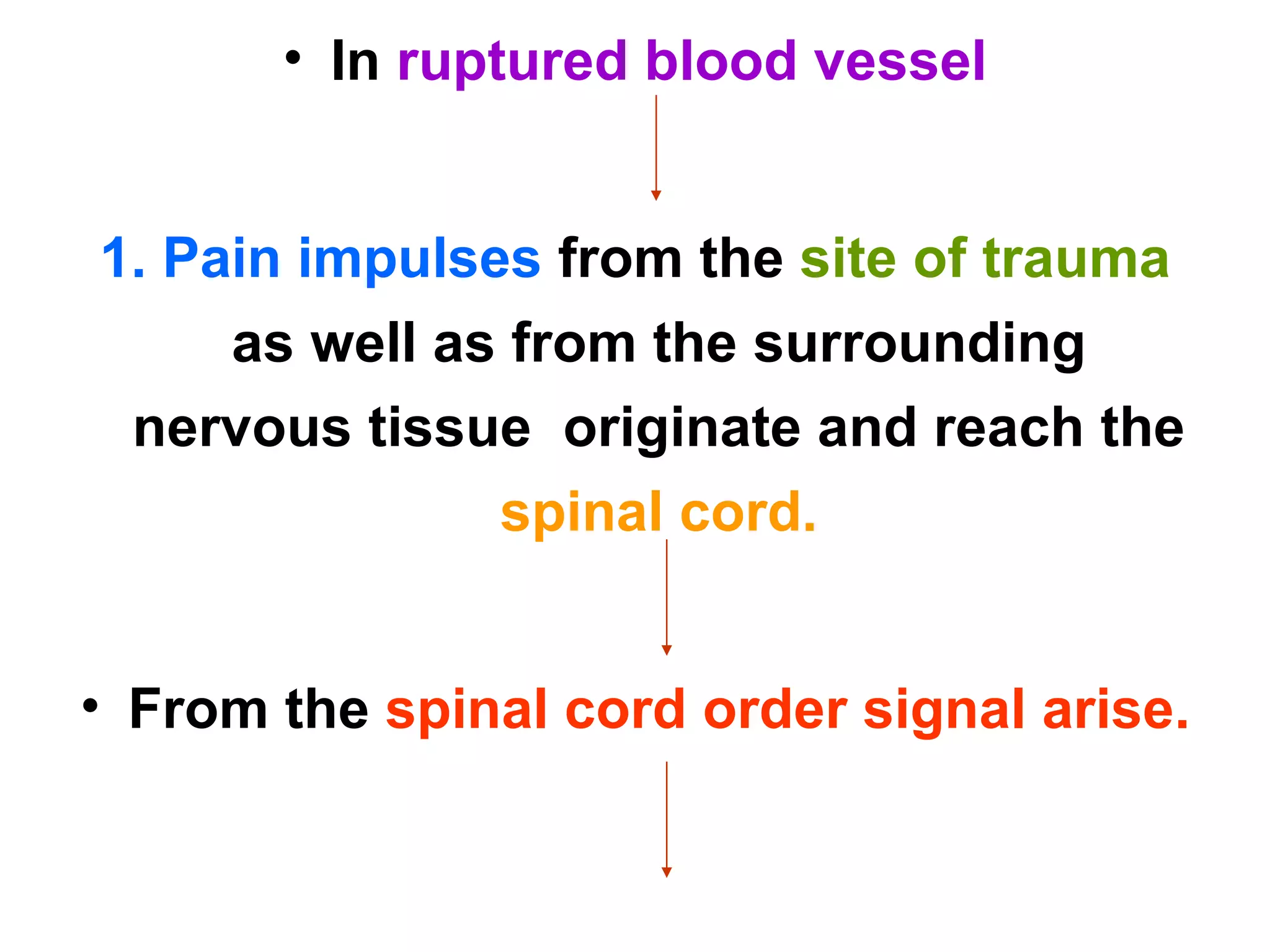
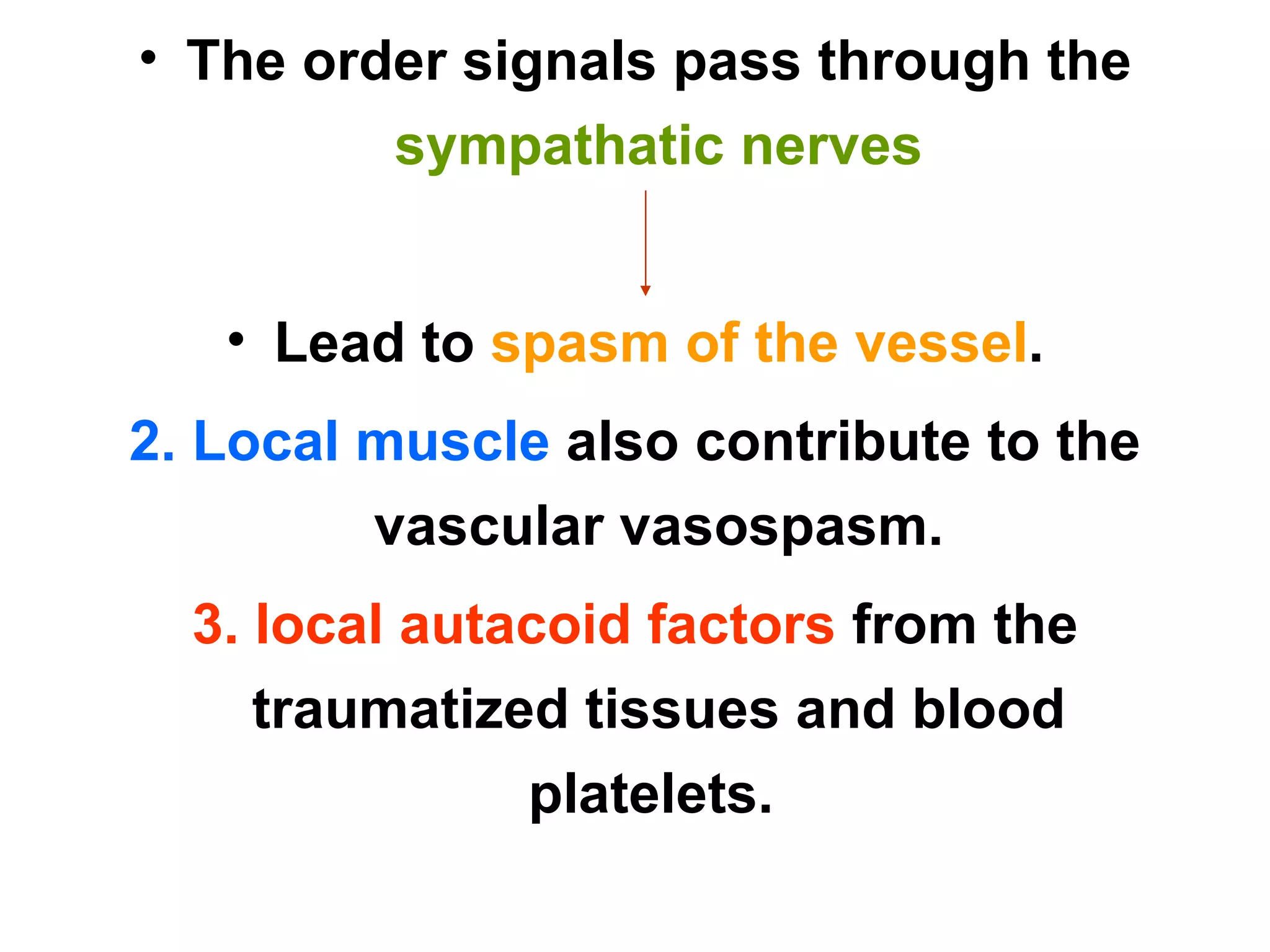
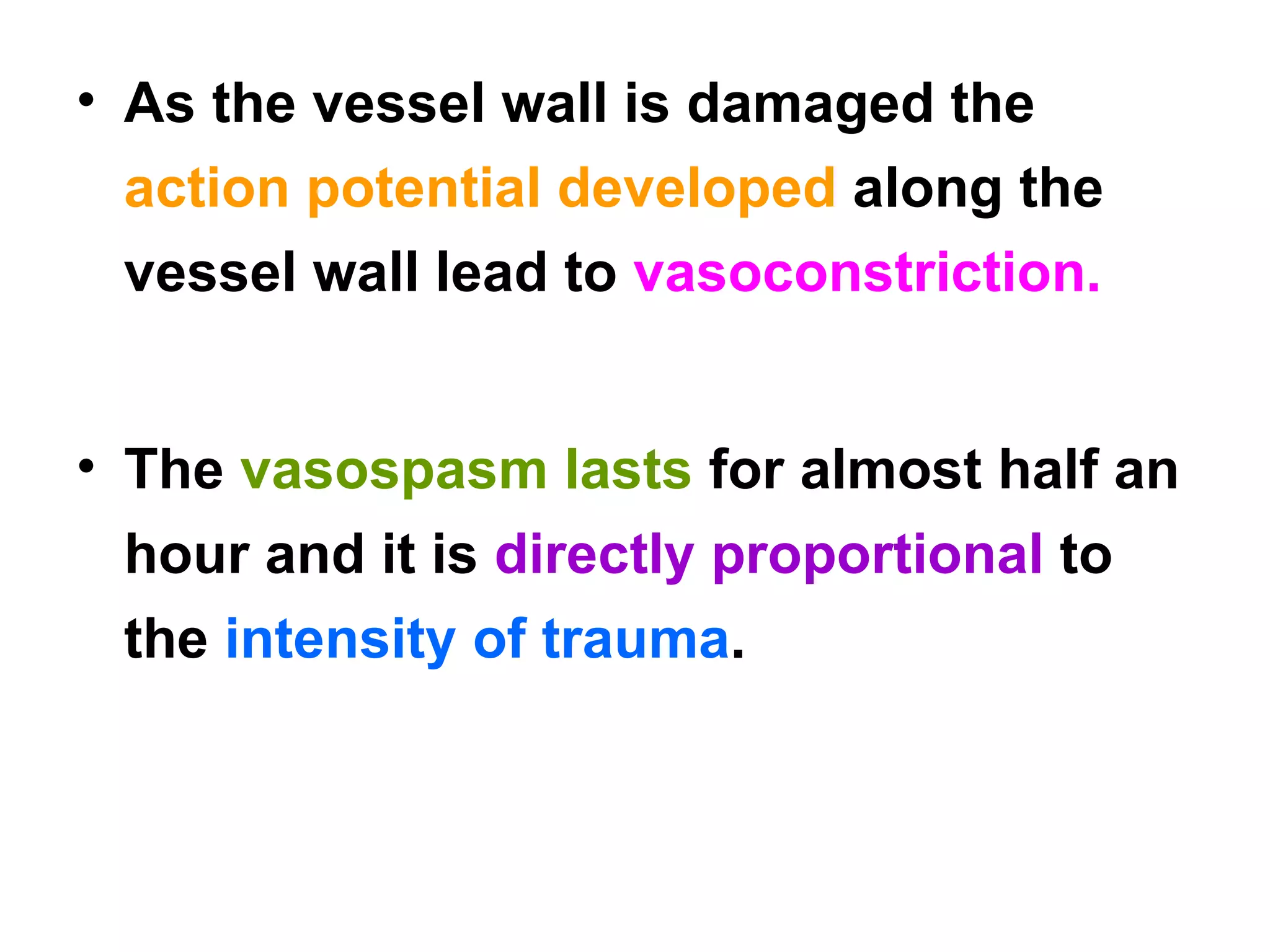
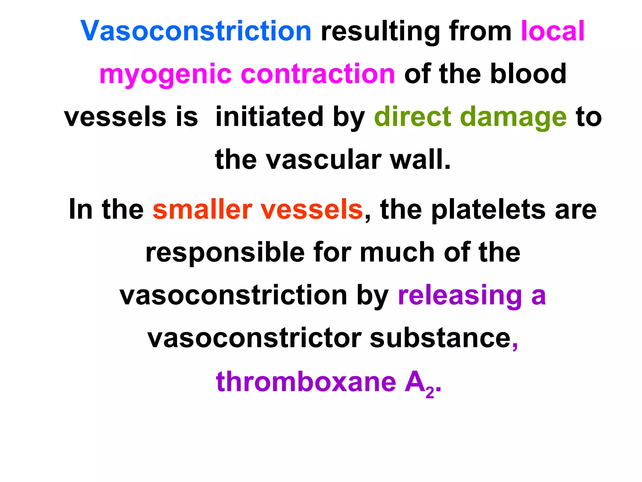
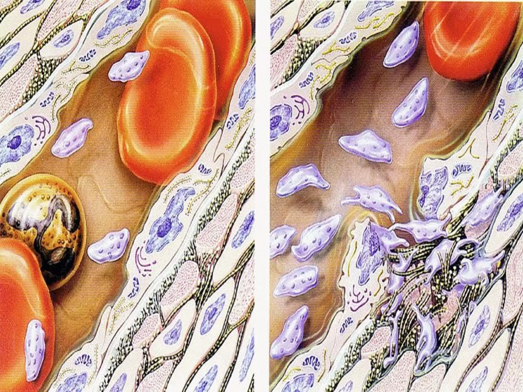
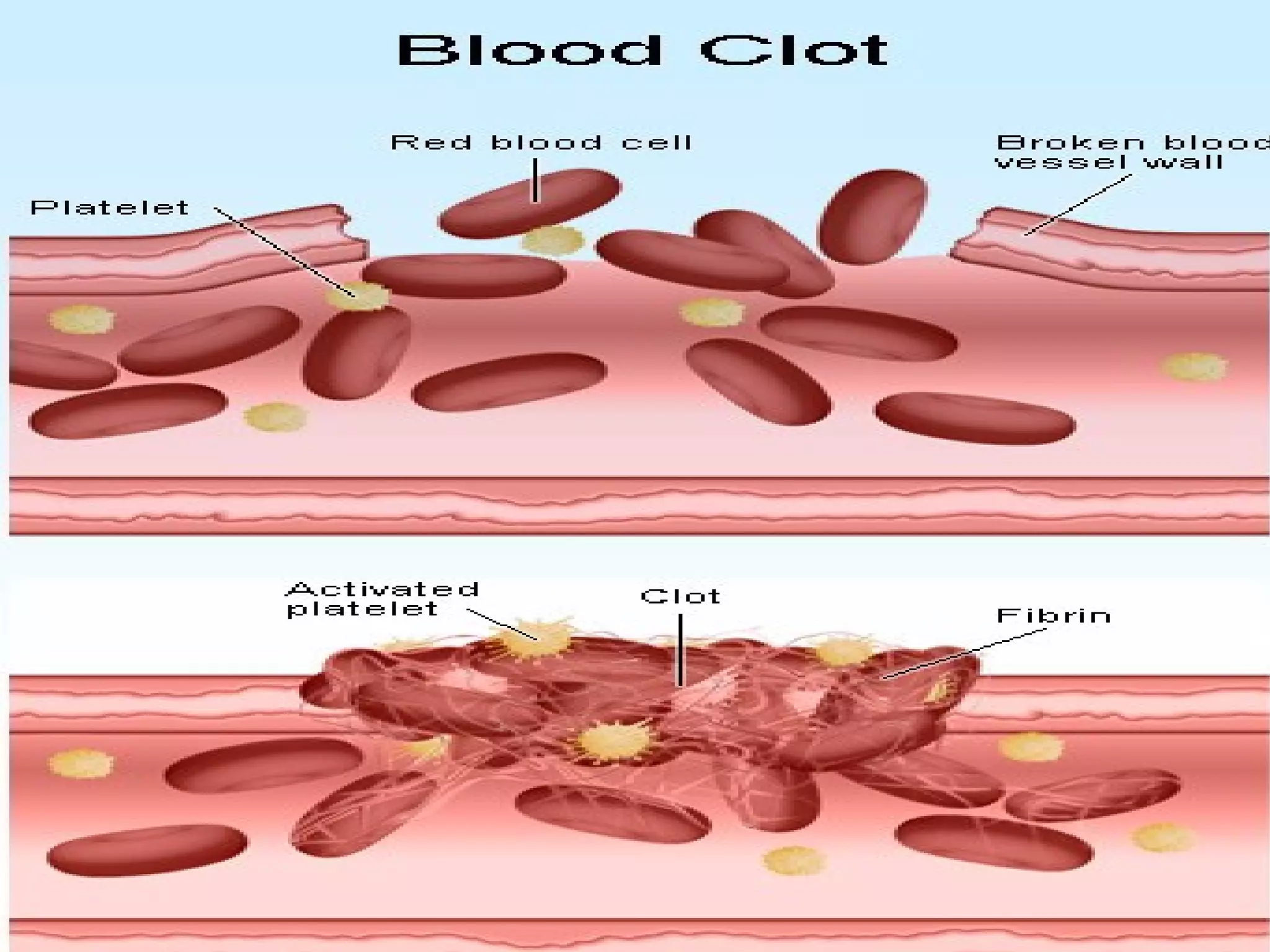
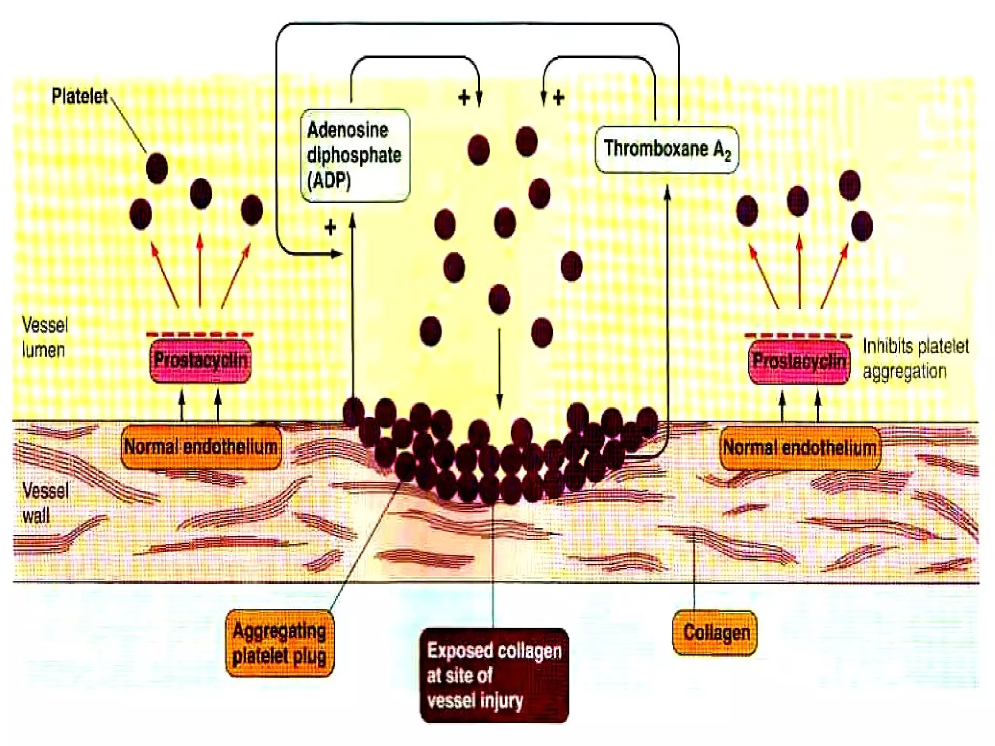
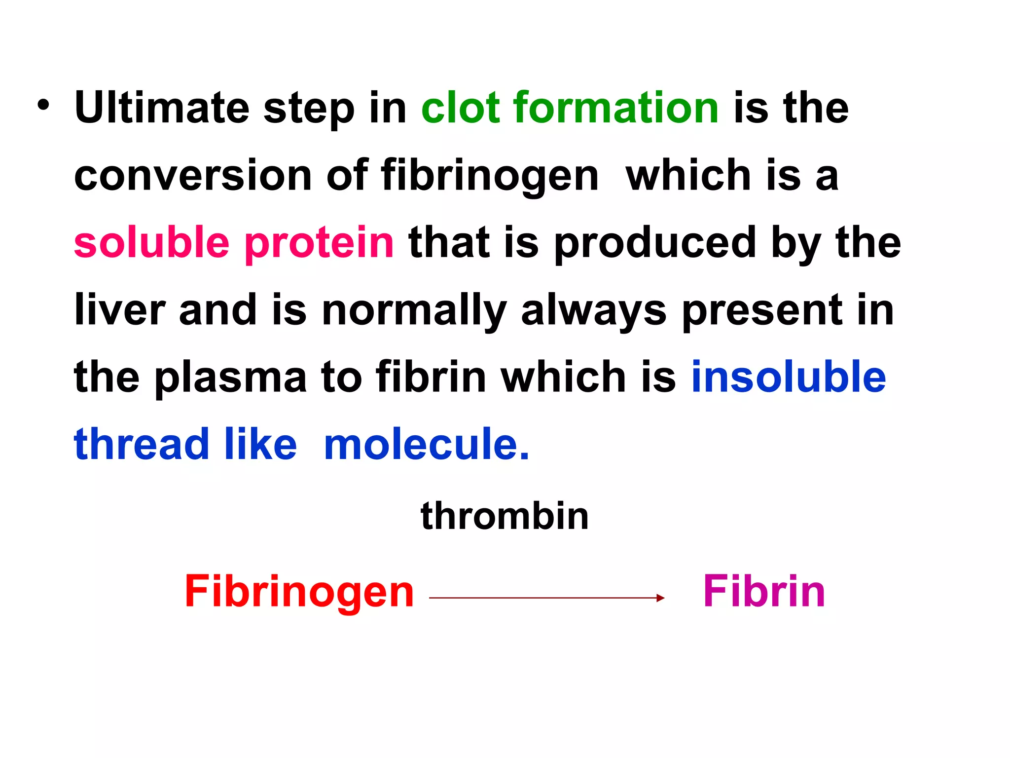
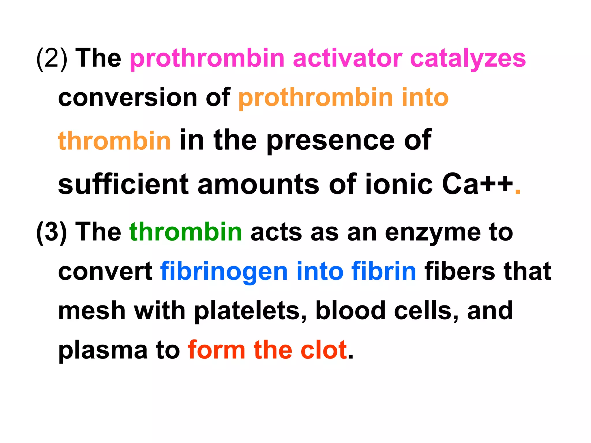
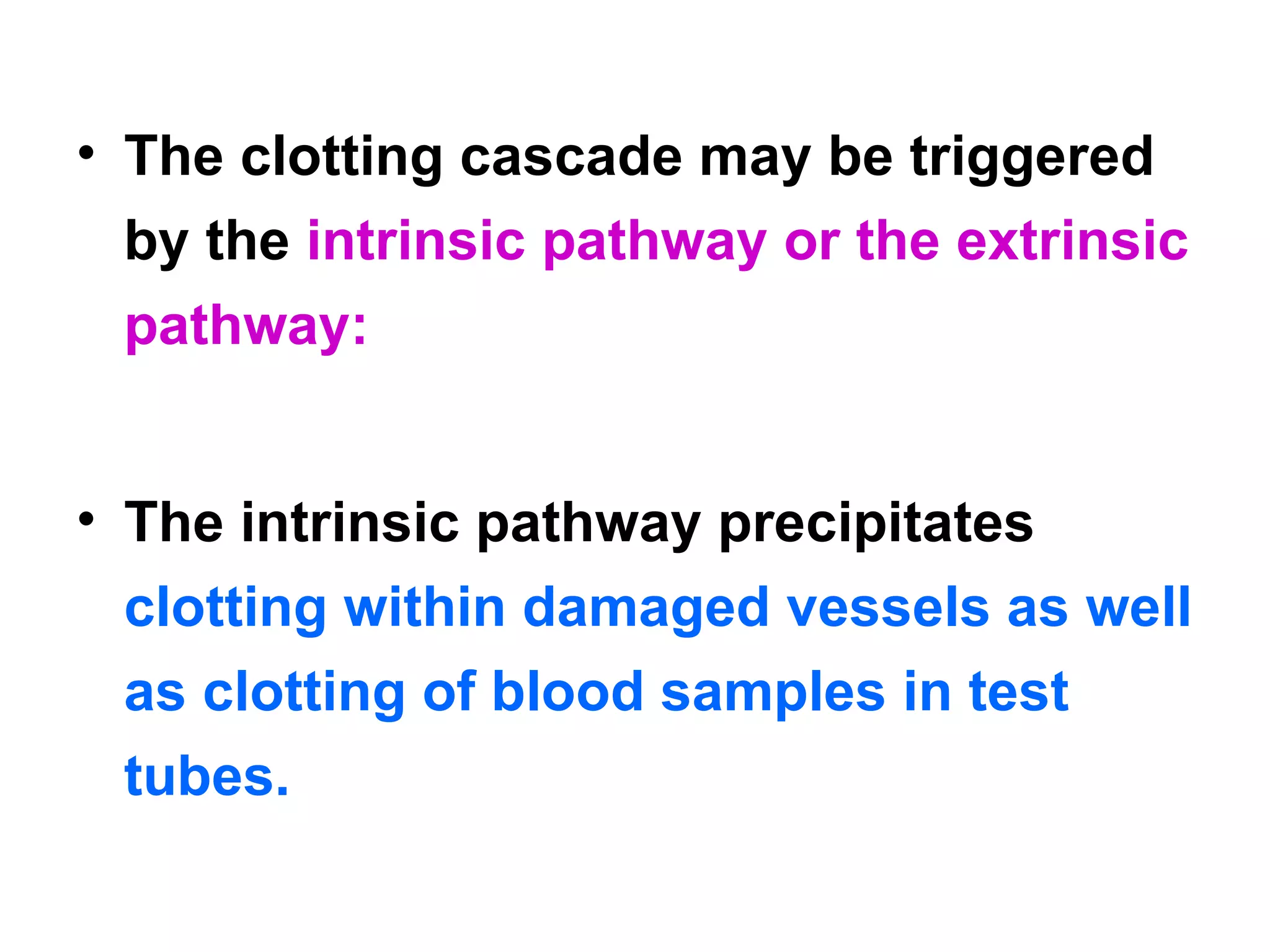
Hemostasis Definition: Prevention of blood loss.
Events Involved In Hemostasis
Whenever a vessel is ruptured, hemostasis is achieved by: Vascular constriction   Formation of a  platelet plug  Formation of a  blood clot  as a result of blood coagulation.  Eventual  growth of fibrous tissue  into the blood clot to  close the hole in the vessel permanently.
Vascular Constriction
In  ruptured blood vessel 1. Pain impulses  from the  site of trauma  as well as from the surrounding nervous tissue  originate and reach the  spinal cord. From the  spinal cord order signal arise.
The order signals pass through the  sympathatic nerves Lead to  spasm of the vessel . 2. Local muscle  also contribute to the vascular vasospasm. 3. local autacoid factors  from the traumatized tissues and blood platelets.
As the vessel wall is damaged the  action potential developed  along the vessel wall lead to  vasoconstriction. The  vasospasm lasts  for almost half an hour and it is  directly proportional  to the  intensity of trauma .
Vasoconstriction  resulting from  local myogenic contraction   of the blood vessels is  initiated by  direct damage  to the vascular wall. In the  smaller vessels , the platelets are responsible for much of the vasoconstriction by  releasing a  vasoconstrictor substance , thromboxane A 2 .
Formation of the Platelet Plug
Platelets
Platelets or thrombocytes are  small colorless ,  non nucleated  cells. Shape is  spherical or  rod shaped  and become  oval or disc shaped when inactivated. Size:   1 to 4 micrometers in diameter.  Life span:   10 - days
 
 
Development:   From the pluripotentstem  cells in the bone marrow. CFU-M Colony forming  megakaryocytes Megakaryoblast Promegakaryoctye Megakaryocytes Platelets
Normal concentration :  150,000 to 300,000 per microliter. Structure: Cell membrane Microtubule Cytoplasm
Cell Membrane of Platelet
It is 6 nm thick and contain  lipids  (phospholipids, cholesterol and glycolipids), Carbohydrates (glycocalyx), Proteins and glycoproteins. Out of all  glycoprotein  and  phospholipids  are functionally  important.
Glycopropteins Prevents the adherence  of platelets to normal endothelium. Accelerates the adherence  of platelets to collagen and damaged endothelium in ruptured blood  vessels. Forms a receptor  for ADP and thrombin.
 
Cytoplasm   The cytoplasm of the platelets  include:  Golgi apparatus Endoplasmic reticulum Mitochondria Microtubule Microvessels Microfilaments Granules
Cytoplasm also contains: Proteins Enzymes Hormones. Chemical substances
Proteins The major proteins present are contractile proteins which are  responsible for the contraction of platelets : Actin   Myosin Thrombosthenin
Chemical substances :  Calcium ions Mg- ions.  Adenosine triphosphate (ATP)  Adenosine diphosphate (ADP)
Function Of Platelets Its surface has glycoprotein coat that adhere  it to injured endothelial cells… …. preventing bleeding . Actin, myosin & thrombosthenin that are contractile proteins…. cause  clot retraction.
Secretes  growth factor  that promotes growth & multiplication of vascular endothelial cells, vascular smooth cells & fibroblasts….  repair damaged vascular wall. Its membrane has  phospholipids  that activate intrinsic system of  blood clotting
Life span O f Platelets Platelets are  eliminated  from the circulation mainly by the  tissue macrophage system  in the spleen.
Mechanism of the Platelet Plug When platelets come in  contact with a damaged vascular surface,  platelets attach to the  exposed collagen fibers  in the vascular wall. Platelets immediately change their own characteristics.
 
Platelets begin to  swell  and assume irregular forms with numerous irradiating  pseudopods protruding  from their surfaces Contractile proteins  in the platelets contract forcefully and cause the release of  granules  that contain  multiple active factors
 
 
Adenosine diphosphate (ADP)  is released which causes surface of nearby  circulating platelets  to become  sticky  and it adheres to the first layer of aggregated platelets
 
 
The  aggregated platelets  adhere to the  von Willebrand factor  that leaks into the traumatized tissue from the plasma  It leads to the  release of more ADP  , which cause  more platelets to pile up  at the defected site.
 
The  aggregating process is reinforced by the  formation of Thromboxane A 2.  It  directly  promotes platelet aggregation  and further enhances  it indirectly by  triggering the release of even more ADP  from the platelet granules. Formation of platelet plug takes place
Thirdly, the platelet  plug release other  chemical substances  that play a role in  blood clotting. Platelet plugging mechanism  alone is  sufficient  to seal tears in the  capillaries and small vessels  but, large holes require  formation of blood clot  to stop  bleeding.
Limitation of Platelet Plug Normal endothelium of the vessel release  Prostacyclin  which  prevents platelet aggregation. So, platelet plug is  limited to the defected part  of the vessel and does not spread to the  normal vascular tissue.
Formation Of Blood Clot If there is a large defect in the vessel then  blood clot + platelet plug  are required to stop bleeding. As clot on the top of platelet plug  supports it and  reinforces the seal over the break in the vessel.
Onset Of Formation Of Blood Clot: 15 – 20 sec …… in severe trauma. 1 – 2 min …… in minor trauma.
Ultimate step in  clot formation  is the conversion of fibrinogen  which is a  soluble protein  that is produced by the liver and is normally always present in the plasma to fibrin which is  insoluble thread like  molecule.  thrombin Fibrinogen   Fibrin
Fibrin molecules adhere to the  damaged vessel  surface  forming a loose  netlike meshwork  that  traps  the cellular elements of blood . The clot  appears red  because of abundance of RBC that are trapped in it.
 
The  original fibrin web is weak   because the fibrin threads are loosely interlaced. Rapidly, various  chemical linkages  are formed  between  adjacent strands   to  strengthen and stabilize  the clot mesh work.
The  cross linkage   process which is  catalyzed  by a clotting factor known as factor  XIII (Fibrin stabilizing factor).
 
Fibrous Organization or Dissolution of the Blood Clot
Once a  blood clot has formed , it can follow one of two courses:  It can become  invaded by fibroblast s , which subsequently form connective tissue all through the clot. It can  dissolve.
The usual course for a clot that forms in a small hole of a vessel wall…… is  invasion by fibroblasts , beginning within a  few hours  after the clot is formed. This event is promoted at least partially by  growth factor   secreted by platelets.
Complete organization  of the clot into fibrous tissue takes place within  1 to 2 weeks.  When  excess blood  has leaked into the tissues and tissue clots have occurred  where they are not needed.
Special substances  within the clot itself usually become activated. These function as  enzymes to dissolve the clot.
Mechanism of Blood Coagulation
Procoagulants: Substances that cause or affect blood coagulation  that have been found in the blood and in the tissues….  promote coagulation Anticoagulants: Substances that  inhibit coagulation  are called Anticoagulants.
Whether  blood will coagulate  depends on the  balance between  these two groups of substances. In the blood stream, the  anticoagulants normally predominate,  so that the blood does not coagulate while it is circulating in the blood vessels.
But when a  vessel is ruptured,   procoagulants  from the area of tissue damage become  “activated”  and override the anticoagulants, and then a  clot does develop.
Three Essential Steps Involved In Clotting:
(1)  In response to  rupture of the vessel or damage to the blood itself,  a complex cascade of chemical reactions occurs in the blood involving more than a dozen blood coagulation factors. Formation of a complex of activated  substances collectively called  prothrombin activator.
(2)  The  prothrombin activator catalyzes  conversion of  prothrombin into thrombin  in the presence of sufficient amounts of ionic Ca++ . (3) The  thrombin  acts as an enzyme to convert  fibrinogen into fibrin   fibers that mesh with platelets, blood cells, and plasma to  form the clot .
 
The clotting cascade may be triggered by the  intrinsic pathway or the extrinsic pathway:  The intrinsic pathway precipitates  clotting within damaged vessels as well as clotting of blood samples in test tubes.
All  elements necessary  to bring about clotting by means of the intrinsic pathway are  present in the blood.
 

Platelets physiology

  • 1.
  • 2.
  • 3.
    Whenever a vesselis ruptured, hemostasis is achieved by: Vascular constriction Formation of a platelet plug Formation of a blood clot as a result of blood coagulation. Eventual growth of fibrous tissue into the blood clot to close the hole in the vessel permanently.
  • 4.
  • 5.
    In rupturedblood vessel 1. Pain impulses from the site of trauma as well as from the surrounding nervous tissue originate and reach the spinal cord. From the spinal cord order signal arise.
  • 6.
    The order signalspass through the sympathatic nerves Lead to spasm of the vessel . 2. Local muscle also contribute to the vascular vasospasm. 3. local autacoid factors from the traumatized tissues and blood platelets.
  • 7.
    As the vesselwall is damaged the action potential developed along the vessel wall lead to vasoconstriction. The vasospasm lasts for almost half an hour and it is directly proportional to the intensity of trauma .
  • 8.
    Vasoconstriction resultingfrom local myogenic contraction of the blood vessels is initiated by direct damage to the vascular wall. In the smaller vessels , the platelets are responsible for much of the vasoconstriction by releasing a vasoconstrictor substance , thromboxane A 2 .
  • 9.
    Formation of thePlatelet Plug
  • 10.
  • 11.
    Platelets or thrombocytesare small colorless , non nucleated cells. Shape is spherical or rod shaped and become oval or disc shaped when inactivated. Size: 1 to 4 micrometers in diameter. Life span: 10 - days
  • 12.
  • 13.
  • 14.
    Development: From the pluripotentstem cells in the bone marrow. CFU-M Colony forming megakaryocytes Megakaryoblast Promegakaryoctye Megakaryocytes Platelets
  • 15.
    Normal concentration : 150,000 to 300,000 per microliter. Structure: Cell membrane Microtubule Cytoplasm
  • 16.
  • 17.
    It is 6nm thick and contain lipids (phospholipids, cholesterol and glycolipids), Carbohydrates (glycocalyx), Proteins and glycoproteins. Out of all glycoprotein and phospholipids are functionally important.
  • 18.
    Glycopropteins Prevents theadherence of platelets to normal endothelium. Accelerates the adherence of platelets to collagen and damaged endothelium in ruptured blood vessels. Forms a receptor for ADP and thrombin.
  • 19.
  • 20.
    Cytoplasm The cytoplasm of the platelets include: Golgi apparatus Endoplasmic reticulum Mitochondria Microtubule Microvessels Microfilaments Granules
  • 21.
    Cytoplasm also contains:Proteins Enzymes Hormones. Chemical substances
  • 22.
    Proteins The majorproteins present are contractile proteins which are responsible for the contraction of platelets : Actin Myosin Thrombosthenin
  • 23.
    Chemical substances : Calcium ions Mg- ions. Adenosine triphosphate (ATP) Adenosine diphosphate (ADP)
  • 24.
    Function Of PlateletsIts surface has glycoprotein coat that adhere it to injured endothelial cells… …. preventing bleeding . Actin, myosin & thrombosthenin that are contractile proteins…. cause clot retraction.
  • 25.
    Secretes growthfactor that promotes growth & multiplication of vascular endothelial cells, vascular smooth cells & fibroblasts…. repair damaged vascular wall. Its membrane has phospholipids that activate intrinsic system of blood clotting
  • 26.
    Life span Of Platelets Platelets are eliminated from the circulation mainly by the tissue macrophage system in the spleen.
  • 27.
    Mechanism of thePlatelet Plug When platelets come in contact with a damaged vascular surface, platelets attach to the exposed collagen fibers in the vascular wall. Platelets immediately change their own characteristics.
  • 28.
  • 29.
    Platelets begin to swell and assume irregular forms with numerous irradiating pseudopods protruding from their surfaces Contractile proteins in the platelets contract forcefully and cause the release of granules that contain multiple active factors
  • 30.
  • 31.
  • 32.
    Adenosine diphosphate (ADP) is released which causes surface of nearby circulating platelets to become sticky and it adheres to the first layer of aggregated platelets
  • 33.
  • 34.
  • 35.
    The aggregatedplatelets adhere to the von Willebrand factor that leaks into the traumatized tissue from the plasma It leads to the release of more ADP , which cause more platelets to pile up at the defected site.
  • 36.
  • 37.
    The aggregatingprocess is reinforced by the formation of Thromboxane A 2. It directly promotes platelet aggregation and further enhances it indirectly by triggering the release of even more ADP from the platelet granules. Formation of platelet plug takes place
  • 38.
    Thirdly, the platelet plug release other chemical substances that play a role in blood clotting. Platelet plugging mechanism alone is sufficient to seal tears in the capillaries and small vessels but, large holes require formation of blood clot to stop bleeding.
  • 39.
    Limitation of PlateletPlug Normal endothelium of the vessel release Prostacyclin which prevents platelet aggregation. So, platelet plug is limited to the defected part of the vessel and does not spread to the normal vascular tissue.
  • 40.
    Formation Of BloodClot If there is a large defect in the vessel then blood clot + platelet plug are required to stop bleeding. As clot on the top of platelet plug supports it and reinforces the seal over the break in the vessel.
  • 41.
    Onset Of FormationOf Blood Clot: 15 – 20 sec …… in severe trauma. 1 – 2 min …… in minor trauma.
  • 42.
    Ultimate step in clot formation is the conversion of fibrinogen which is a soluble protein that is produced by the liver and is normally always present in the plasma to fibrin which is insoluble thread like molecule. thrombin Fibrinogen Fibrin
  • 43.
    Fibrin molecules adhereto the damaged vessel surface forming a loose netlike meshwork that traps the cellular elements of blood . The clot appears red because of abundance of RBC that are trapped in it.
  • 44.
  • 45.
    The originalfibrin web is weak because the fibrin threads are loosely interlaced. Rapidly, various chemical linkages are formed between adjacent strands to strengthen and stabilize the clot mesh work.
  • 46.
    The crosslinkage process which is catalyzed by a clotting factor known as factor XIII (Fibrin stabilizing factor).
  • 47.
  • 48.
    Fibrous Organization orDissolution of the Blood Clot
  • 49.
    Once a blood clot has formed , it can follow one of two courses: It can become invaded by fibroblast s , which subsequently form connective tissue all through the clot. It can dissolve.
  • 50.
    The usual coursefor a clot that forms in a small hole of a vessel wall…… is invasion by fibroblasts , beginning within a few hours after the clot is formed. This event is promoted at least partially by growth factor secreted by platelets.
  • 51.
    Complete organization of the clot into fibrous tissue takes place within 1 to 2 weeks. When excess blood has leaked into the tissues and tissue clots have occurred where they are not needed.
  • 52.
    Special substances within the clot itself usually become activated. These function as enzymes to dissolve the clot.
  • 53.
    Mechanism of BloodCoagulation
  • 54.
    Procoagulants: Substances thatcause or affect blood coagulation that have been found in the blood and in the tissues…. promote coagulation Anticoagulants: Substances that inhibit coagulation are called Anticoagulants.
  • 55.
    Whether bloodwill coagulate depends on the balance between these two groups of substances. In the blood stream, the anticoagulants normally predominate, so that the blood does not coagulate while it is circulating in the blood vessels.
  • 56.
    But when a vessel is ruptured, procoagulants from the area of tissue damage become “activated” and override the anticoagulants, and then a clot does develop.
  • 57.
    Three Essential StepsInvolved In Clotting:
  • 58.
    (1) Inresponse to rupture of the vessel or damage to the blood itself, a complex cascade of chemical reactions occurs in the blood involving more than a dozen blood coagulation factors. Formation of a complex of activated substances collectively called prothrombin activator.
  • 59.
    (2) The prothrombin activator catalyzes conversion of prothrombin into thrombin in the presence of sufficient amounts of ionic Ca++ . (3) The thrombin acts as an enzyme to convert fibrinogen into fibrin fibers that mesh with platelets, blood cells, and plasma to form the clot .
  • 60.
  • 61.
    The clotting cascademay be triggered by the intrinsic pathway or the extrinsic pathway: The intrinsic pathway precipitates clotting within damaged vessels as well as clotting of blood samples in test tubes.
  • 62.
    All elementsnecessary to bring about clotting by means of the intrinsic pathway are present in the blood.
  • 63.