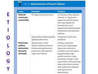
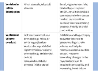
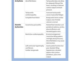
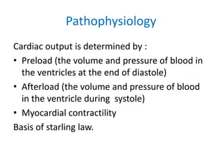
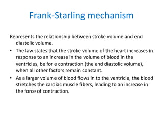
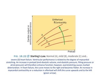
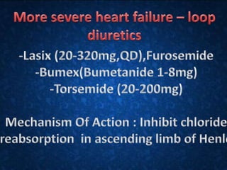
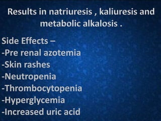
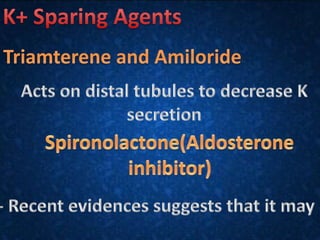
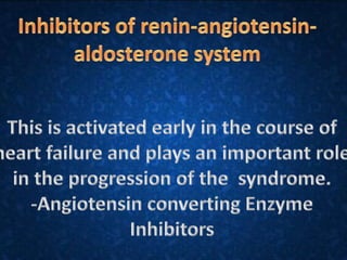
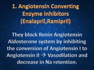
Congestive Cardiac Failure
Presented by: 88-2013
113-2013
135-2013
167-2013
173-2013
Under the guidance of DR.Medha Gupta
Content
• Definition
• Etiology
• Pathophysiology
• Clinical Features
• Bedside Examination
• Investigation
• Management
Congestive Cardiac Failure
Definition:
CCF or Heart Failure in a clinical syndrome that
develops when the heart muscle is weakened
and cannot maintain the adequate cardiac
output or can do so only at an expence of
elevated ventricular filling pressure.
Pathophysiology
Cardiac output is determined by :
• Preload (the volume and pressure of blood in
the ventricles at the end of diastole)
• Afterload (the volume and pressure of blood
in the ventricle during systole)
• Myocardial contractility
Basis of starling law.
Frank-Starling mechanism
Represents the relationship between stroke volume and end
diastolic volume.
• The law states that the stroke volume of the heart increases in
response to an increase in the volume of blood in the
ventricles, be for e contraction (the end diastolic volume),
when all other factors remain constant.
• As a larger volume of blood flows in to the ventricle, the blood
stretches the cardiac muscle fibers, leading to an increase in
the force of contraction.
severe (D) heart failure. Ventricular performance is related to the degree of myocardial
stretching. An increase in preload (end-diastolic volume, end-diastolic pressure, filling pressure or
atrial pressure) will therefore enhance function; however, overstretching causes marked
deterioration. In heart failure, the curve moves to the right and becomes flatter. An increase in
myocardial contractility or a reduction in afterload will shift the curve upwards and to the left
(green arrow).
• Heart failure is characterised by: a decrease in cardiac output. Cardiac
output in turn is a function of preload, after load and myocardial
contractility. In heart failure, preload is increased, afterload is increased
and myocardial contractility is decreased.
• In initial stages of heart failure with the reduction in CO, certain
compensatory mechanisms come into operation. These compensatory
mechanisms are initially beneficial, but later become counterproductive
and account for most of the manifestations of heart failure.
• These compensatory mechanisms are renin-angiotensin system and
autonomic nervous system.
• Increased myocardial contractility: As the ventricles dilates (resulting in
increased preload), the ventricular contractility increases resulting in
relatively increased volume of blood ejected (Frank-Starling law).
• The increase left ventricular filling pressures are transmitted to the
pulmonary veins which results in alveolar transduction of fluid and
pulmonary congestion. Finally, oxygen demand is enhanced due to
increased contractility.
• Myocardial hypertophy occurs due to volume and pressure overload in the
ventricles and helps in enhanced contractility and hence increased CO.
• However, it produces the increased oxygen demand and reduced
compliance of the ventricle that results in increased preload which is again
transmitted to the pulmonary vasculature.
• Sympathetic stimulation results in peripheral vascularisation which in turn
leads to Na+-water retention due to direct and indirect effects of RAS.
• Sympathetic stimulation also produces increased heart rate and enhances
contractility. Increased afterload maybe deleterious as it tends to reduce
cardiac output and increases oxygen demand of the heart.
• Myocardial remodelling: Changes in structure and function of myocardium
and includes hypertrophy and apoptosis of myocytes and alterations in the
quantity and composition of extracellular matrix. Remodelling involves not
only the ischaemic area, but also the viable myocardium resulting in
gradual loss of contractility over a period of time.
Types of heart failure
• Left, right and biventricular heart failure
• The left side of the heart comprises the functional unit of the LA and LV,
together with the mitral and aortic valves; the right heart comprises the
RA, RV, and tricuspid and pulmonary valves.
• Left-sided heart failure. There is a reduction in left ventricular output and
an increase in left atrial and pulmonary venous pressure. An acute
increase in left atrial pressure causes pulmonary congestion or pulmonary
oedema; a more gradual increase in left atrial pressure, as occurs with
mitral stenosis, leads to reflex pulmonary vasoconstriction, which protects
the patient from pulmonary oedema. This increases pulmonary vascular
resistance and causes pulmonary hypertension, which can, in turn, impair
right ventricular function.
• Right-sided heart failure. There is a reduction in right
ventricular output and an increase in right atrial and
systemic venous pressure. Causes of isolated right
heart failure include chronic lung disease (cor
pulmonale), pulmonary embolism and pulmonary
valvular stenosis.
• Biventricular heart failure. Failure of the left and right
heart may develop because the disease process, such
as dilated cardiomyopathy or ischaemic heart disease,
affects both ventricles or because disease of the left
heart leads to chronic elevation of the left atrial
pressure, pulmonary hypertension and right heart
failure.
Clinical features
Clinical Features
Left Sided Heart Failure:
1) Pulmonary Edema
a) Dyspnea (SOB)
b)Orthopnea
c)Paroxysmal Nocturnal Dyspnea
2)Decreased forward Perfusion
Activated Renin Angiotensin Aldosterone System, which
cause fluid retention
and worsenen CHF.
Clinical Features
Right Sided Heart Failure:
Most common cause of cause of Right Heart Failure is
Left side
Heart Failure.
a) Dyspnea(SOB)
b)Jugular Venous Distention
c)Pitting Edema
d)Ascitis
e)Nutmeg Liver “Hepatomegaly”
General Examination
• General appearance
Cool Peripheral extrimities
Cynosis of lips and nail beds
Dyspnea:-
Early stage of HF-while exertion
Orthopnea- later manifestation of HF
Chronic or severe stages-even at rest
• Pulmonary examination
Pulmonary crackles (Rales or crepitations) can be
heard.
Rales are frequently absent in patients with chronic
HF
But patients without concomitant lung diseases,
rales are specific
For HF
• Cardiac examination
S3(gallop rhythm) is audible and palpable at apex
Murmurs of mitral and tricuspid regurgitation are
frequently
present in patients with advanced HF
• Abdomen and extrimities
Hepatomegaly is seen (frequently tender
and pulsating)
Ascities and jaundice are late signs of HF
Peripheral edema is cardinal
manifestation of HF
(Usually symmetric) but is also
nonspecific
• Jugular Veins
JVP-Usually elevated
• Cardiac Cachexia
With severe chronic HF there is marked
weight loss and cachexia
Investigations
• BNP>100pg/mL
• Electrocardiogram may be normal or it could show numerous
abnormalities including acute ST-T–wave changes from
myocardial ischemia, atrial fibrillation, bradycardia, left
ventricular hypertrophy
• Serum creatinine may be increased because of hypoperfusion.
Pre existing renal dysfunction can contribute to volume
overload.
• Complete blood count useful to determine if heart failure is a
result of reduced oxygen-carrying capacity
• Chest radiography is useful for detection of cardiac
enlargement, pulmonary edema, and pleural
effusions
• Echocardiogram assesses left ventricle size, valve
function, pericardial effusion, and ejection fraction
• Hyponatremia, serum sodium <130mEq/L, is
associated with reduced survival and may indicate
worsening volume overload and/or disease
progression
Thiazides are ineffective with
GFR<30--/min.
Side Effects –
-Pre renal azotemia
-Skin rashes
-Neutropenia
-Thrombocytopenia
-Hyperglycemia
-Increased uric acid
Congestive cardiac failure

Congestive cardiac failure

  • 1.
    Congestive Cardiac Failure Presentedby: 88-2013 113-2013 135-2013 167-2013 173-2013 Under the guidance of DR.Medha Gupta
  • 2.
    Content • Definition • Etiology •Pathophysiology • Clinical Features • Bedside Examination • Investigation • Management
  • 3.
    Congestive Cardiac Failure Definition: CCFor Heart Failure in a clinical syndrome that develops when the heart muscle is weakened and cannot maintain the adequate cardiac output or can do so only at an expence of elevated ventricular filling pressure.
  • 7.
    Pathophysiology Cardiac output isdetermined by : • Preload (the volume and pressure of blood in the ventricles at the end of diastole) • Afterload (the volume and pressure of blood in the ventricle during systole) • Myocardial contractility Basis of starling law.
  • 8.
    Frank-Starling mechanism Represents therelationship between stroke volume and end diastolic volume. • The law states that the stroke volume of the heart increases in response to an increase in the volume of blood in the ventricles, be for e contraction (the end diastolic volume), when all other factors remain constant. • As a larger volume of blood flows in to the ventricle, the blood stretches the cardiac muscle fibers, leading to an increase in the force of contraction.
  • 9.
    severe (D) heartfailure. Ventricular performance is related to the degree of myocardial stretching. An increase in preload (end-diastolic volume, end-diastolic pressure, filling pressure or atrial pressure) will therefore enhance function; however, overstretching causes marked deterioration. In heart failure, the curve moves to the right and becomes flatter. An increase in myocardial contractility or a reduction in afterload will shift the curve upwards and to the left (green arrow).
  • 10.
    • Heart failureis characterised by: a decrease in cardiac output. Cardiac output in turn is a function of preload, after load and myocardial contractility. In heart failure, preload is increased, afterload is increased and myocardial contractility is decreased. • In initial stages of heart failure with the reduction in CO, certain compensatory mechanisms come into operation. These compensatory mechanisms are initially beneficial, but later become counterproductive and account for most of the manifestations of heart failure. • These compensatory mechanisms are renin-angiotensin system and autonomic nervous system. • Increased myocardial contractility: As the ventricles dilates (resulting in increased preload), the ventricular contractility increases resulting in relatively increased volume of blood ejected (Frank-Starling law). • The increase left ventricular filling pressures are transmitted to the pulmonary veins which results in alveolar transduction of fluid and pulmonary congestion. Finally, oxygen demand is enhanced due to increased contractility.
  • 11.
    • Myocardial hypertophyoccurs due to volume and pressure overload in the ventricles and helps in enhanced contractility and hence increased CO. • However, it produces the increased oxygen demand and reduced compliance of the ventricle that results in increased preload which is again transmitted to the pulmonary vasculature. • Sympathetic stimulation results in peripheral vascularisation which in turn leads to Na+-water retention due to direct and indirect effects of RAS. • Sympathetic stimulation also produces increased heart rate and enhances contractility. Increased afterload maybe deleterious as it tends to reduce cardiac output and increases oxygen demand of the heart. • Myocardial remodelling: Changes in structure and function of myocardium and includes hypertrophy and apoptosis of myocytes and alterations in the quantity and composition of extracellular matrix. Remodelling involves not only the ischaemic area, but also the viable myocardium resulting in gradual loss of contractility over a period of time.
  • 13.
    Types of heartfailure • Left, right and biventricular heart failure • The left side of the heart comprises the functional unit of the LA and LV, together with the mitral and aortic valves; the right heart comprises the RA, RV, and tricuspid and pulmonary valves. • Left-sided heart failure. There is a reduction in left ventricular output and an increase in left atrial and pulmonary venous pressure. An acute increase in left atrial pressure causes pulmonary congestion or pulmonary oedema; a more gradual increase in left atrial pressure, as occurs with mitral stenosis, leads to reflex pulmonary vasoconstriction, which protects the patient from pulmonary oedema. This increases pulmonary vascular resistance and causes pulmonary hypertension, which can, in turn, impair right ventricular function.
  • 14.
    • Right-sided heartfailure. There is a reduction in right ventricular output and an increase in right atrial and systemic venous pressure. Causes of isolated right heart failure include chronic lung disease (cor pulmonale), pulmonary embolism and pulmonary valvular stenosis. • Biventricular heart failure. Failure of the left and right heart may develop because the disease process, such as dilated cardiomyopathy or ischaemic heart disease, affects both ventricles or because disease of the left heart leads to chronic elevation of the left atrial pressure, pulmonary hypertension and right heart failure.
  • 15.
  • 17.
    Clinical Features Left SidedHeart Failure: 1) Pulmonary Edema a) Dyspnea (SOB) b)Orthopnea c)Paroxysmal Nocturnal Dyspnea 2)Decreased forward Perfusion Activated Renin Angiotensin Aldosterone System, which cause fluid retention and worsenen CHF.
  • 18.
    Clinical Features Right SidedHeart Failure: Most common cause of cause of Right Heart Failure is Left side Heart Failure. a) Dyspnea(SOB) b)Jugular Venous Distention c)Pitting Edema d)Ascitis e)Nutmeg Liver “Hepatomegaly”
  • 20.
  • 21.
    • General appearance CoolPeripheral extrimities Cynosis of lips and nail beds Dyspnea:- Early stage of HF-while exertion Orthopnea- later manifestation of HF Chronic or severe stages-even at rest
  • 22.
    • Pulmonary examination Pulmonarycrackles (Rales or crepitations) can be heard. Rales are frequently absent in patients with chronic HF But patients without concomitant lung diseases, rales are specific For HF • Cardiac examination S3(gallop rhythm) is audible and palpable at apex Murmurs of mitral and tricuspid regurgitation are frequently present in patients with advanced HF
  • 23.
    • Abdomen andextrimities Hepatomegaly is seen (frequently tender and pulsating) Ascities and jaundice are late signs of HF Peripheral edema is cardinal manifestation of HF (Usually symmetric) but is also nonspecific
  • 24.
    • Jugular Veins JVP-Usuallyelevated • Cardiac Cachexia With severe chronic HF there is marked weight loss and cachexia
  • 25.
    Investigations • BNP>100pg/mL • Electrocardiogrammay be normal or it could show numerous abnormalities including acute ST-T–wave changes from myocardial ischemia, atrial fibrillation, bradycardia, left ventricular hypertrophy • Serum creatinine may be increased because of hypoperfusion. Pre existing renal dysfunction can contribute to volume overload. • Complete blood count useful to determine if heart failure is a result of reduced oxygen-carrying capacity
  • 26.
    • Chest radiographyis useful for detection of cardiac enlargement, pulmonary edema, and pleural effusions • Echocardiogram assesses left ventricle size, valve function, pericardial effusion, and ejection fraction • Hyponatremia, serum sodium <130mEq/L, is associated with reduced survival and may indicate worsening volume overload and/or disease progression
  • 34.
    Thiazides are ineffectivewith GFR<30--/min.
  • 36.
    Side Effects – -Prerenal azotemia -Skin rashes -Neutropenia -Thrombocytopenia -Hyperglycemia -Increased uric acid