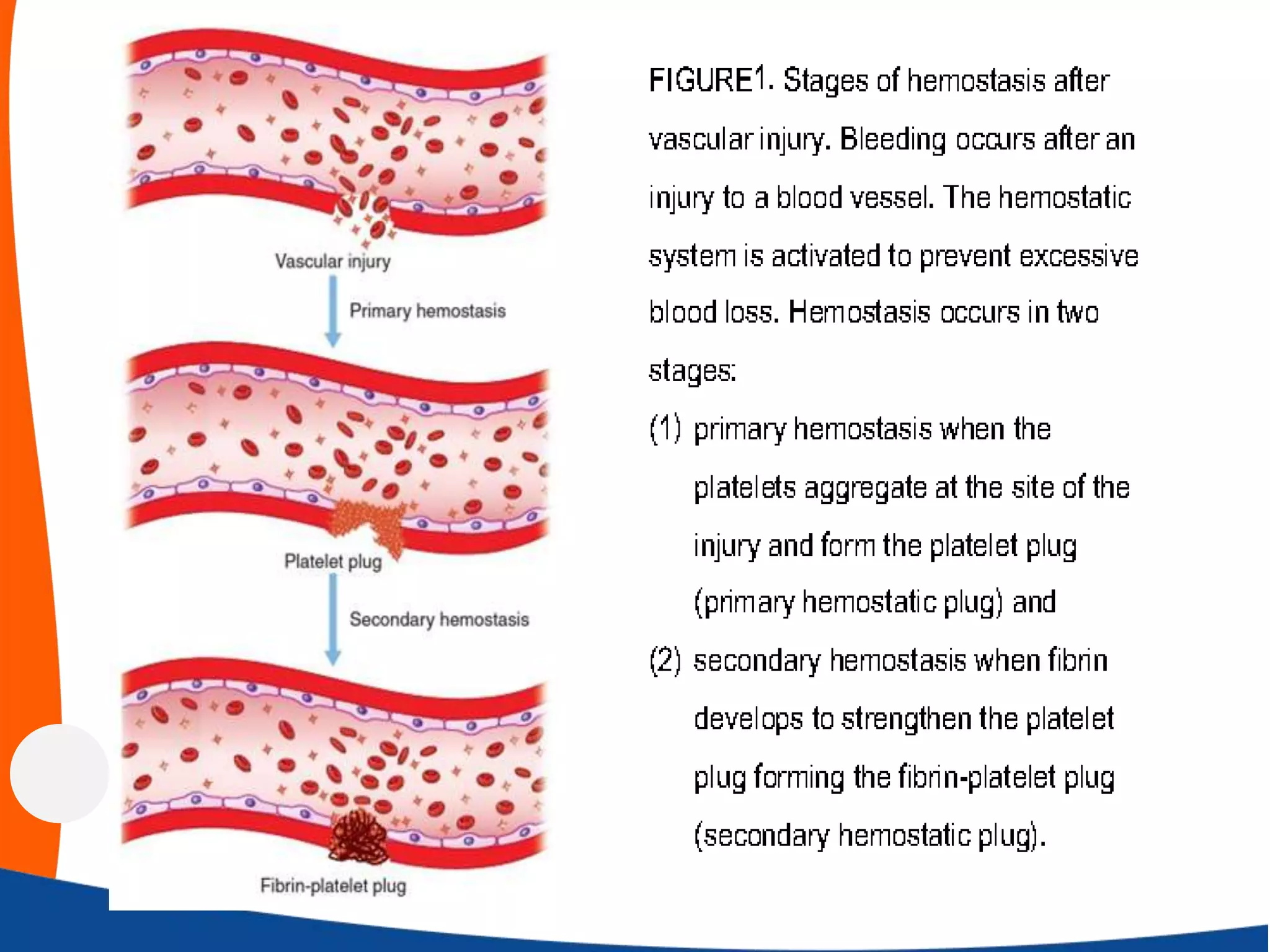
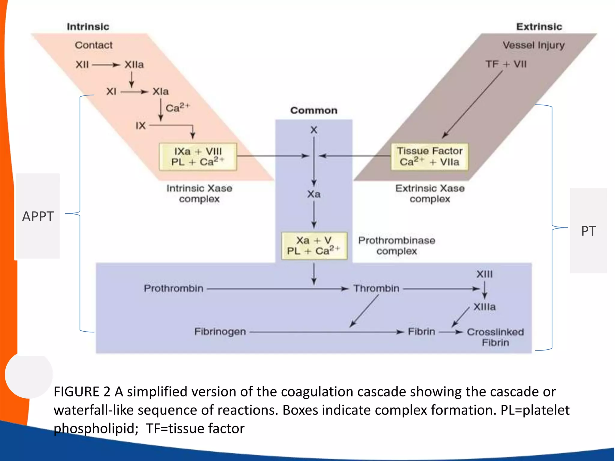
Haemostasis-Introduction
By
Ahmed Riyadh Abdul Rahman
Al-Noor University College
Haemostasis
Haemostasis is the process by which haemorrhage
following vascular injury is arrested (Fig.1). It depends
on closely linked-interaction between:
• The vessel wall,
• Platelets and
• Coagulation factors.
The fibrinolytic system and inhibitors of coagulation
ensure coagulation is limited to the site of injury.
The vessel wall
The intact vessel wall has an important role in
preventing thrombosis. Endothelial cells produce:
• Prostacyclin, which causes vasodilatation and
inhibits platelet aggregation;
• Nitric oxide, which causes vasodilatation and
inhibits platelet aggregation;
• Protein C activator (thrombomodulin), which,
when bound to protein C, inhibits coagulation
• Tissue plasminogen activator (TPA), which
activates fibrinolysis;
Injury to the vessel wall:
(a) activates membrane bound tissue factor, which
initiates coagulation cascade.
(b) exposes subendothelial connective tissue allowing
binding of platelets Von Willebrand factor (vWF), a
large multimeric protein made by endothelial cells.
ADAMTS13 is a plasma protease that cleaves high
molecular weight vWF complexes to smaller active
molecules.
VWF mediates platelet adhesion to endothelium and
carries clotting factor VIII in plasma.
Platelets
Platelets are non-nucleated cells required for normal
haemostasis. They circulate for 7–10 days. Their lifespan
is reduced when there is increased platelet consumption
(thrombosis, infection and splenic enlargement).
Platelets appear in peripheral blood films as granular
basophilic forms with a mean diameter of 1–2 μm. The
normal concentration is 140–400 ×109/L.
Thrombopoietin (TPO) is produced mainly in the liver
and stimulates megakaryocyte and platelet production.
Platelets
Platelet Roles in Hemostasis
•Perform surveillance of blood vessel continuity
•Platelet-platelet interactions (primary hemostatic plug)
•Platelet-coagulation protein interactions (secondary
hemostatic plug)
•Aid in healing injured tissue
Coagulation factors
The proteins of the coagulation cascade are pro-enzymes
and pro-cofactors, which are activated sequentially.
The cascade has been divided on the basis of laboratory
tests into intrinsic, extrinsic and common pathways
(Fig.2).This division is useful in understanding results of in-
vitro coagulation tests.
In vivo, however, these pathways are closely interlinked.
Coagulation begins in vivo when tissue factor is activated
on the surface of injured cells and binds and activates factor
VII, which ends in the generation of thrombin; this, in
turn, converts soluble plasma fibrinogen into fibrin.
Coagulation factors
Fibrin enmeshes the platelet aggregates at the sites of
vascular injury and converts the unstable primary
platelet plugs to firm, definitive and stable haemostatic
plugs.
Factor XIII is also activated by thrombin and stabilizes
the fibrin polymers with the formation of covalent bond
cross‐links.
FIGURE 2 A simplified version of the coagulation cascade showing the cascade or
waterfall-like sequence of reactions. Boxes indicate complex formation. PL=platelet
phospholipid; TF=tissue factor
PT
APPT
Tests of haemostatic function
Defective haemostasis with abnormal bleeding may result
from:
1. A vascular disorder;
2. Thrombocytopenia or a disorder of platelet function; or
3. Defective blood coagulation.
Tests that involved in the assessment of haemostasis are:
A. Blood count and blood film examination
B. Screening tests of blood coagulation
C. Specific assays of coagulation factors
D. Bleeding time
E. Tests of platelet function
F. Tests of fibrinolysis
Screening tests of blood coagulation

Haemostasis introduction

  • 1.
    Haemostasis-Introduction By Ahmed Riyadh AbdulRahman Al-Noor University College
  • 2.
    Haemostasis Haemostasis is theprocess by which haemorrhage following vascular injury is arrested (Fig.1). It depends on closely linked-interaction between: • The vessel wall, • Platelets and • Coagulation factors. The fibrinolytic system and inhibitors of coagulation ensure coagulation is limited to the site of injury.
  • 4.
    The vessel wall Theintact vessel wall has an important role in preventing thrombosis. Endothelial cells produce: • Prostacyclin, which causes vasodilatation and inhibits platelet aggregation; • Nitric oxide, which causes vasodilatation and inhibits platelet aggregation; • Protein C activator (thrombomodulin), which, when bound to protein C, inhibits coagulation • Tissue plasminogen activator (TPA), which activates fibrinolysis;
  • 5.
    Injury to thevessel wall: (a) activates membrane bound tissue factor, which initiates coagulation cascade. (b) exposes subendothelial connective tissue allowing binding of platelets Von Willebrand factor (vWF), a large multimeric protein made by endothelial cells. ADAMTS13 is a plasma protease that cleaves high molecular weight vWF complexes to smaller active molecules. VWF mediates platelet adhesion to endothelium and carries clotting factor VIII in plasma.
  • 6.
    Platelets Platelets are non-nucleatedcells required for normal haemostasis. They circulate for 7–10 days. Their lifespan is reduced when there is increased platelet consumption (thrombosis, infection and splenic enlargement). Platelets appear in peripheral blood films as granular basophilic forms with a mean diameter of 1–2 μm. The normal concentration is 140–400 ×109/L. Thrombopoietin (TPO) is produced mainly in the liver and stimulates megakaryocyte and platelet production.
  • 7.
    Platelets Platelet Roles inHemostasis •Perform surveillance of blood vessel continuity •Platelet-platelet interactions (primary hemostatic plug) •Platelet-coagulation protein interactions (secondary hemostatic plug) •Aid in healing injured tissue
  • 8.
    Coagulation factors The proteinsof the coagulation cascade are pro-enzymes and pro-cofactors, which are activated sequentially. The cascade has been divided on the basis of laboratory tests into intrinsic, extrinsic and common pathways (Fig.2).This division is useful in understanding results of in- vitro coagulation tests. In vivo, however, these pathways are closely interlinked. Coagulation begins in vivo when tissue factor is activated on the surface of injured cells and binds and activates factor VII, which ends in the generation of thrombin; this, in turn, converts soluble plasma fibrinogen into fibrin.
  • 9.
    Coagulation factors Fibrin enmeshesthe platelet aggregates at the sites of vascular injury and converts the unstable primary platelet plugs to firm, definitive and stable haemostatic plugs. Factor XIII is also activated by thrombin and stabilizes the fibrin polymers with the formation of covalent bond cross‐links.
  • 10.
    FIGURE 2 Asimplified version of the coagulation cascade showing the cascade or waterfall-like sequence of reactions. Boxes indicate complex formation. PL=platelet phospholipid; TF=tissue factor PT APPT
  • 11.
    Tests of haemostaticfunction Defective haemostasis with abnormal bleeding may result from: 1. A vascular disorder; 2. Thrombocytopenia or a disorder of platelet function; or 3. Defective blood coagulation. Tests that involved in the assessment of haemostasis are: A. Blood count and blood film examination B. Screening tests of blood coagulation C. Specific assays of coagulation factors D. Bleeding time E. Tests of platelet function F. Tests of fibrinolysis
  • 12.
    Screening tests ofblood coagulation