Phagocytosis is the process by which immune cells called phagocytes engulf and destroy microorganisms and foreign particles. The most important phagocytes are neutrophils, macrophages, and dendritic cells.
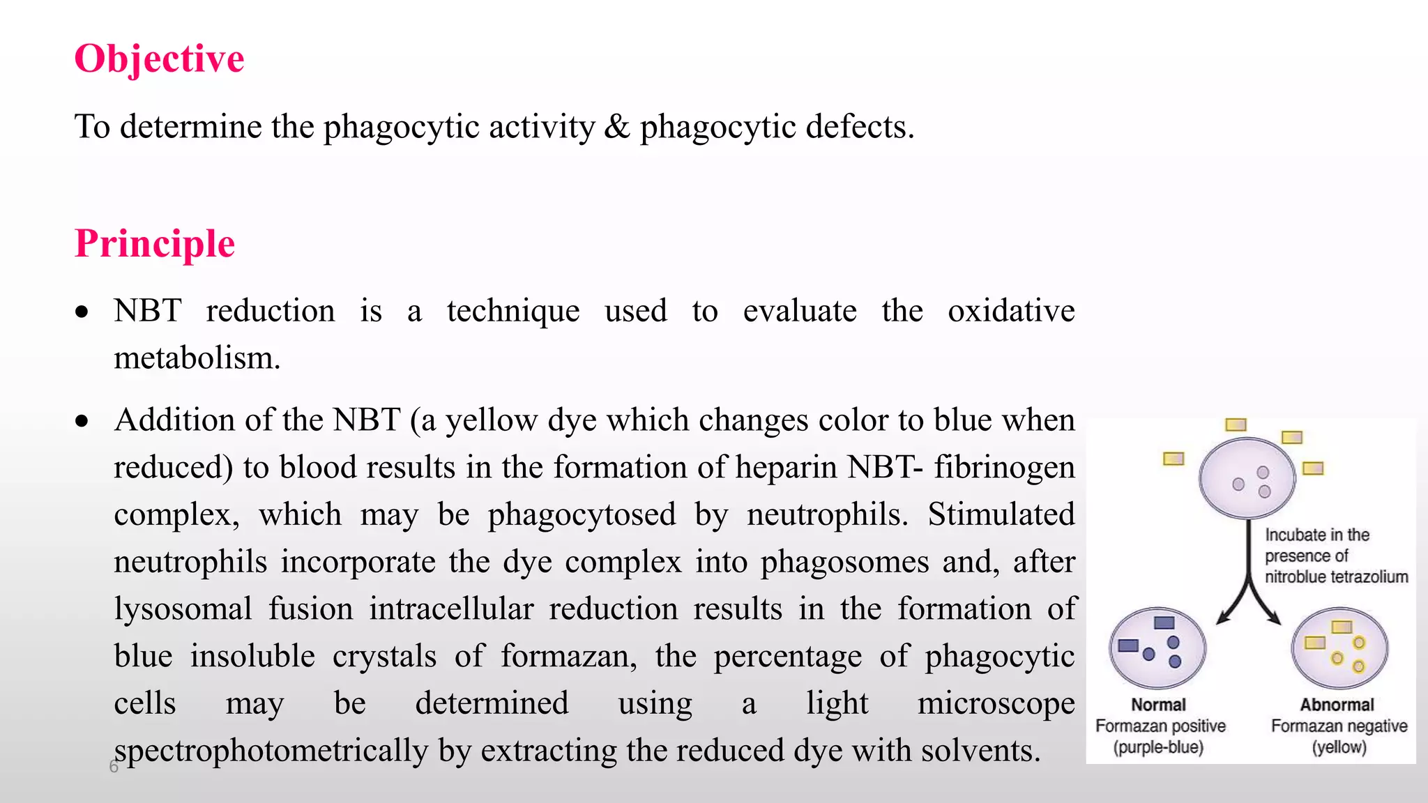
The nitroblue tetrazolium (NBT) assay is a microscopic method used to determine the phagocytic activity of cells by measuring their ability to reduce yellow-colored NBT to a blue insoluble form through the production of superoxide during phagocytosis. The assay involves incubating blood or cells with NBT, then examining stained smears under a microscope to count the percentage of cells containing blue deposits, indicating NBT reduction and normal phagocytic function.
The NBT assay can be used to