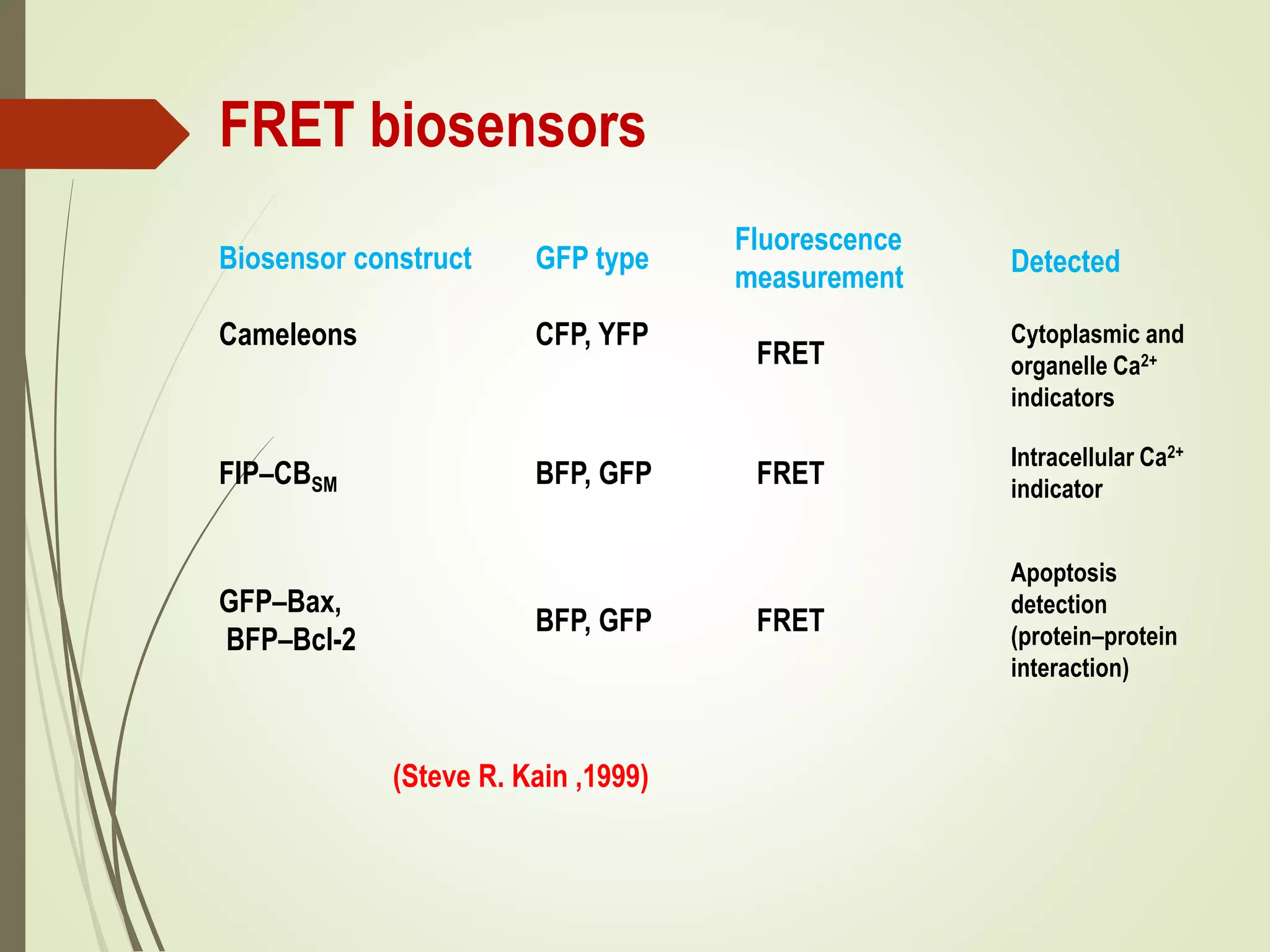
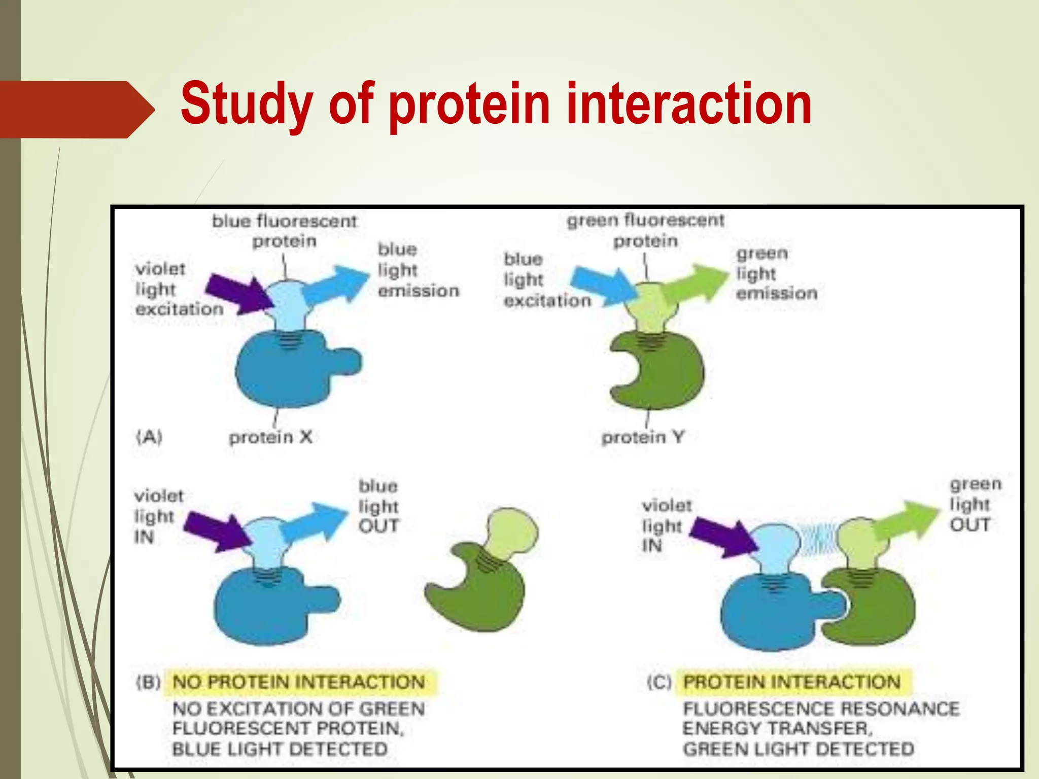
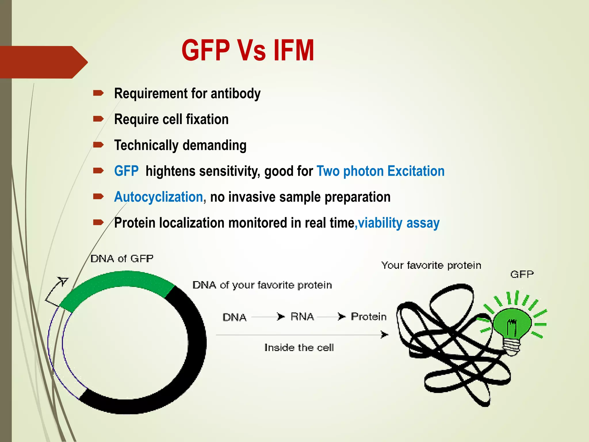
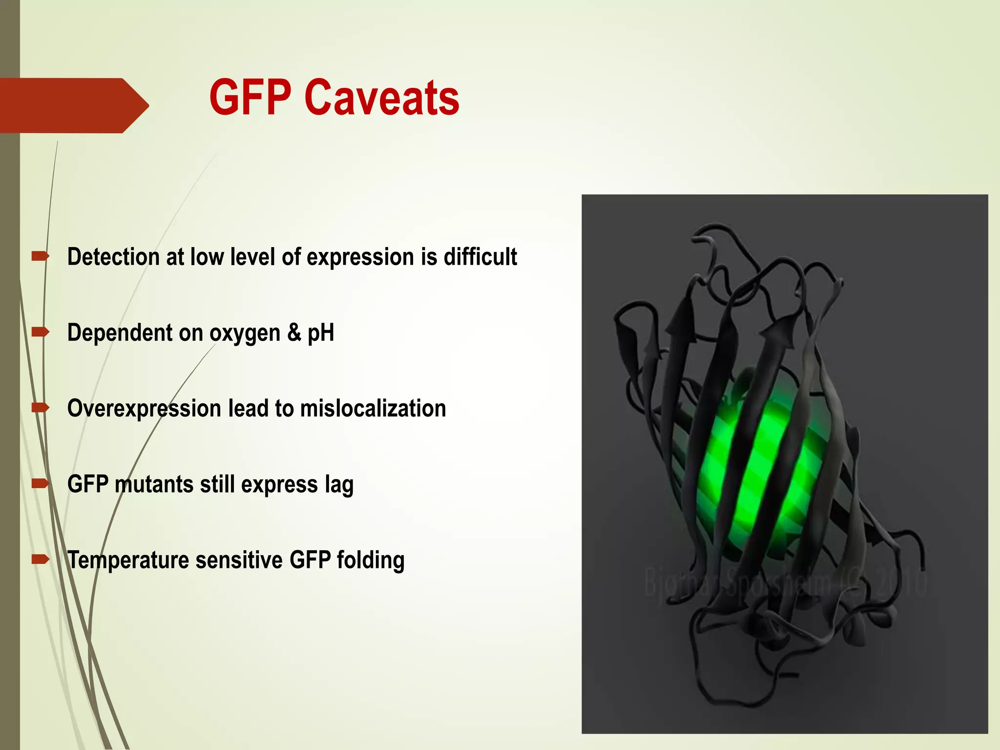
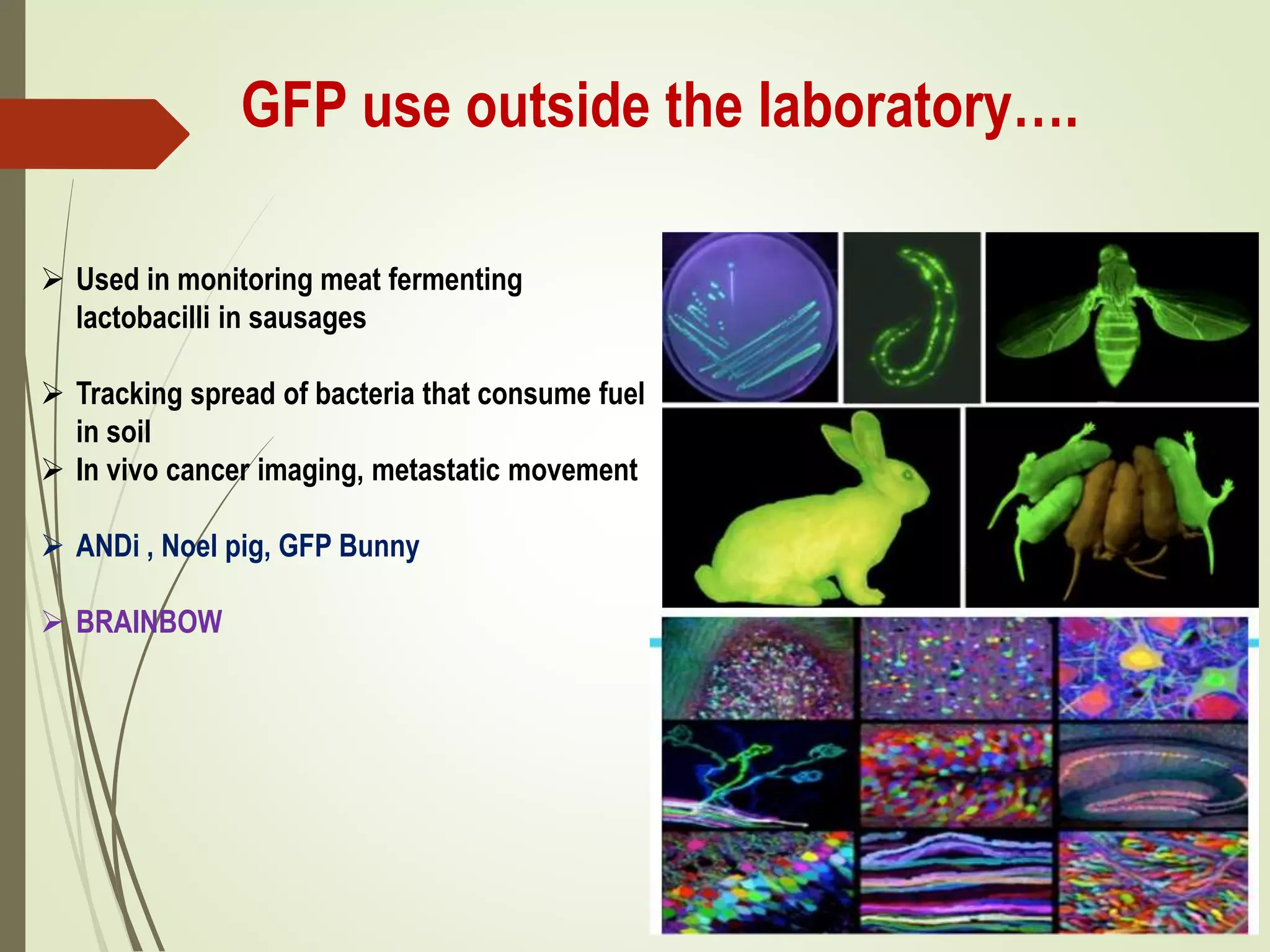
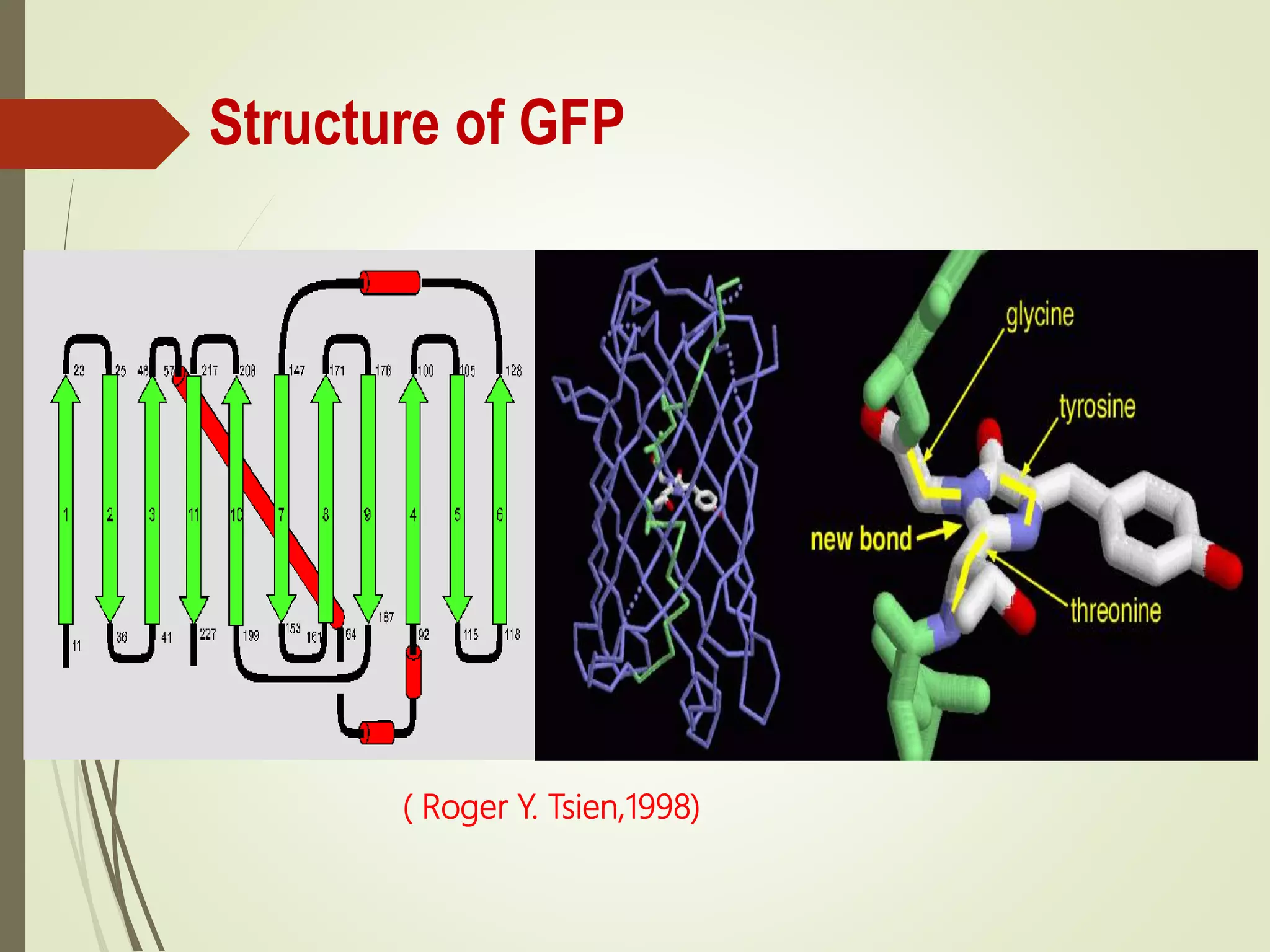
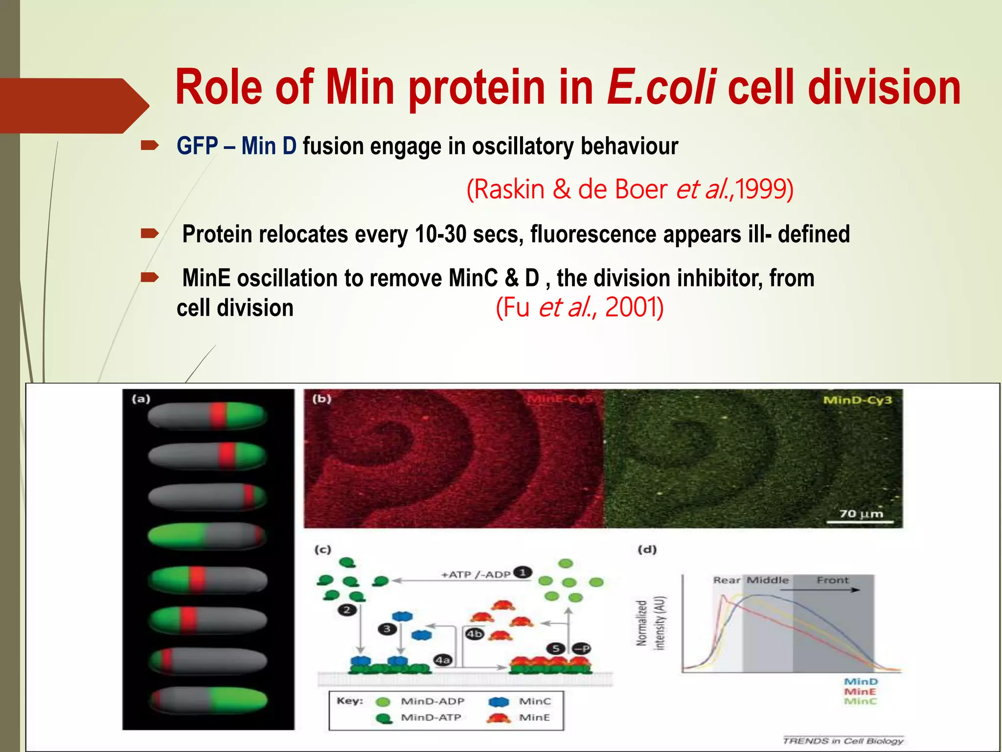
GFP has numerous applications for visualizing bacterial dynamics and disease diagnosis. It can be used to track gene expression, protein localization, and cell processes in real-time without disrupting bacterial viability. Fusion of GFP to proteins of interest allows visualization of processes like cell division, sporulation, and biofilm formation. GFP reporters also help identify genes expressed during bacterial infection and pathogenesis. Advances like FRET biosensors now enable monitoring of intracellular calcium and protein-protein interactions. Overall, GFP has revolutionized the study of bacterial physiology, gene regulation, and host-pathogen interactions.

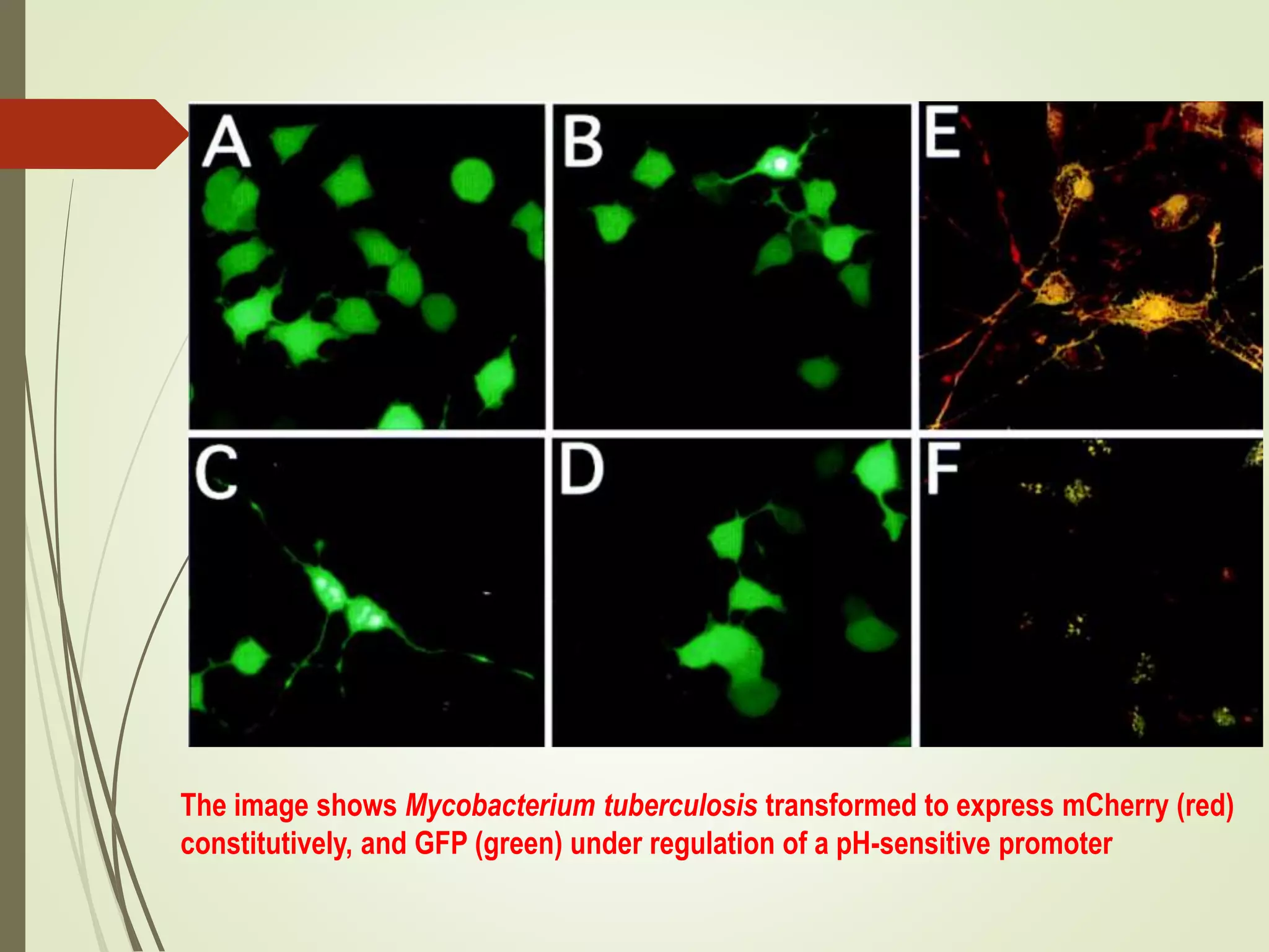
![Mycobacterium tuberculosis reporter
strain
Mycobacterium tuberculosis senses chloride & pH
(Tan, S., Sukumar, N., Abramovitch & Russell, D.G.
2013)
rv2390c: [Cl-] and low pH
rv2390c’::GFP a Sensor for phagosome maturation
hspX: Hypoxia and NO
HIV /TB coinfection](https://image.slidesharecdn.com/gfpapplicationinbacterialdynamicsanddiseasediagnosis-190909112528/75/Gfp-application-in-bacterial-dynamics-and-disease-diagnosis-24-2048.jpg)