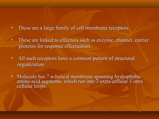
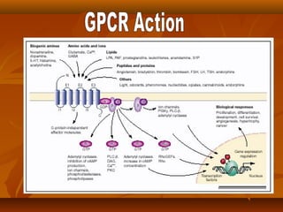
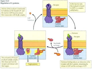
G PROTEIN COUPLEDG PROTEIN COUPLED
RECEPTORSRECEPTORS
 These are a large family of cell membrane receptors.These are a large family of cell membrane receptors.
 These are linked to effectors such as enzyme, channel, carrierThese are linked to effectors such as enzyme, channel, carrier
proteins for response effectuation.proteins for response effectuation.
 All such receptors have a common pattern of structuralAll such receptors have a common pattern of structural
organization.organization.
 Molecule has 7Molecule has 7 αα-helical membrane spanning hydrophobic-helical membrane spanning hydrophobic
amino acid segments, which run into 3 extra cellular 3 intraamino acid segments, which run into 3 extra cellular 3 intra
cellular loops.cellular loops.
 The agonist binding site is located some were between the helicesThe agonist binding site is located some were between the helices
on the extra cellular face, while another recognition site formedon the extra cellular face, while another recognition site formed
by cytosolic segments.by cytosolic segments.
 In the inactive state GDP is bound to their exposed domainIn the inactive state GDP is bound to their exposed domain
 Displacement of GDP by GTP occurs.Displacement of GDP by GTP occurs.
 The activeThe active αα-subunit carring GTP dissociates from the other two-subunit carring GTP dissociates from the other two
subunits and either activates or inhibits the effector.subunits and either activates or inhibits the effector.
 ββ-subunits also been shown to modulate certain effectors like-subunits also been shown to modulate certain effectors like
adenylylcyclase and phospholipase C.adenylylcyclase and phospholipase C.
 TheThe αα-subunit has GTPase activity.-subunit has GTPase activity.
.
There are 3 major effector path ways,through which GPCRs Functions
 Adenylylcylase (AC) :-
cyclic AMP pathway:
Activation of AC
Intracellular accumulation of second messenger cAMP
Protein kinase (PKA)
Alters the functions of many enzymes, ion channels.
 Phospholipase C: IP3-DAG pathway :-
Activation of phospholipase C
Hydrolyses of the membrane phopholipid
phosphatidyl inositol 4,5-bis-phosphate
Generate second messenger IP3 andDAG
Protein kinase (PKC )
Ca 2+
Modulates Contraction, secretion, metabolism
Channel regulation :-
Activated G-proteins can also open or close ionic channels
specipic for Ca2+
or K+
or Na+
with out intervention of cAMP
or IP3
Hyper polarization / depolarization
Changes in Physiological responses
Maribissen & Gutkind, 2001. G-protein coupled receptors and signaling
networks: emerging paradigms. Trend Pharm. Sci. 22:368-376.
Luttrel, et al., 1999. Regulation of tyrosine kinase cacades by G protein coupled
receptors. Curr. Opin.Cell Biol. 11:177-183.
Schonberg, T, et al., 1999. Structural basis of G protein-coupled receptor
function. Mol. Cell. Endocrin. 151:181-193.
Hamm, H. 1998. The many faces of G protein signaling. JBC 273:669-672.
Ji et al., 1998. G protein coupled receptors I. Diversity of receptor-ligand
interactions. JBC 273:17299-17302.
Gether and Koblikas, 1998. G protein coupled receptors: II. Mechansim of
agonist actiavtion. JBC 273:17979-17982.
Lefkowitz, RJ. 1998. G protein coupled receptors III: New roles for receptor
kinases and b-arrestins in receptor signaling and desensitization. JBC 273:18677.
Gutkind, S. 1998. The pathways connecting G protien coupled receptors to the
nucleus through divergent mitogen-activated protein kinase cascades. JBC
273:1839.
Fukuhara et al., 2000. Signaling from G p receptors to the nucleus, text.
Gether & Koblikas, 1998. JBC 273:17979-17982.
Palczewski, et al., 2000. Crystal
structure of Rhodopsin
Science 289:739-745.
Gutkind, S. 1998. JBC 23:1839-1842.
Hamm,
1998. JBC
273:669
Silver: G subunit
Magenta: bound nucleotides; A,
GTPS; B, GDP
G contact sites: Pink, polar
residues; yellow, hydrophobic
residues; blue, basic residues;
red, acidic residues
Orange: loops in receptor that
interact with G protein
Blue: Galpha
Pink: G beta
Dark blue: G gamma
Magenta: Bound GDP
Red: receptor contact sites
Hurley, 1999. JBC 274:7599.
Binding sites for:
Gsalpha, black
Gialpha, blue
Gbg, yellow
cAMP activates protein kinase A
cAMP activates
Protein kinase A, which
phosphorylates CREB
protein and initiates gene
transcription.
Fig. 15-29 Inositol phospholipid signaling
PI Phosphoinositide
PIC Phosphoinositidase C
PI3-Kinase (PI3K) Phosphoinositide 3-kinase
PtdIns (PtdI, PI) Phosphatidylinositol
PtdIns(3)P (PtInsP, PIP) Phosphatidylinositol 3-phosphate
PtdIns(4,5)P2 (PI(4,5)P2, PIP2) Phosphatidylinositol (4,5)-bis
phosphate
PtdIns(3,4,5) P3 (PI(3,4,5)P3, PIP3) Phosphatidylinositol (3,4,5)-
phosphate
Ins(1,4)P2 (IP2) Inositol (1,4) bisphosphate
Ins(1,4,5)P3 (IP3) Inositol (1,4,5) trisphosphate
DAG Diacylglycerol
PLC Phospholipase C
Fig. 15-32
Activation of
gene
transcription
by PKC
11 PKC isozymes fall into 3 groups:
1. Conventional (      and )
Activated by DG and Ca2+
2. Novel (      )
Activated by DG not Ca2+
3. Atypical (,  DG and Ca2+
independent
Fig. 15-27 Controls on Cytosolic Calcium
Fukuhara, et al. 2000.
Signaling from G protien-
coupled receptors to the
nucleus. From: signaling
networks and cell cycle
control: The molecular
basis of cancer and other
diseases, Ed. JS Gutkind,
Humana Press, NJ.
Maribissen & Gutkind, 2001. G-protein coupled receptors and signaling
networks: emerging paradigms. Trend Pharm. Sci. 22:368-376.
Miller & Lefkowitz, 2001.
Expanding roles for -
arrestins as scaffolds and
adaptors in GPCR
signaling and trafficking.
Curr. Opin. Cell Biol.
13:139-145.
GPCR

GPCR

  • 1.
    G PROTEIN COUPLEDGPROTEIN COUPLED RECEPTORSRECEPTORS
  • 2.
     These area large family of cell membrane receptors.These are a large family of cell membrane receptors.  These are linked to effectors such as enzyme, channel, carrierThese are linked to effectors such as enzyme, channel, carrier proteins for response effectuation.proteins for response effectuation.  All such receptors have a common pattern of structuralAll such receptors have a common pattern of structural organization.organization.  Molecule has 7Molecule has 7 αα-helical membrane spanning hydrophobic-helical membrane spanning hydrophobic amino acid segments, which run into 3 extra cellular 3 intraamino acid segments, which run into 3 extra cellular 3 intra cellular loops.cellular loops.
  • 3.
     The agonistbinding site is located some were between the helicesThe agonist binding site is located some were between the helices on the extra cellular face, while another recognition site formedon the extra cellular face, while another recognition site formed by cytosolic segments.by cytosolic segments.  In the inactive state GDP is bound to their exposed domainIn the inactive state GDP is bound to their exposed domain  Displacement of GDP by GTP occurs.Displacement of GDP by GTP occurs.  The activeThe active αα-subunit carring GTP dissociates from the other two-subunit carring GTP dissociates from the other two subunits and either activates or inhibits the effector.subunits and either activates or inhibits the effector.  ββ-subunits also been shown to modulate certain effectors like-subunits also been shown to modulate certain effectors like adenylylcyclase and phospholipase C.adenylylcyclase and phospholipase C.  TheThe αα-subunit has GTPase activity.-subunit has GTPase activity. .
  • 4.
    There are 3major effector path ways,through which GPCRs Functions  Adenylylcylase (AC) :- cyclic AMP pathway: Activation of AC Intracellular accumulation of second messenger cAMP Protein kinase (PKA) Alters the functions of many enzymes, ion channels.
  • 5.
     Phospholipase C:IP3-DAG pathway :- Activation of phospholipase C Hydrolyses of the membrane phopholipid phosphatidyl inositol 4,5-bis-phosphate Generate second messenger IP3 andDAG Protein kinase (PKC ) Ca 2+ Modulates Contraction, secretion, metabolism
  • 6.
    Channel regulation :- ActivatedG-proteins can also open or close ionic channels specipic for Ca2+ or K+ or Na+ with out intervention of cAMP or IP3 Hyper polarization / depolarization Changes in Physiological responses
  • 7.
    Maribissen & Gutkind,2001. G-protein coupled receptors and signaling networks: emerging paradigms. Trend Pharm. Sci. 22:368-376. Luttrel, et al., 1999. Regulation of tyrosine kinase cacades by G protein coupled receptors. Curr. Opin.Cell Biol. 11:177-183. Schonberg, T, et al., 1999. Structural basis of G protein-coupled receptor function. Mol. Cell. Endocrin. 151:181-193. Hamm, H. 1998. The many faces of G protein signaling. JBC 273:669-672. Ji et al., 1998. G protein coupled receptors I. Diversity of receptor-ligand interactions. JBC 273:17299-17302. Gether and Koblikas, 1998. G protein coupled receptors: II. Mechansim of agonist actiavtion. JBC 273:17979-17982. Lefkowitz, RJ. 1998. G protein coupled receptors III: New roles for receptor kinases and b-arrestins in receptor signaling and desensitization. JBC 273:18677. Gutkind, S. 1998. The pathways connecting G protien coupled receptors to the nucleus through divergent mitogen-activated protein kinase cascades. JBC 273:1839. Fukuhara et al., 2000. Signaling from G p receptors to the nucleus, text.
  • 8.
    Gether & Koblikas,1998. JBC 273:17979-17982.
  • 9.
    Palczewski, et al.,2000. Crystal structure of Rhodopsin Science 289:739-745.
  • 10.
    Gutkind, S. 1998.JBC 23:1839-1842.
  • 13.
    Hamm, 1998. JBC 273:669 Silver: Gsubunit Magenta: bound nucleotides; A, GTPS; B, GDP G contact sites: Pink, polar residues; yellow, hydrophobic residues; blue, basic residues; red, acidic residues
  • 14.
    Orange: loops inreceptor that interact with G protein Blue: Galpha Pink: G beta Dark blue: G gamma Magenta: Bound GDP Red: receptor contact sites
  • 17.
    Hurley, 1999. JBC274:7599. Binding sites for: Gsalpha, black Gialpha, blue Gbg, yellow
  • 20.
  • 21.
    cAMP activates Protein kinaseA, which phosphorylates CREB protein and initiates gene transcription.
  • 22.
    Fig. 15-29 Inositolphospholipid signaling
  • 23.
    PI Phosphoinositide PIC PhosphoinositidaseC PI3-Kinase (PI3K) Phosphoinositide 3-kinase PtdIns (PtdI, PI) Phosphatidylinositol PtdIns(3)P (PtInsP, PIP) Phosphatidylinositol 3-phosphate PtdIns(4,5)P2 (PI(4,5)P2, PIP2) Phosphatidylinositol (4,5)-bis phosphate PtdIns(3,4,5) P3 (PI(3,4,5)P3, PIP3) Phosphatidylinositol (3,4,5)- phosphate Ins(1,4)P2 (IP2) Inositol (1,4) bisphosphate Ins(1,4,5)P3 (IP3) Inositol (1,4,5) trisphosphate DAG Diacylglycerol PLC Phospholipase C
  • 27.
  • 28.
    11 PKC isozymesfall into 3 groups: 1. Conventional (      and ) Activated by DG and Ca2+ 2. Novel (      ) Activated by DG not Ca2+ 3. Atypical (,  DG and Ca2+ independent
  • 29.
    Fig. 15-27 Controlson Cytosolic Calcium
  • 32.
    Fukuhara, et al.2000. Signaling from G protien- coupled receptors to the nucleus. From: signaling networks and cell cycle control: The molecular basis of cancer and other diseases, Ed. JS Gutkind, Humana Press, NJ.
  • 33.
    Maribissen & Gutkind,2001. G-protein coupled receptors and signaling networks: emerging paradigms. Trend Pharm. Sci. 22:368-376.
  • 38.
    Miller & Lefkowitz,2001. Expanding roles for - arrestins as scaffolds and adaptors in GPCR signaling and trafficking. Curr. Opin. Cell Biol. 13:139-145.