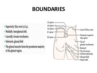
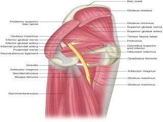
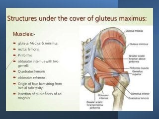
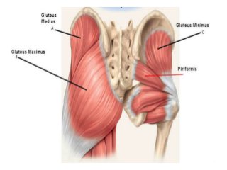
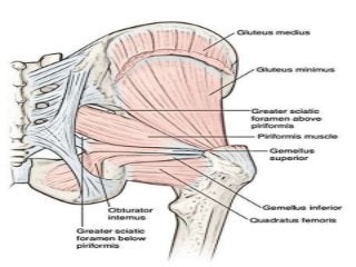
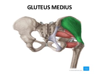
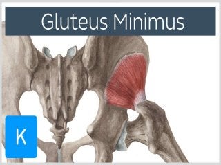
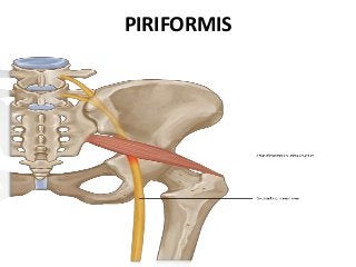
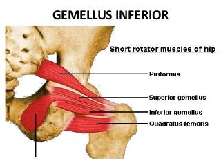
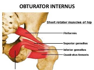
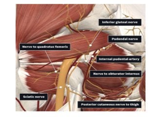
GLUTEAL REGION
DR. SUNDIP CHARMODE
ASSOCIATE PROFESSOR
DEPARTMENT OF ANATOMY
AIIMS RAJKOT
BOUNDARIES
BONY
FRAMEWORK
• Dorsal surface of Sacrum and
Coccyx
• Gluteal surface of Ilium, dorsal
surface of Ischium with ischial
tuberosity, ischial spine, greater
and lesser sciatic notches.
• Posterior surface of upper part of
femur – neck, greater and lesser
trochanter & gluteal tuberosity.
LIGAMENTS - BONY FRAMEWORK
1. Sacro-tuberous ligament
2. Sacro-spinous ligament
3. Dorsal Sacro-iliac ligament
4. Posterior part of the capsular ligaments of hip
joint
SURFACE/BONY LANDMARKS
1. Highest point of iliac crest: corresponds to 4th
Lumbar vertebra
2. Posterior Superior Iliac Spine: Skin dimple. The line
joining PSIS passes through 2nd sacral spine.
3. Tip of Greater trochanter of Femur
4. Ischial tuberosity
5. Iliac crest
6. Anterior Superior Iliac Spine
7. Sacrum and Coccyx
SURFACE/BONY LANDMARKS
7. Sacro-tuberous ligaments
8. Buttock
9. Natal cleft
10. Gluteal fold.
SUPERFICIAL FASCIA
• Thick and fatty, and the subcutaneous fat is abundant
in adult female.
• Contains cutaneous nerves, vessels and lymphatics.
• Cutaneous nerve supply :
– Upper anterior part
– Lower anterior part
– Upper posterior part
– Lower posterior part
SUPERFICIAL FASCIA
 Cutaneous blood supply :
 Lymphatics
DEEP FASCIA
• Attached above to the iliac crest, and behind to the
sacrum.
• The fascia splits twice along the iliac crest, to
enclose the tensor fascia latae and gluteus maximus
and forms a thickened sheet between them, the
Gluteal Aponeurosis, which covers the gluteus
medius.
DEEP FASCIA
• The layers which enclose the Gluteus maximus are
connected to each other by numerous fibrous septa
which pass through the fasciculi of the muscle.
DEEP FASCIA
• Fascia over Gluteus
Medius is thick,
opaque and white.
• Fascia over the Gluteus
Maximus muscle is
thin and transparent.
• Fascia splits and
encloses GM muscle.
CONTENTS / FEATURES
• Gluteus Maximus
• Structures under the cover of Gluteus
Maximus
STRUCTURES UNDER THE COVER OF
GLUTEUS MAXIMUS
1. Gluteus Medius
2. Gluteus Minimus
3. Reflected head of Rectus Femoris
4. Piriformis
5. Obturator Internus with two gemelli
6. Quadratus Femoris
7. Obturator Externus
8. Origin of four Hamstrings from the Ischial tuberosity
9. Insertion of upper/ pubic fibres of Adductor
Magnus.
GLUTEUS MAXIMUS
 Origin :
• Outer slope of Dorsal segment of iliac crest
• Posterior gluteal line
• Gluteal surface of ileum behind posterior
gluteal line
• Erector Spinae aponeurosis
• Dorsal surface of lower part of Sacrum
• Side of Coccyx
• Sacro-tuberous ligament
• Fascia over the Gluteus Medius
 Insertion :
 Greater part ¾ : Ilio-tibial tract
 Deep fibres ¼ : Gluteal tuberosity
 Nerve supply : Inferior Gluteal nerve L5,S1,S2
 Action :
1. Chief extensor of thigh at hip joint
2. Lateral rotator of thigh
3. Abduction of thigh ( upper fibres )
4. Stabilizes knee through Ilio-tibial tract.
GLUTEUS MEDIUS
• Origin : Gluteal surface of Ileum between
anterior and posterior gluteal lines.
• Insertion : Into Greater Trochanter of femur
on oblique ridge on lateral surface
• Nerve supply : Superior Gluteal nerve L4,L5,S1
• Action :
1. Powerful abductor of thigh
2. Medial Rotator (anterior fibres)
GLUTEUS MINIMUS
 Origin :
• Gluteal surface of Ileum between anterior and
Inferior gluteal lines.
 Insertion :
• Into Greater Trochanter of femur on a ridge on
anterior surface
 Nerve supply : Superior Gluteal nerve
L4,L5,S1
 Action :
• Powerful abductor of thigh
• Medial Rotator (anterior fibres)
PIRIFORMIS
PIRIFORMIS
 Origin :
• Pelvic surface of the middle three pieces of
sacrum by three digitations.
• Upper margin of Greater Sciatic notch
 Insertion :
• Into Apex of Greater Trochanter of femur.
 Nerve supply : Ventral rami of S1,S2.
 Action : Lateral rotator of thigh at Hip joint.
GEMELLUS SUPERIOR
 Origin :
• Upper part of Lesser sciatic notch
 Insertion :
• Blends with tendon of Obturator Internus &
inserts in medial surface of Greater Trochanter
of femur.
 Nerve supply : Nerve to Obturator Internus
L5, S1,S2
 Action : Lateral rotator of thigh at Hip joint.
GEMELLUS INFERIOR
 Origin :
• Lower part of Lesser sciatic notch
 Insertion :
• Blends with tendon of Obturator Internus &
inserts in medial surface of Greater Trochanter
of femur.
 Nerve supply : Nerve to Quadratus Femoris
L4, L 5, S1.
 Action : Lateral rotator of thigh at Hip joint.
OBTURATOR INTERNUS
 Origin :
• Pelvic surface of Obturator Membrane
• Pelvic surface of body of ischium, ischial
tuberosity, ischiopubic rami.
• Obturator fascia.
• Insertion :
• Forms a tendon passes through Lesser Sciatic
foramen, bends at right angle around Lesser
Sciatic notch & runs laterally to insert on medial
surface of Gr. Trochanter.
• Nerve supply : Nerve to Obturator Internus L5,
S1,S2
 Action : Lateral rotator of thigh at Hip joint.
QUADRATUS FEMORIS
 Origin :
• Upper part of outer border of Ischial
Tuberosity.
 Insertion :
• Quadrate tubercle and the area below it.
 Nerve supply : Nerve to Quadratus Femoris
L4, L 5, S1.
 Action : Lateral rotator of thigh at Hip joint.
OBTURATOR EXTERNUS
OBTURATOR EXTERNUS
 Origin :
• Outer surface of Obturator Membrane
• Outer surface of bony margins of Obturator
Foramen
• Insertion :
• Forms a tendon runs up & laterally behind neck
of femur to reach gluteal region to insert in
trochanteric fossa on medial surface of Gr.
Trochanter.
• Nerve supply : Posterior division of obturator
nerve L2, L 3, L 4.
 Action : Lateral rotator of thigh at Hip joint.
TENSOR FASCIA LATA
 Origin :
• Anterior 5 cm of Outer Lip Of Iliac Crest up to
the tubercle of iliac crest.
 Insertion : Ilio-tibial tract 3-5 cm below the
level of greater trochanter.
 Nerve supply : Superior Gluteal nerve L4, L 5,
S1.
 Action :
• Abductor and Medial Rotator of thigh
• Extensor of Knee joint.
VESSELS UNDER THE COVER OF
GLUTEUS MAXIMUS
1. Superior gluteal vessels
2. Inferior gluteal vessels
3. Internal pudendal vessels
4. Ascending branch of Medial Circumflex
femoral artery
5. Trochanteric anastomoses
6. Cruciate anastomoses
7. First perforating artery
NERVES UNDER THE COVER OF
GLUTEUS MAXIMUS
1. Superior gluteal nerve L4, L5, S1.
2. Inferior gluteal nerve L5,S1,S2.
3. Sciatic nerve L 4,5,S 1,2,3.
4. Posterior cutaneous nerve of thigh S 1,2,3.
5. Nerve to Quadratus Femoris L 4, L 5, S 1.
6. Pudendal nerve S 2,3,4.
7. Nerve to Obturator Internus L 5,S 1,S 2.
8. Perforating cutaneous nerves. S 2,3.
BONES / JOINTS UNDER THE COVER
OF GLUTEUS MAXIMUS
1. Ilium
2. Ischium with ischial tuberosity
3. Upper end of femur with the greater
trochanter
4. Sacrum and coccyx
5. Hip joint
6. Sacroiliac joint
LIGAMENTS / BURSAE UNDER THE
COVER OF GLUTEUS MAXIMUS
1. Sacrotuberous
2. Sacrospinous
3. Ischiofemoral
 Bursae :
• Trochanteric bursae of gluteus maximus
• Bursa over ischial tuberosity
• Bursa between gluteus maximus and vastus
lateralis.
STRUCTURES UNDER THE COVER OF
GLUTEUS MEDIUS
1. Superior gluteal nerve
2. Deep branch of Superior gluteal artery
3. Gluteus minimus
4. Trochanteric bursa of Gluteus medius.
STRUCTURES UNDER THE COVER OF
GLUTEUS MINIMUS
1. Reflected head of Rectus Femoris
2. Capsule of Hip joint
LIGAMENTS
1. Sacro-tuberous ligament – long and strong.
2. Saro-spinous ligament – short, thick ,
triangular band deep to Sacro-tuberous
ligament.
STRUCTURES PASSING THROUGH
GREATER SCIATIC FORAMEN
 Structures passing above Piriformis :
1. Superior Gluteal vessels
2. Superior Gluteal nerve
STRUCTURES PASSING THROUGH
GREATER SCIATIC FORAMEN
 Structures passing below Piriformis :
1. Inferior Gluteal nerve
2. Inferior Gluteal vessels
3. Sciatic nerve
4. Posterior cutaneous nerve of thigh
5. Nerve to Quadratus Femoris
6. Pudendal nerve
7. Internal Pudendal vessels
8. Nerve to Obturator Internus
STRUCTURES PASSING THROUGH
LESSER SCIATIC FORAMEN
1. Pudendal nerve
2. Internal Pudendal vessels
3. Tendon of Obturator Internus
4. Nerve to Obturator Internus
NERVES OF GLUTEAL REGION
1. Superior Gluteal nerve
2. Inferior Gluteal nerve
3. Sciatic nerve
4. Posterior cutaneous nerve of thigh
5. Nerve to Quadratus Femoris
6. Pudendal nerve
7. Nerve to Obturator Internus
8. Perforating cutaneous nerve
SUPERIOR
GLUTEAL
NERVE
• Formed by dorsal branches
of ventral rami of L4, L5, S1
• Curves upward and forward
between gluteus medius
and gluteus minimus.
• Supplies G. Medius, G.
Minimus and Tensor fascia
latae.
• Provides an articular branch
to hip joint and gives no
cutaneous branch.
INFERIOR GLUTEAL
NERVE
• Formed by dorsal branches of
ventral rami of L5, S1, S2.
• Curves upward to supply Gluteus
maximus.
• This nerve gives no cutaneous
branch.
POSTERIOR
CUTANEOUS NERVE
• Formed by fibers from dorsal
branches of S1,S2 and ventral
branches of S2 and S3.
• It passes superficial or medial to
sciatic nerve.
• It gives 3-4 gluteal branches which
curl around the lower border of G.
Maximus and supplies the skin
over lower and lateral part of
gluteal region.
SCIATIC NERVE
• Widest nerve of the body
• 2 cm broad and have
tibial and common
peroneal components
• Tibial component:
ventral branches of
ventral; rami of L4, L5, S1,
S2, S3.
• Common peroneal
component: dorsal
branches of the ventral
rami of L4,L5, S1, S2
PUDENDAL
NERVE
• Formed by ventral
branches of ventral rami
of S2, S3, S4
• Crosses just medial to
ischial spine around
sacro-spinous ligament
and winds forward
through lesser sciatic
foramen to enter
pudendal canal and
supply perineum.
NERVE TO OBTURATOR INTERNUS
• Formed by ventral branches of ventral rami of
L5 ,S1, S2.
• Rests on the dorsal surface of base of ischial
spine and pass forward through lesser sciatic
foramen.
• Supplies Obturator internus and gemellus
superior.
NERVE TO QUADRATUS FEMORIS
• Formed by ventral
branches of ventral rami
of L4, L5 ,S1.
• It rests on ischium,
descends deep to tendon
of OBT. Internus and two
gemelli.
• Supplies gemellus
inferior, Quadratus
femoris and articular
branch to hip joint.
VESSELS OF GLUTEAL REGION
1. Superior Gluteal vessels
2. Inferior Gluteal vessels
3. Internal Pudendal vessels
4. Trochanteric anastomoses
5. Cruciate anastomoses
SUPERIOR GLUTEAL VESSELS
• Branch of posterior division of Internal iliac
artery, appears above the piriformis along with
superior gluteal nerve.
• Superficial branch: pass between G. Max. and G.
Med. And supplies both.
• Deep branch: pass laterally between G. Med. And
G. Min. and subdivides into Upper and lower
branches.
SUPERIOR GLUTEAL VESSELS
• Upper branch: forms a spinous
anastomoses over to ASIS along with:
– Superficial circumflex iliac artery
– Deep circumflex iliac artery
– Ascending branch of lateral circumflex femoral
artery
– Iliac branch of ilio-lumbar artery
• Lower branch: joins trochanteric
anastomoses
INFERIOR GLUTEAL VESSELS
• Branch of anterior division of Internal iliac
artery, appears below the piriformis.
• Gives three sets of arteries:
– Muscular branches to adj. muscles
– Anastomotic branches
– Arteria Nervi Ischiadici which accompanies and
supplies sciatic nerve
INTERNAL PUDENDAL VESSELS
• Branch of anterior division of Internal iliac
artery, appears below piriformis.
• It crosses the tip of ischial spine accompanied
by a pair of venae comitantes on each side.
• Related laterally to nerve to obturator
internus and medially to pudendal nerve.
• Pass through lesser sciatic notch, then
pudendal canal along with pudendal nerve
and supply structures of perineum.
TROCHANTERIC ANASTOMOSES
• Located near the trochanteric fossa of femur
1. Descending branch of superior gluteal artery
2. Inferior gluteal artery
3. Ascending branch of Lateral circumflex
femoral artery
4. Ascending branch of Medial circumflex
femoral artery
SUPPLY
Branches from this ring,
the retinacular vessels,
pierce the capsule and
ascend along the femoral
neck to give the main
blood supply to the head
of the femur.
CRUCIATE ANASTOMOSES
• Lies at the level of the
lesser trochanter, near the
lower edge of the femoral
attachment of quadratus
femoris.
• This establishes a
collateral circulation
between the branches of
the Internal iliac artery
and profunda femoris
artery.
CRUCIATE ANASTOMOSES
• Above: Anastomotic branch of Inferior gluteal
artery
• Below: Ascending branch of first perforating
artery, a branch of Profunda femoris artery
• Medially: Transverse branch of Medial
circumflex femoral artery
• Laterally: Transverse branch of Lateral
circumflex femoral artery
CLINICAL CORRELATES
1. Testing Gluteus Maximus muscle
2. In paralyses, there is difficulty to ?
3. Intramuscular injections
4. Mechanism of normal gait
5. Lurching gait
6. Waddling gait
7. Trendelenburg test
8. Piriformis syndrome
9. Sciatica
CLINICAL CORRELATES
10.To control hemorrhage in the perineum,
internal pudendal artery may be compressed
against the ischial spine.
THANK YOU

Gluteal region.pptx

  • 1.
    GLUTEAL REGION DR. SUNDIPCHARMODE ASSOCIATE PROFESSOR DEPARTMENT OF ANATOMY AIIMS RAJKOT
  • 3.
  • 4.
    BONY FRAMEWORK • Dorsal surfaceof Sacrum and Coccyx • Gluteal surface of Ilium, dorsal surface of Ischium with ischial tuberosity, ischial spine, greater and lesser sciatic notches. • Posterior surface of upper part of femur – neck, greater and lesser trochanter & gluteal tuberosity.
  • 7.
    LIGAMENTS - BONYFRAMEWORK 1. Sacro-tuberous ligament 2. Sacro-spinous ligament 3. Dorsal Sacro-iliac ligament 4. Posterior part of the capsular ligaments of hip joint
  • 9.
    SURFACE/BONY LANDMARKS 1. Highestpoint of iliac crest: corresponds to 4th Lumbar vertebra 2. Posterior Superior Iliac Spine: Skin dimple. The line joining PSIS passes through 2nd sacral spine. 3. Tip of Greater trochanter of Femur 4. Ischial tuberosity 5. Iliac crest 6. Anterior Superior Iliac Spine 7. Sacrum and Coccyx
  • 10.
    SURFACE/BONY LANDMARKS 7. Sacro-tuberousligaments 8. Buttock 9. Natal cleft 10. Gluteal fold.
  • 11.
    SUPERFICIAL FASCIA • Thickand fatty, and the subcutaneous fat is abundant in adult female. • Contains cutaneous nerves, vessels and lymphatics. • Cutaneous nerve supply : – Upper anterior part – Lower anterior part – Upper posterior part – Lower posterior part
  • 14.
    SUPERFICIAL FASCIA  Cutaneousblood supply :  Lymphatics
  • 15.
    DEEP FASCIA • Attachedabove to the iliac crest, and behind to the sacrum. • The fascia splits twice along the iliac crest, to enclose the tensor fascia latae and gluteus maximus and forms a thickened sheet between them, the Gluteal Aponeurosis, which covers the gluteus medius.
  • 17.
    DEEP FASCIA • Thelayers which enclose the Gluteus maximus are connected to each other by numerous fibrous septa which pass through the fasciculi of the muscle.
  • 18.
    DEEP FASCIA • Fasciaover Gluteus Medius is thick, opaque and white. • Fascia over the Gluteus Maximus muscle is thin and transparent. • Fascia splits and encloses GM muscle.
  • 20.
    CONTENTS / FEATURES •Gluteus Maximus • Structures under the cover of Gluteus Maximus
  • 21.
    STRUCTURES UNDER THECOVER OF GLUTEUS MAXIMUS 1. Gluteus Medius 2. Gluteus Minimus 3. Reflected head of Rectus Femoris 4. Piriformis 5. Obturator Internus with two gemelli 6. Quadratus Femoris 7. Obturator Externus 8. Origin of four Hamstrings from the Ischial tuberosity 9. Insertion of upper/ pubic fibres of Adductor Magnus.
  • 26.
  • 28.
     Origin : •Outer slope of Dorsal segment of iliac crest • Posterior gluteal line • Gluteal surface of ileum behind posterior gluteal line • Erector Spinae aponeurosis • Dorsal surface of lower part of Sacrum • Side of Coccyx • Sacro-tuberous ligament • Fascia over the Gluteus Medius
  • 29.
     Insertion : Greater part ¾ : Ilio-tibial tract  Deep fibres ¼ : Gluteal tuberosity  Nerve supply : Inferior Gluteal nerve L5,S1,S2  Action : 1. Chief extensor of thigh at hip joint 2. Lateral rotator of thigh 3. Abduction of thigh ( upper fibres ) 4. Stabilizes knee through Ilio-tibial tract.
  • 30.
  • 32.
    • Origin :Gluteal surface of Ileum between anterior and posterior gluteal lines. • Insertion : Into Greater Trochanter of femur on oblique ridge on lateral surface • Nerve supply : Superior Gluteal nerve L4,L5,S1 • Action : 1. Powerful abductor of thigh 2. Medial Rotator (anterior fibres)
  • 33.
  • 35.
     Origin : •Gluteal surface of Ileum between anterior and Inferior gluteal lines.  Insertion : • Into Greater Trochanter of femur on a ridge on anterior surface  Nerve supply : Superior Gluteal nerve L4,L5,S1  Action : • Powerful abductor of thigh • Medial Rotator (anterior fibres)
  • 36.
  • 37.
  • 38.
     Origin : •Pelvic surface of the middle three pieces of sacrum by three digitations. • Upper margin of Greater Sciatic notch  Insertion : • Into Apex of Greater Trochanter of femur.  Nerve supply : Ventral rami of S1,S2.  Action : Lateral rotator of thigh at Hip joint.
  • 39.
  • 40.
     Origin : •Upper part of Lesser sciatic notch  Insertion : • Blends with tendon of Obturator Internus & inserts in medial surface of Greater Trochanter of femur.  Nerve supply : Nerve to Obturator Internus L5, S1,S2  Action : Lateral rotator of thigh at Hip joint.
  • 41.
  • 42.
     Origin : •Lower part of Lesser sciatic notch  Insertion : • Blends with tendon of Obturator Internus & inserts in medial surface of Greater Trochanter of femur.  Nerve supply : Nerve to Quadratus Femoris L4, L 5, S1.  Action : Lateral rotator of thigh at Hip joint.
  • 43.
  • 44.
     Origin : •Pelvic surface of Obturator Membrane • Pelvic surface of body of ischium, ischial tuberosity, ischiopubic rami. • Obturator fascia. • Insertion : • Forms a tendon passes through Lesser Sciatic foramen, bends at right angle around Lesser Sciatic notch & runs laterally to insert on medial surface of Gr. Trochanter. • Nerve supply : Nerve to Obturator Internus L5, S1,S2  Action : Lateral rotator of thigh at Hip joint.
  • 45.
  • 46.
     Origin : •Upper part of outer border of Ischial Tuberosity.  Insertion : • Quadrate tubercle and the area below it.  Nerve supply : Nerve to Quadratus Femoris L4, L 5, S1.  Action : Lateral rotator of thigh at Hip joint.
  • 47.
  • 48.
  • 49.
     Origin : •Outer surface of Obturator Membrane • Outer surface of bony margins of Obturator Foramen • Insertion : • Forms a tendon runs up & laterally behind neck of femur to reach gluteal region to insert in trochanteric fossa on medial surface of Gr. Trochanter. • Nerve supply : Posterior division of obturator nerve L2, L 3, L 4.  Action : Lateral rotator of thigh at Hip joint.
  • 50.
  • 52.
     Origin : •Anterior 5 cm of Outer Lip Of Iliac Crest up to the tubercle of iliac crest.  Insertion : Ilio-tibial tract 3-5 cm below the level of greater trochanter.  Nerve supply : Superior Gluteal nerve L4, L 5, S1.  Action : • Abductor and Medial Rotator of thigh • Extensor of Knee joint.
  • 54.
    VESSELS UNDER THECOVER OF GLUTEUS MAXIMUS 1. Superior gluteal vessels 2. Inferior gluteal vessels 3. Internal pudendal vessels 4. Ascending branch of Medial Circumflex femoral artery 5. Trochanteric anastomoses 6. Cruciate anastomoses 7. First perforating artery
  • 56.
    NERVES UNDER THECOVER OF GLUTEUS MAXIMUS 1. Superior gluteal nerve L4, L5, S1. 2. Inferior gluteal nerve L5,S1,S2. 3. Sciatic nerve L 4,5,S 1,2,3. 4. Posterior cutaneous nerve of thigh S 1,2,3. 5. Nerve to Quadratus Femoris L 4, L 5, S 1. 6. Pudendal nerve S 2,3,4. 7. Nerve to Obturator Internus L 5,S 1,S 2. 8. Perforating cutaneous nerves. S 2,3.
  • 57.
    BONES / JOINTSUNDER THE COVER OF GLUTEUS MAXIMUS 1. Ilium 2. Ischium with ischial tuberosity 3. Upper end of femur with the greater trochanter 4. Sacrum and coccyx 5. Hip joint 6. Sacroiliac joint
  • 58.
    LIGAMENTS / BURSAEUNDER THE COVER OF GLUTEUS MAXIMUS 1. Sacrotuberous 2. Sacrospinous 3. Ischiofemoral  Bursae : • Trochanteric bursae of gluteus maximus • Bursa over ischial tuberosity • Bursa between gluteus maximus and vastus lateralis.
  • 59.
    STRUCTURES UNDER THECOVER OF GLUTEUS MEDIUS 1. Superior gluteal nerve 2. Deep branch of Superior gluteal artery 3. Gluteus minimus 4. Trochanteric bursa of Gluteus medius.
  • 60.
    STRUCTURES UNDER THECOVER OF GLUTEUS MINIMUS 1. Reflected head of Rectus Femoris 2. Capsule of Hip joint
  • 61.
    LIGAMENTS 1. Sacro-tuberous ligament– long and strong. 2. Saro-spinous ligament – short, thick , triangular band deep to Sacro-tuberous ligament.
  • 64.
    STRUCTURES PASSING THROUGH GREATERSCIATIC FORAMEN  Structures passing above Piriformis : 1. Superior Gluteal vessels 2. Superior Gluteal nerve
  • 65.
    STRUCTURES PASSING THROUGH GREATERSCIATIC FORAMEN  Structures passing below Piriformis : 1. Inferior Gluteal nerve 2. Inferior Gluteal vessels 3. Sciatic nerve 4. Posterior cutaneous nerve of thigh 5. Nerve to Quadratus Femoris 6. Pudendal nerve 7. Internal Pudendal vessels 8. Nerve to Obturator Internus
  • 66.
    STRUCTURES PASSING THROUGH LESSERSCIATIC FORAMEN 1. Pudendal nerve 2. Internal Pudendal vessels 3. Tendon of Obturator Internus 4. Nerve to Obturator Internus
  • 67.
    NERVES OF GLUTEALREGION 1. Superior Gluteal nerve 2. Inferior Gluteal nerve 3. Sciatic nerve 4. Posterior cutaneous nerve of thigh 5. Nerve to Quadratus Femoris 6. Pudendal nerve 7. Nerve to Obturator Internus 8. Perforating cutaneous nerve
  • 68.
    SUPERIOR GLUTEAL NERVE • Formed bydorsal branches of ventral rami of L4, L5, S1 • Curves upward and forward between gluteus medius and gluteus minimus. • Supplies G. Medius, G. Minimus and Tensor fascia latae. • Provides an articular branch to hip joint and gives no cutaneous branch.
  • 69.
    INFERIOR GLUTEAL NERVE • Formedby dorsal branches of ventral rami of L5, S1, S2. • Curves upward to supply Gluteus maximus. • This nerve gives no cutaneous branch.
  • 70.
    POSTERIOR CUTANEOUS NERVE • Formedby fibers from dorsal branches of S1,S2 and ventral branches of S2 and S3. • It passes superficial or medial to sciatic nerve. • It gives 3-4 gluteal branches which curl around the lower border of G. Maximus and supplies the skin over lower and lateral part of gluteal region.
  • 71.
    SCIATIC NERVE • Widestnerve of the body • 2 cm broad and have tibial and common peroneal components • Tibial component: ventral branches of ventral; rami of L4, L5, S1, S2, S3. • Common peroneal component: dorsal branches of the ventral rami of L4,L5, S1, S2
  • 72.
    PUDENDAL NERVE • Formed byventral branches of ventral rami of S2, S3, S4 • Crosses just medial to ischial spine around sacro-spinous ligament and winds forward through lesser sciatic foramen to enter pudendal canal and supply perineum.
  • 73.
    NERVE TO OBTURATORINTERNUS • Formed by ventral branches of ventral rami of L5 ,S1, S2. • Rests on the dorsal surface of base of ischial spine and pass forward through lesser sciatic foramen. • Supplies Obturator internus and gemellus superior.
  • 75.
    NERVE TO QUADRATUSFEMORIS • Formed by ventral branches of ventral rami of L4, L5 ,S1. • It rests on ischium, descends deep to tendon of OBT. Internus and two gemelli. • Supplies gemellus inferior, Quadratus femoris and articular branch to hip joint.
  • 76.
    VESSELS OF GLUTEALREGION 1. Superior Gluteal vessels 2. Inferior Gluteal vessels 3. Internal Pudendal vessels 4. Trochanteric anastomoses 5. Cruciate anastomoses
  • 77.
    SUPERIOR GLUTEAL VESSELS •Branch of posterior division of Internal iliac artery, appears above the piriformis along with superior gluteal nerve. • Superficial branch: pass between G. Max. and G. Med. And supplies both. • Deep branch: pass laterally between G. Med. And G. Min. and subdivides into Upper and lower branches.
  • 78.
    SUPERIOR GLUTEAL VESSELS •Upper branch: forms a spinous anastomoses over to ASIS along with: – Superficial circumflex iliac artery – Deep circumflex iliac artery – Ascending branch of lateral circumflex femoral artery – Iliac branch of ilio-lumbar artery • Lower branch: joins trochanteric anastomoses
  • 80.
    INFERIOR GLUTEAL VESSELS •Branch of anterior division of Internal iliac artery, appears below the piriformis. • Gives three sets of arteries: – Muscular branches to adj. muscles – Anastomotic branches – Arteria Nervi Ischiadici which accompanies and supplies sciatic nerve
  • 81.
    INTERNAL PUDENDAL VESSELS •Branch of anterior division of Internal iliac artery, appears below piriformis. • It crosses the tip of ischial spine accompanied by a pair of venae comitantes on each side. • Related laterally to nerve to obturator internus and medially to pudendal nerve. • Pass through lesser sciatic notch, then pudendal canal along with pudendal nerve and supply structures of perineum.
  • 82.
    TROCHANTERIC ANASTOMOSES • Locatednear the trochanteric fossa of femur 1. Descending branch of superior gluteal artery 2. Inferior gluteal artery 3. Ascending branch of Lateral circumflex femoral artery 4. Ascending branch of Medial circumflex femoral artery
  • 84.
    SUPPLY Branches from thisring, the retinacular vessels, pierce the capsule and ascend along the femoral neck to give the main blood supply to the head of the femur.
  • 85.
    CRUCIATE ANASTOMOSES • Liesat the level of the lesser trochanter, near the lower edge of the femoral attachment of quadratus femoris. • This establishes a collateral circulation between the branches of the Internal iliac artery and profunda femoris artery.
  • 86.
    CRUCIATE ANASTOMOSES • Above:Anastomotic branch of Inferior gluteal artery • Below: Ascending branch of first perforating artery, a branch of Profunda femoris artery • Medially: Transverse branch of Medial circumflex femoral artery • Laterally: Transverse branch of Lateral circumflex femoral artery
  • 88.
    CLINICAL CORRELATES 1. TestingGluteus Maximus muscle 2. In paralyses, there is difficulty to ? 3. Intramuscular injections 4. Mechanism of normal gait 5. Lurching gait 6. Waddling gait 7. Trendelenburg test 8. Piriformis syndrome 9. Sciatica
  • 89.
    CLINICAL CORRELATES 10.To controlhemorrhage in the perineum, internal pudendal artery may be compressed against the ischial spine.
  • 90.