Download as PDF, PPTX

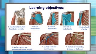
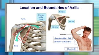
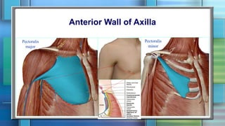
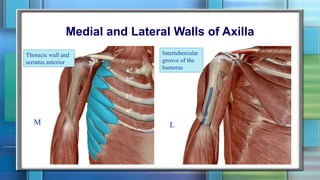

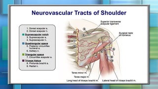
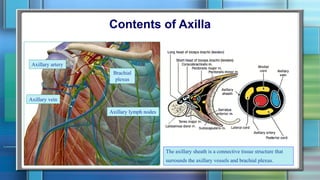
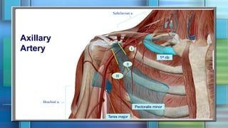
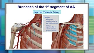
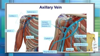
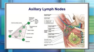

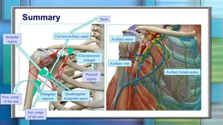


The document provides an overview of the anatomy of the axilla region. It discusses the boundaries and walls of the axilla, including the anterior, medial, posterior and lateral walls. It describes the main neurovascular structures in the axilla, including the axillary artery and its branches, the axillary vein and its tributaries, and the axillary lymph nodes. It also discusses the contents of the axilla, noting the axillary vessels, brachial plexus, and axillary lymph nodes are surrounded by the axillary sheath.


![Lecture 25 Intermuscular sapces and axilla [Autosaved].pptx](https://cdn.slidesharecdn.com/ss_thumbnails/lecture25intermuscularsapcesandaxillaautosaved-251110002658-47b36c78-thumbnail.jpg?width=640&height=640&fit=bounds)


