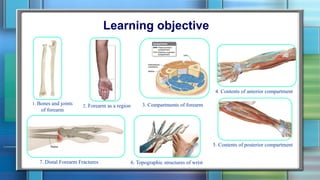
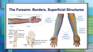
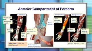
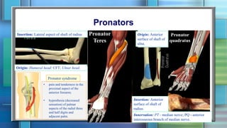
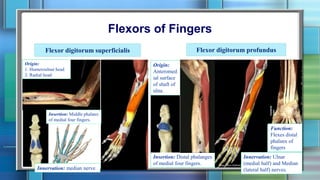
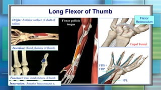
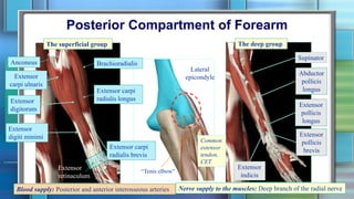
UL: Forearm
Lecture 5
Maryna Kornieieva, PhD, MD
Asst. of Clinical Anatomy
Learning objective
1. Bones and joints
of forearm
2. Forearm as a region 3. Compartments of forearm
4. Contents of anterior compartment
5. Contents of posterior compartment
6. Topographic structures of wrist7. Distal Forearm Fractures
Bony Framework of Forearm
Supination
Pronation
Axis of
rotation
Anular
ligament
The Forearm: Borders, Superficial Structures
Medial
cutaneous n.
of forearm
Lateral
cutaneous n.
of forearm
Lateral
cutaneous n.
of forearm
Posterior
cutaneous n.
of forearm
Elbow Wrist
Forearm Hand
Deep Fascia of Forearm
Attachment of deep
fascia to the ulna
Intermuscular
septae
Volkmann’s ischemic contractureCompartment
Syndrome of the
Forearm
Fasciotomy for forearm
compartment syndrome.
Supracondylar
fracture
When the wrist is flexed to a right angle it
is possible to extend the fingers.
Anterior Compartment of Forearm
Nerve s.: Median + UlnarBlood supply: Ulnar and
Radial arteries
Palmaris
longus
Flexor
carpi
ulnaris
Flexor carpi
radialis
Pronator teres
4 superficial 1 intermediate 3 deep muscles
Flexor
digitorum
superficialis
Flexor
digitorum
profundus
Flexor
policis
longus
Pronator
tquadratus
Medial
epicondyle
Common
flexor tendon,
CFT
“Golfer’s elbow”
(Medial epicondylitis)
Pronators
Origin: Humeral head: CFT; Ulnar head.
Insertion: Lateral aspect of shaft of radius Pronator
Teres
Pronator
quadratus
Origin: Anterior
surface of shaft of
ulna.
Innervation: PT - median nerve; PQ - anterior
interosseous branch of median nerve.
Insertion: Anterior
surface of shaft of
radius.
Pronator syndrome
• pain and tenderness in the
proximal aspect of the
anterior forearm;
• hypesthesia (decreased
sensation) of palmar
aspects of the radial three
and half digits and
adjacent palm.
Flexors of Fingers
Flexor digitorum profundus
Origin:
Anteromed
ial surface
of shaft of
ulna.
Insertion: Distal phalanges
of medial four fingers.
Innervation: Ulnar
(medial half) and Median
(lateral half) nerves.
Function:
Flexes distal
phalanx of
fingers
Flexor digitorum superficialis
Origin:
1. Humeroulnar head
2. Radial head
Insertion: Middle phalanx
of medial four fingers.
Innervation: median nerve
Long Flexor of Thumb
Insertion: Distal phalanx of thumb.
Function: Flexes distal phalanx of thumb
Origin: Anterior surface of shaft of
radius.
Flexor pollicis
longus
Carpal Tunnel
Flexor
Retinaculum
FPL
FDS +
FDP
Innervation: Anterior interosseous n.
Flexors of Wrist
Origin: CFO
Insertion: Bases of second
and third metacarpal bones
Flexor
carpi
radialis
Nerve s.: Ulnar nerve
Origin:
1) humeral head CFO
2) ulnar head
Insertion: Pisiform bone, hook of the
hamate, base at fifth metacarpal bone
Flexor
carpi
ulnaris
Palmaris
Longus
Insertion:
palmar
aponeurosis
“Cubital tunnel
syndrome”
Ulnar n.
Nerve s.: Median nerve
LRadial a.
L
L
Median n.
Ulnar
a +n
Radial and Ulnar aa.
Radial artery
5
4
6
7
Branches of ulnar a.:
4. Common interosseous artery;
5. Anterior and posterior ulnar
recurrent arteries;
6. Muscular arteries;
7. Dorsal carpal branch and
Palmar carpal branch.
Ulnar Artery
Common interosseous a.(4)
Anterior
i/o (9)
Wrist
AC
PC
4
8
9
Branches of radial a.:
1. Radial recurrent a.
2. Muscular aa.
3. Superficial palmar a.
Recurrent
i/o (10)
Anterior
ulnar
recurrent
(5)
Posterior
ulnar
recurrent
(5)
Posterior
i/o (8)
Superficial palmar archDeep palmar arch
Ulnar artery
1
2
3
Median Nerve in Forearm
Course:
• Enters the forearm passing
between two heads of the
pronator teres (★);
• Descends to the wrist
laying b/w flexors
digitorun superficialis and
profundus;
• Enters the hand via the
carpal tunel.
Carpal tunel
syndrome
Common places
of injury:
At the wrist
Upper
forearm
FDP
FDS
Supracondylar
fracture of the
humerus
At the
elbow
Pronator
teres
syndrome
+ Lunate
dislocation
• at the wrist
(superficial to flexor retinaculum)
• at the elbow
(posterior to medial epicondyle)
Common places
of injury:
Guyon's canal
syndrome
Cubital tunnel
syndrome
Course:
Ulnar Nerve
Flexor
Carpi
Ulnaris
Flexor
digitorum
profundus
Guyon’s canal
Palmar
cutaneous
branch
Dorsal
cutaneous
branch
Posterior Compartment of Forearm
Blood supply: Posterior and anterior interosseous arteries
The superficial group
Extensor
carpi ulnaris
Extensor
digitorum
Extensor
digiti minimi
Anconeus
Nerve supply to the muscles: Deep branch of the radial nerve
Supinator
Abductor
pollicis
longus
Extensor
pollicis
brevis
Extensor
pollicis
longus
Extensor
indicis
The deep group
Brachioradialis
Extensor carpi
radialis longus
Extensor carpi
radialis brevis
Lateral
epicondyle
Common
extensor
tendon,
CET
Extensor
retinaculum
“Tenis elbow”
Supinator of Forearm
Origin: Lateral
epicondyle of
humerus, anular
ligament of proximal
radioulnar joint, and
ulna.
Insertion: Neck and shaft of radius
Innervation: Deep branch of radial nerve – posterior interosseous nerve
Arcade of Frohse
(supinator arch)
“Radial tunnel syndrome” /
“Posterior interosseous nerve syndrome”/
“Supinator syndrome”
Sensory loss will not present because PIN is purely motor.
Radial Nerve in Forearm
Common places of injury:
1) Radial (spiral) groove of the humerus
2) Entering the posterior compartment of
forearm b/w 2 heads of supunator
Radial tunnel syndrome
2
1
Course
Deep branch
of radial n.
Brachioradialis
(faded)
Extensor Carpi
Radialis Longus
Superficial branch
of radial n.
Extensors of Fingers
Origin: Posterior surface of
shaft of ulna
Origin: CEO
Extensor
digitorum
Insertion: Extensor
expansion of 2nd -5th fingers
Extensor
digiti minimi
Extensor
indicis
Extensors of Wrist
Origin: Insertion:
Extensor carpi radialis longus (2)
Extensor carpi radialis brevis (3)
Extensor carpi ulnaris (1)
Brachioradialis
Insertion: Styloid
process of radius
Origin: Lateral
supracondylar
ridge
Action: Flexes
forearm at elbow
joint; rotates
forearm to the
midprone position.
1
2 3
Thumb Muscles
Extensor
Pollicis Brevis
Abductor
Pollicis Longus
Extensors
Pollicis
Origin: Posterior surface of shafts of
radius/ulna/interosseous membrane
Insertion:
Base of first
metacarpal bone
Insertion:
Base of
proximal
phalanx
Insertion:
Base of distal
phalanx
“Anatomical snuffbox”
the lateral border:
- abductor pollicis longus;
- extensor pollicis brevis;
the medial border
- extensor pollicis longus;
the floor
- scaphoid and trapezium.
Borders:
Palpable structures:
• Radial artery;
• Scaphoid and trapezium in
the floor;
• Radial styloid process
proximally;
• Base of the first metacarpal
distally.
Distal Forearm Fractures
Scaphoid fracture
Scaphoid fracture
in two weeks:
Summary
Posterior compartment Anterior compartment
Radial n:
• Brachioradialis
• Extensor carpi
radialis longus
Posterior interosseous n:
• all the rest muscles
Anterior interosseous n:
• all the deep muscles (exception:
medial half of FDP)
Median n:
• all muscles of the
superficial and
intermediate groups
Ulnar n:
1 and ½ –
flexor carpi
ulnaris +
medial half
of FDP
Thank you for your attention!!!

Forearm

  • 1.
    UL: Forearm Lecture 5 MarynaKornieieva, PhD, MD Asst. of Clinical Anatomy
  • 2.
    Learning objective 1. Bonesand joints of forearm 2. Forearm as a region 3. Compartments of forearm 4. Contents of anterior compartment 5. Contents of posterior compartment 6. Topographic structures of wrist7. Distal Forearm Fractures
  • 3.
    Bony Framework ofForearm Supination Pronation Axis of rotation Anular ligament
  • 4.
    The Forearm: Borders,Superficial Structures Medial cutaneous n. of forearm Lateral cutaneous n. of forearm Lateral cutaneous n. of forearm Posterior cutaneous n. of forearm Elbow Wrist Forearm Hand
  • 5.
    Deep Fascia ofForearm Attachment of deep fascia to the ulna Intermuscular septae Volkmann’s ischemic contractureCompartment Syndrome of the Forearm Fasciotomy for forearm compartment syndrome. Supracondylar fracture When the wrist is flexed to a right angle it is possible to extend the fingers.
  • 6.
    Anterior Compartment ofForearm Nerve s.: Median + UlnarBlood supply: Ulnar and Radial arteries Palmaris longus Flexor carpi ulnaris Flexor carpi radialis Pronator teres 4 superficial 1 intermediate 3 deep muscles Flexor digitorum superficialis Flexor digitorum profundus Flexor policis longus Pronator tquadratus Medial epicondyle Common flexor tendon, CFT “Golfer’s elbow” (Medial epicondylitis)
  • 7.
    Pronators Origin: Humeral head:CFT; Ulnar head. Insertion: Lateral aspect of shaft of radius Pronator Teres Pronator quadratus Origin: Anterior surface of shaft of ulna. Innervation: PT - median nerve; PQ - anterior interosseous branch of median nerve. Insertion: Anterior surface of shaft of radius. Pronator syndrome • pain and tenderness in the proximal aspect of the anterior forearm; • hypesthesia (decreased sensation) of palmar aspects of the radial three and half digits and adjacent palm.
  • 8.
    Flexors of Fingers Flexordigitorum profundus Origin: Anteromed ial surface of shaft of ulna. Insertion: Distal phalanges of medial four fingers. Innervation: Ulnar (medial half) and Median (lateral half) nerves. Function: Flexes distal phalanx of fingers Flexor digitorum superficialis Origin: 1. Humeroulnar head 2. Radial head Insertion: Middle phalanx of medial four fingers. Innervation: median nerve
  • 9.
    Long Flexor ofThumb Insertion: Distal phalanx of thumb. Function: Flexes distal phalanx of thumb Origin: Anterior surface of shaft of radius. Flexor pollicis longus Carpal Tunnel Flexor Retinaculum FPL FDS + FDP Innervation: Anterior interosseous n.
  • 10.
    Flexors of Wrist Origin:CFO Insertion: Bases of second and third metacarpal bones Flexor carpi radialis Nerve s.: Ulnar nerve Origin: 1) humeral head CFO 2) ulnar head Insertion: Pisiform bone, hook of the hamate, base at fifth metacarpal bone Flexor carpi ulnaris Palmaris Longus Insertion: palmar aponeurosis “Cubital tunnel syndrome” Ulnar n. Nerve s.: Median nerve LRadial a. L L Median n. Ulnar a +n
  • 11.
    Radial and Ulnaraa. Radial artery 5 4 6 7 Branches of ulnar a.: 4. Common interosseous artery; 5. Anterior and posterior ulnar recurrent arteries; 6. Muscular arteries; 7. Dorsal carpal branch and Palmar carpal branch. Ulnar Artery Common interosseous a.(4) Anterior i/o (9) Wrist AC PC 4 8 9 Branches of radial a.: 1. Radial recurrent a. 2. Muscular aa. 3. Superficial palmar a. Recurrent i/o (10) Anterior ulnar recurrent (5) Posterior ulnar recurrent (5) Posterior i/o (8) Superficial palmar archDeep palmar arch Ulnar artery 1 2 3
  • 12.
    Median Nerve inForearm Course: • Enters the forearm passing between two heads of the pronator teres (★); • Descends to the wrist laying b/w flexors digitorun superficialis and profundus; • Enters the hand via the carpal tunel. Carpal tunel syndrome Common places of injury: At the wrist Upper forearm FDP FDS Supracondylar fracture of the humerus At the elbow Pronator teres syndrome + Lunate dislocation
  • 13.
    • at thewrist (superficial to flexor retinaculum) • at the elbow (posterior to medial epicondyle) Common places of injury: Guyon's canal syndrome Cubital tunnel syndrome Course: Ulnar Nerve Flexor Carpi Ulnaris Flexor digitorum profundus Guyon’s canal Palmar cutaneous branch Dorsal cutaneous branch
  • 14.
    Posterior Compartment ofForearm Blood supply: Posterior and anterior interosseous arteries The superficial group Extensor carpi ulnaris Extensor digitorum Extensor digiti minimi Anconeus Nerve supply to the muscles: Deep branch of the radial nerve Supinator Abductor pollicis longus Extensor pollicis brevis Extensor pollicis longus Extensor indicis The deep group Brachioradialis Extensor carpi radialis longus Extensor carpi radialis brevis Lateral epicondyle Common extensor tendon, CET Extensor retinaculum “Tenis elbow”
  • 15.
    Supinator of Forearm Origin:Lateral epicondyle of humerus, anular ligament of proximal radioulnar joint, and ulna. Insertion: Neck and shaft of radius Innervation: Deep branch of radial nerve – posterior interosseous nerve Arcade of Frohse (supinator arch) “Radial tunnel syndrome” / “Posterior interosseous nerve syndrome”/ “Supinator syndrome” Sensory loss will not present because PIN is purely motor.
  • 16.
    Radial Nerve inForearm Common places of injury: 1) Radial (spiral) groove of the humerus 2) Entering the posterior compartment of forearm b/w 2 heads of supunator Radial tunnel syndrome 2 1 Course Deep branch of radial n. Brachioradialis (faded) Extensor Carpi Radialis Longus Superficial branch of radial n.
  • 17.
    Extensors of Fingers Origin:Posterior surface of shaft of ulna Origin: CEO Extensor digitorum Insertion: Extensor expansion of 2nd -5th fingers Extensor digiti minimi Extensor indicis
  • 18.
    Extensors of Wrist Origin:Insertion: Extensor carpi radialis longus (2) Extensor carpi radialis brevis (3) Extensor carpi ulnaris (1) Brachioradialis Insertion: Styloid process of radius Origin: Lateral supracondylar ridge Action: Flexes forearm at elbow joint; rotates forearm to the midprone position. 1 2 3
  • 19.
    Thumb Muscles Extensor Pollicis Brevis Abductor PollicisLongus Extensors Pollicis Origin: Posterior surface of shafts of radius/ulna/interosseous membrane Insertion: Base of first metacarpal bone Insertion: Base of proximal phalanx Insertion: Base of distal phalanx
  • 20.
    “Anatomical snuffbox” the lateralborder: - abductor pollicis longus; - extensor pollicis brevis; the medial border - extensor pollicis longus; the floor - scaphoid and trapezium. Borders: Palpable structures: • Radial artery; • Scaphoid and trapezium in the floor; • Radial styloid process proximally; • Base of the first metacarpal distally.
  • 21.
    Distal Forearm Fractures Scaphoidfracture Scaphoid fracture in two weeks:
  • 22.
    Summary Posterior compartment Anteriorcompartment Radial n: • Brachioradialis • Extensor carpi radialis longus Posterior interosseous n: • all the rest muscles Anterior interosseous n: • all the deep muscles (exception: medial half of FDP) Median n: • all muscles of the superficial and intermediate groups Ulnar n: 1 and ½ – flexor carpi ulnaris + medial half of FDP
  • 23.
    Thank you foryour attention!!!