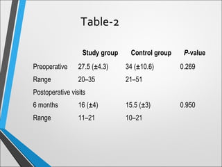
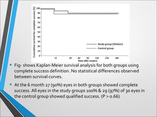
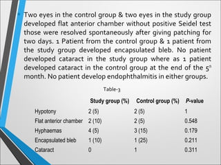
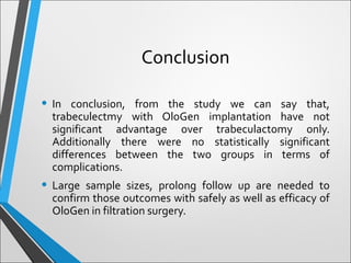
Good Morning
©FAi
• Horizon; Volume: 8, September , 2013
• Presented By:
Dr. Farhadul Alam
Chittagong Eye Infirmary and Training Complex
Filtration Surgery – Trabeculectomy
versus Trabeculectomy with OloGen
for the treatment of Glaucoma : a
Pilot Study
-By Dr. Shams Md Noman
• Trabeculectomy was introduced in 1968 and is now the most
common operation for the treatment of glaucoma worldwide
(Cairns 1968).
• However, wound healing and scar formation may result in
fibrosis of the bleb and obstruction of the drainage fistula,
eventually leading to bleb failure.
• Hence, the inhibition of scar formation during the wound-
healing process should promote greater success.
Introduction
• Fibroblast growth beneath the conjunctiva (between 3rd
-5th
post operative day) plays an important role in bleb failure.
• Adjunctive antimetabolites like 5-fluoro- uracil (5-FU) and
mitomycin-C may enhance the success rate by preventing
fibrosis.
• Antimetabolites increases the risk of post operative wound
leak, hypotony & endophthalmitis.
• Recent Studies in animal models show, the uses of
biodegredable collagen matrix implant beneath the
conjunctiva helps in controlling wound healing process &
maintain space for drainage without post operative
complications those are common with antimetablites use.
• The purpose of this study was to compare the outcomes of
trabeculectomy with OloGen (Collagen matrix) implant and
trabeculectomy without implant in patients requiring
glaucoma surgery for uncontrolled IOP.
• This is a prospective randomized pilot clinical trial that was
done in the glaucoma department of Chittagong Eye
Infirmary and Training Complex, Bangladesh.
• Randomly patients are divided into trabeculectomy (control
group) and trabeculectomy & collagen (study group)
• 6x1 mm biodegradable, porous collagen matrix
(atelocollagen plus glycosaminoglycans) were used for the
implant.
Materials and Methods
Fig. . OloGen implant: a drug-free, biodegradable, porous
collagen matrix of 1% collagen ⁄ C-6-S copolymer with a
diameter of
6 mm and a thickness of 1 mm.
• CEITC hospital review board approved the study following
Helsinki declaration. Informed consent was taken after detail
explanation about the implant and operation. Explanation
was done to Muslim people as OloGen is not Halal origin.
• 2 Patients refused due to religious binding. No patient lost for
follow up.
• Young age group (<18 yrs), neovasular glaucoma, history of
previous surgery or laser & absolute complicated glaucoma
were excluded form the study.
• Preoperative following data were collected age, gender,
diagnosis, level of intraocular pressure (day before surgery),
numbers of medications. All patients were under went single
future trabeculectomy.
• Post operative IOP, bleb condition & number of glaucoma
medications, were also recorded in each follow up. IOP
measurement was done with Goldmann applanation
tonometry.
• Success was defined with some criteria : (1) complete success
when IOP of 21mmHg or less without antiglaucoma
mediations & (2) qualified success when IOP of 21 mmHg with
the use of antiglaucoma medication.
• Hyptony was defined as an IOP <6mmHg.
• Flat anterior chamber can be defined as peripheral
iridocorneal touch with central at least 1 corneal thickness
depth.
• All Surgeries were done by same surgeon (Dr. Shams Md
Noman, Glaucoma Consultant, CEITC).
• After peribulbar anesthesia & proper drapping, fornix based
incision was given around 12’O Clock. Triangular, superficial
scleral flap (4x4mm) was produced facing apex towards 12 O’
clock position. 2x1 mm deep sclerectomy & a peripheral
iridectomy was performed thereafter.
• Scleral flap was closed with a single suture (10/0 nylon).
Surgical Technique
• Ologen was placed over the apex of the triangular flap
beneath the conjunctiva after making the operation area dry.
• Conjunctiva was closed like a wing with 2 sutures by 10/O
nylon.
• Post operatively all patients were treated with Atropine 1%, 3
times daily for 2 weeks, Moxifloxacin 4 times daily for 1 week
& Prednisolne acetate eye drop 6 times daily for 3 weeks then
tapered gradually.
• 6O eyes were enrolled in the study & randomly divided into
two groups of Trabeculectomy with or without OloGen
implant.
• Table-1 describes demographic & diagnostic data before
operation. There were no significant differences between the
groups in terms of age, gender, eye laterality, diagnosis, pre
operative IOP and number of topical & systemic anti
glaucoma medications.
• 8 patients from the study group and 9 patients from control
group needed systemic carbonic anhydrase inhibitors.
• Operations were uncomplicated in both groups.
Results
  Study group Control group P-value
No. of eyes 30 30
Age (years)
Mean (±SD) 61.3 (±18.5) 70.9 (±12.9) 0.188
Range 20–80 32–86
Median 65 74.5
Gender
Male 17 (55%) 18 (60%)
Female 13 (45%) 12 (40%) 0.759
Eye laterality
Right 12 (40%) 17 (55%)
Left 18 (60%) 13 (45%) 0.17
Diagnosis
POAG 15 (50%) 15 (50%)
PXG 6 (20%) 6 (20%)
PACG 3 (10%) 6 (20%)
IG 6 (20%) 3 (10%) 0.838
Preoperative IOP (mmHg)
Mean (±SD) 27.5 (±4.3) 34 (±10.6) 0.289
Range 20–35 21–51
No. of preoperative medications
Mean (±SD) 3 (±0.5) 3.5 (±0.7) 0.613
Range 2–4 1–4
Time of preoperative medications (months)
Mean (±SD) 45.15 (±37.34) 43.35 (±35.96) 0.180
Table-1
** SD, standard deviation; IOP, intraocular pressure; POAG, primary open-angle glaucoma; PXG, pseudoexfoliative glaucoma; PACG, primary angle closure
glaucoma; IG, inflammatory glaucoma.
• Mean IOPs for both groups are listed in Table-2. No difference
is observed in IOP measurement 6 month after operation
between two groups. Post operative IOP levels in both groups
is significantly lower than preoperative one. (P<0.05) None of
the eyes in the study group developed high IOP>21mmHg
post operatively where 2 eyes of control group developed so
at the six month visit.
• Post operatively in control group mean number of
antiglaucoma medications was dropped from (3.5 ± 0.7) to
(0.4 ± 0.1) (P < 0.001). Where in the study group from (3.5 ±
0.5) to (0.2 ± 0.5) (P < 0.001). There is no significant difference
in their reduction between & groups.
Study group Control group P-value
Preoperative 27.5 (±4.3) 34 (±10.6) 0.269
Range 20–35 21–51
Postoperative visits
6 months 16 (±4) 15.5 (±3) 0.950
Range 11–21 10–21
Table-2
• Fig- shows Kaplan-Meier survival analysis for both groups using
complete success definition. No statistical differences observed
between survival curves.
• At the 6 month 27 (90%) eyes in both groups showed complete
success. All eyes in the study groups 100% & 29 (97%) of 30 eyes in
the control group showed qualified success. (P > 0.66)
• At the 6 month 27 (90%) eyes in both groups showed
complete success. All eyes in the study groups 100% & 29
(97%) of 30 eyes in the control group showed qualified
success. (P > 0.66)
• No statistical significance difference was observed in post
operative patients in terms of post operative complication
(Table-3).
• In the first post operative day 2 eyes in the control group & 2
eyes in the study group developed hypotony those were
improved within one month.
• Two eyes in the control group & two eyes in the study group
developed flat anterior chamber without positive Seidel test
those were resolved spontaneously after giving patching for
two days. 1 Patient from the control group & 1 patient from
the study group developed encapsulated bleb. No patient
developed cataract in the study group where as 1 patient
developed cataract in the control group at the end of the 5th
month. No patient develop endophthalmitis in either groups.
Study group (%) Control group (%) P-value
Hypotony 2 (5) 2 (5) 1
Flat anterior chamber 2 (10) 2 (5) 0.548
Hyphaemas 4 (5) 3 (15) 0.179
Encapsulated bleb 1 (10) 1 (25) 0.211
Cataract 0 1 0.311
Table-3
•Management plan for Glaucoma
Discussion
Glaucoma Surgery
• 1.Procedures that facilitate outflow of aqueous humor
-Laser trabeculoplasty (ALT,SLT)
-Iridotomy(LPI)
-Iridectomy
-Filtering procedures:
penetrating –
Full thickness Procedures –sclerectomy, trephination, iridenclesis, and
sclerostomy
Guarded (Partial thickness) filtering procedures -Trabeculectomy
non-penetrating - Bleb-forming and viscocanalostomy
-Other surgical procedures –
Goniotomy, trabeculotomy, Canaloplasty,Tube-shunt
surgery or drainage valve implant surgery
• 2.Procedures that decrease production of aqueous humor
-ciliary destructive or cyclodestructive procedure
Cyclocryotherapy
Cyclophotocoagulation
Cyclodiathermy
Cycloelectrolysis
Trabeculectomy
• Trabeculectomy, a guarded filtration procedure remains
the ‘gold standard’ for long lasting intraocular pressure
reduction in uncontrolled glaucoma
• Popularized by Cairns (1968)
Introduction
• Creation of a fistula at the limbus which allows a direct
communication between anterior chamber and
subconjunctival space bypassing the trabecular
meshwork, schlemm canal and collecting channels
Mechanism of action
Indications
• Intraocular pressure too high to prevent future glaucoma
damage and functional visual loss
• Documented progression of glaucoma damage at current
level of intraocular pressure with treatment
• Presumed rapid rate of progression of glaucoma damage
without intervention
• Poor compliance with medical therapy : cost ,
inconvenience, understanding of disease
• Intolerance to medical therapy due to side effects
Complications
Intra-operative complications
1.Conjunctival tear
2.Hemorrhage
3.Scleral flap damage
4.Supra-choroidal Hemorrhage
5.Decompression Retinopathy
Post operative complications
Early post-operative complications:
1)Hypotony and shallow anterior chamber
2)Hypotony and deep anterior chamber
3)Elevated intraocular pressure and flat anterior chamber
4)Elevated intraocular pressure and deep anterior chamber
Other early post-operative
complications
• Uveitis
• Hyphaema
• Dellen: Adjacent to large filtering bleb
• Loss of central vision:
“Snuff-out syndrome”- common in old age, hypotony,
macular splitting(visual field loss within 10° of fixation)
Late post operative complication
a) Late failure of filtration :
• Fibrosis of scleral flap
• Scarring of conjunctiva
• Poor response to drugs or digital pressure
Late post operative complication
b) Leaking filtering bleb
• Thin walled large avascular blebs are at risk
• Use of adjunctive anti-metabolite
c) Bleb related infections
• Blebitis and endophthalmitis are potentially blinding
emergencies
• Tends to occur months or years after surgery
Conclusion
• In conclusion, from the study we can say that,
trabeculectmy with OloGen implantation have not
significant advantage over trabeculactomy only.
Additionally there were no statistically significant
differences between the two groups in terms of
complications.
• Large sample sizes, prolong follow up are needed to
confirm those outcomes with safely as well as efficacy of
OloGen in filtration surgery.
Glaucoma Filtration Surgery Study

Glaucoma Filtration Surgery Study

  • 1.
  • 2.
    • Horizon; Volume:8, September , 2013 • Presented By: Dr. Farhadul Alam Chittagong Eye Infirmary and Training Complex Filtration Surgery – Trabeculectomy versus Trabeculectomy with OloGen for the treatment of Glaucoma : a Pilot Study -By Dr. Shams Md Noman
  • 3.
    • Trabeculectomy wasintroduced in 1968 and is now the most common operation for the treatment of glaucoma worldwide (Cairns 1968). • However, wound healing and scar formation may result in fibrosis of the bleb and obstruction of the drainage fistula, eventually leading to bleb failure. • Hence, the inhibition of scar formation during the wound- healing process should promote greater success. Introduction
  • 4.
    • Fibroblast growthbeneath the conjunctiva (between 3rd -5th post operative day) plays an important role in bleb failure. • Adjunctive antimetabolites like 5-fluoro- uracil (5-FU) and mitomycin-C may enhance the success rate by preventing fibrosis. • Antimetabolites increases the risk of post operative wound leak, hypotony & endophthalmitis. • Recent Studies in animal models show, the uses of biodegredable collagen matrix implant beneath the conjunctiva helps in controlling wound healing process & maintain space for drainage without post operative complications those are common with antimetablites use.
  • 5.
    • The purposeof this study was to compare the outcomes of trabeculectomy with OloGen (Collagen matrix) implant and trabeculectomy without implant in patients requiring glaucoma surgery for uncontrolled IOP.
  • 6.
    • This isa prospective randomized pilot clinical trial that was done in the glaucoma department of Chittagong Eye Infirmary and Training Complex, Bangladesh. • Randomly patients are divided into trabeculectomy (control group) and trabeculectomy & collagen (study group) • 6x1 mm biodegradable, porous collagen matrix (atelocollagen plus glycosaminoglycans) were used for the implant. Materials and Methods
  • 7.
    Fig. . OloGenimplant: a drug-free, biodegradable, porous collagen matrix of 1% collagen ⁄ C-6-S copolymer with a diameter of 6 mm and a thickness of 1 mm.
  • 8.
    • CEITC hospitalreview board approved the study following Helsinki declaration. Informed consent was taken after detail explanation about the implant and operation. Explanation was done to Muslim people as OloGen is not Halal origin. • 2 Patients refused due to religious binding. No patient lost for follow up. • Young age group (<18 yrs), neovasular glaucoma, history of previous surgery or laser & absolute complicated glaucoma were excluded form the study.
  • 9.
    • Preoperative followingdata were collected age, gender, diagnosis, level of intraocular pressure (day before surgery), numbers of medications. All patients were under went single future trabeculectomy. • Post operative IOP, bleb condition & number of glaucoma medications, were also recorded in each follow up. IOP measurement was done with Goldmann applanation tonometry.
  • 10.
    • Success wasdefined with some criteria : (1) complete success when IOP of 21mmHg or less without antiglaucoma mediations & (2) qualified success when IOP of 21 mmHg with the use of antiglaucoma medication. • Hyptony was defined as an IOP <6mmHg. • Flat anterior chamber can be defined as peripheral iridocorneal touch with central at least 1 corneal thickness depth.
  • 11.
    • All Surgerieswere done by same surgeon (Dr. Shams Md Noman, Glaucoma Consultant, CEITC). • After peribulbar anesthesia & proper drapping, fornix based incision was given around 12’O Clock. Triangular, superficial scleral flap (4x4mm) was produced facing apex towards 12 O’ clock position. 2x1 mm deep sclerectomy & a peripheral iridectomy was performed thereafter. • Scleral flap was closed with a single suture (10/0 nylon). Surgical Technique
  • 12.
    • Ologen wasplaced over the apex of the triangular flap beneath the conjunctiva after making the operation area dry. • Conjunctiva was closed like a wing with 2 sutures by 10/O nylon. • Post operatively all patients were treated with Atropine 1%, 3 times daily for 2 weeks, Moxifloxacin 4 times daily for 1 week & Prednisolne acetate eye drop 6 times daily for 3 weeks then tapered gradually.
  • 14.
    • 6O eyeswere enrolled in the study & randomly divided into two groups of Trabeculectomy with or without OloGen implant. • Table-1 describes demographic & diagnostic data before operation. There were no significant differences between the groups in terms of age, gender, eye laterality, diagnosis, pre operative IOP and number of topical & systemic anti glaucoma medications. • 8 patients from the study group and 9 patients from control group needed systemic carbonic anhydrase inhibitors. • Operations were uncomplicated in both groups. Results
  • 15.
      Study groupControl group P-value No. of eyes 30 30 Age (years) Mean (±SD) 61.3 (±18.5) 70.9 (±12.9) 0.188 Range 20–80 32–86 Median 65 74.5 Gender Male 17 (55%) 18 (60%) Female 13 (45%) 12 (40%) 0.759 Eye laterality Right 12 (40%) 17 (55%) Left 18 (60%) 13 (45%) 0.17 Diagnosis POAG 15 (50%) 15 (50%) PXG 6 (20%) 6 (20%) PACG 3 (10%) 6 (20%) IG 6 (20%) 3 (10%) 0.838 Preoperative IOP (mmHg) Mean (±SD) 27.5 (±4.3) 34 (±10.6) 0.289 Range 20–35 21–51 No. of preoperative medications Mean (±SD) 3 (±0.5) 3.5 (±0.7) 0.613 Range 2–4 1–4 Time of preoperative medications (months) Mean (±SD) 45.15 (±37.34) 43.35 (±35.96) 0.180 Table-1 ** SD, standard deviation; IOP, intraocular pressure; POAG, primary open-angle glaucoma; PXG, pseudoexfoliative glaucoma; PACG, primary angle closure glaucoma; IG, inflammatory glaucoma.
  • 16.
    • Mean IOPsfor both groups are listed in Table-2. No difference is observed in IOP measurement 6 month after operation between two groups. Post operative IOP levels in both groups is significantly lower than preoperative one. (P<0.05) None of the eyes in the study group developed high IOP>21mmHg post operatively where 2 eyes of control group developed so at the six month visit. • Post operatively in control group mean number of antiglaucoma medications was dropped from (3.5 ± 0.7) to (0.4 ± 0.1) (P < 0.001). Where in the study group from (3.5 ± 0.5) to (0.2 ± 0.5) (P < 0.001). There is no significant difference in their reduction between & groups.
  • 17.
    Study group Controlgroup P-value Preoperative 27.5 (±4.3) 34 (±10.6) 0.269 Range 20–35 21–51 Postoperative visits 6 months 16 (±4) 15.5 (±3) 0.950 Range 11–21 10–21 Table-2
  • 18.
    • Fig- showsKaplan-Meier survival analysis for both groups using complete success definition. No statistical differences observed between survival curves. • At the 6 month 27 (90%) eyes in both groups showed complete success. All eyes in the study groups 100% & 29 (97%) of 30 eyes in the control group showed qualified success. (P > 0.66)
  • 19.
    • At the6 month 27 (90%) eyes in both groups showed complete success. All eyes in the study groups 100% & 29 (97%) of 30 eyes in the control group showed qualified success. (P > 0.66) • No statistical significance difference was observed in post operative patients in terms of post operative complication (Table-3). • In the first post operative day 2 eyes in the control group & 2 eyes in the study group developed hypotony those were improved within one month.
  • 20.
    • Two eyesin the control group & two eyes in the study group developed flat anterior chamber without positive Seidel test those were resolved spontaneously after giving patching for two days. 1 Patient from the control group & 1 patient from the study group developed encapsulated bleb. No patient developed cataract in the study group where as 1 patient developed cataract in the control group at the end of the 5th month. No patient develop endophthalmitis in either groups. Study group (%) Control group (%) P-value Hypotony 2 (5) 2 (5) 1 Flat anterior chamber 2 (10) 2 (5) 0.548 Hyphaemas 4 (5) 3 (15) 0.179 Encapsulated bleb 1 (10) 1 (25) 0.211 Cataract 0 1 0.311 Table-3
  • 21.
    •Management plan forGlaucoma Discussion
  • 23.
    Glaucoma Surgery • 1.Proceduresthat facilitate outflow of aqueous humor -Laser trabeculoplasty (ALT,SLT) -Iridotomy(LPI) -Iridectomy -Filtering procedures: penetrating – Full thickness Procedures –sclerectomy, trephination, iridenclesis, and sclerostomy Guarded (Partial thickness) filtering procedures -Trabeculectomy non-penetrating - Bleb-forming and viscocanalostomy -Other surgical procedures – Goniotomy, trabeculotomy, Canaloplasty,Tube-shunt surgery or drainage valve implant surgery
  • 24.
    • 2.Procedures thatdecrease production of aqueous humor -ciliary destructive or cyclodestructive procedure Cyclocryotherapy Cyclophotocoagulation Cyclodiathermy Cycloelectrolysis
  • 25.
  • 26.
    • Trabeculectomy, aguarded filtration procedure remains the ‘gold standard’ for long lasting intraocular pressure reduction in uncontrolled glaucoma • Popularized by Cairns (1968) Introduction
  • 27.
    • Creation ofa fistula at the limbus which allows a direct communication between anterior chamber and subconjunctival space bypassing the trabecular meshwork, schlemm canal and collecting channels Mechanism of action
  • 28.
    Indications • Intraocular pressuretoo high to prevent future glaucoma damage and functional visual loss • Documented progression of glaucoma damage at current level of intraocular pressure with treatment • Presumed rapid rate of progression of glaucoma damage without intervention • Poor compliance with medical therapy : cost , inconvenience, understanding of disease • Intolerance to medical therapy due to side effects
  • 29.
    Complications Intra-operative complications 1.Conjunctival tear 2.Hemorrhage 3.Scleralflap damage 4.Supra-choroidal Hemorrhage 5.Decompression Retinopathy
  • 30.
    Post operative complications Earlypost-operative complications: 1)Hypotony and shallow anterior chamber 2)Hypotony and deep anterior chamber 3)Elevated intraocular pressure and flat anterior chamber 4)Elevated intraocular pressure and deep anterior chamber
  • 31.
    Other early post-operative complications •Uveitis • Hyphaema • Dellen: Adjacent to large filtering bleb • Loss of central vision: “Snuff-out syndrome”- common in old age, hypotony, macular splitting(visual field loss within 10° of fixation)
  • 32.
    Late post operativecomplication a) Late failure of filtration : • Fibrosis of scleral flap • Scarring of conjunctiva • Poor response to drugs or digital pressure
  • 33.
    Late post operativecomplication b) Leaking filtering bleb • Thin walled large avascular blebs are at risk • Use of adjunctive anti-metabolite
  • 34.
    c) Bleb relatedinfections • Blebitis and endophthalmitis are potentially blinding emergencies • Tends to occur months or years after surgery
  • 35.
    Conclusion • In conclusion,from the study we can say that, trabeculectmy with OloGen implantation have not significant advantage over trabeculactomy only. Additionally there were no statistically significant differences between the two groups in terms of complications. • Large sample sizes, prolong follow up are needed to confirm those outcomes with safely as well as efficacy of OloGen in filtration surgery.

Editor's Notes

  • #25 Open angle glaucoma-No internal flow block and IOP remains too high despite medical therapy, surgery is needed to relieve outflow block
  • #32 Hyphaema- bleeding frm iris, anterior ciliary body, corneoscleral wound
  • #33 Ab externo /interno.??