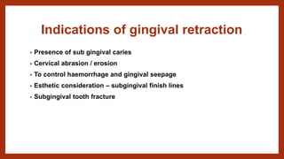
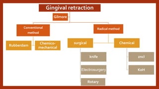
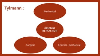
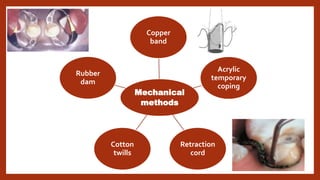
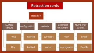
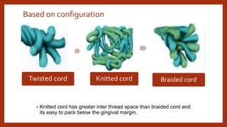
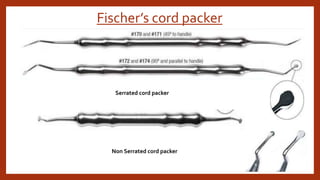
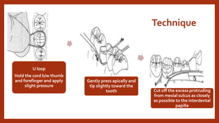
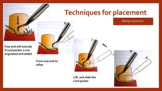
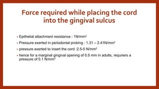
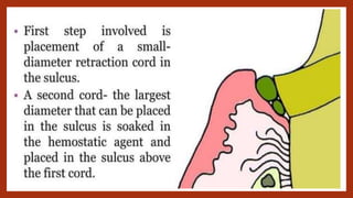
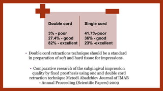
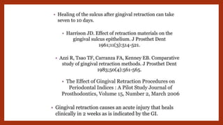
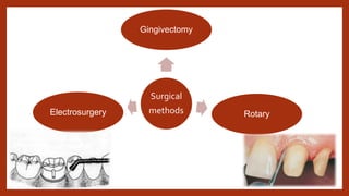
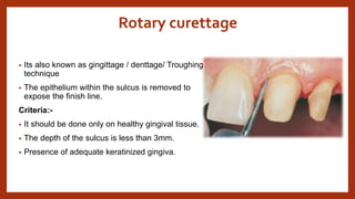
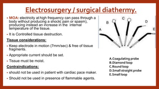
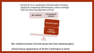
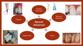
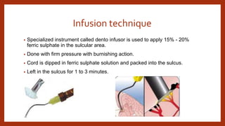
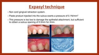
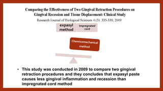
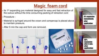
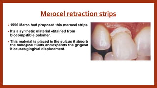
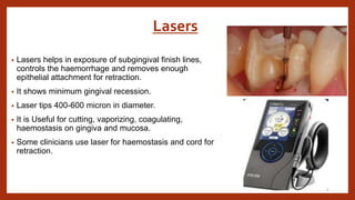
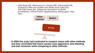
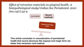
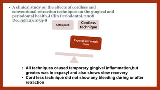
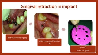
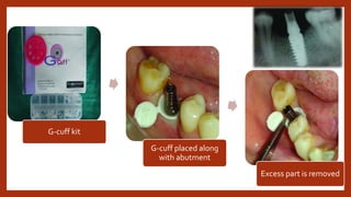
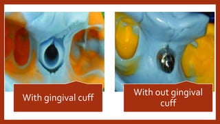
GINGIVAL RETRACTION
Dr .shiva sai vemula
3rd year MDS
Contents
• Definition
• Criteria for gingival retraction
• Indications
• Classification of gingival retraction
methods
• Mechanical
• Chemico-mechanical
• surgical
• Classification of retraction cords
• techniques for placement
• Surgical methods
• Recent advances
• Gingival retraction in implant
• Conclusion
• references
DEFINITION
• Its defined as the procedure of temporary eversion / resection of
gingiva away from the tooth surface / deepening of gingival
sulcus to expose the cervical portion of the tooth in order to have
proper marginal finish to the restoration and recording the
preparation accurately.(GPT-9)
• Gingival tissue displacement is the deflection of the marginal
gingiva away from a tooth.
• Barmuda triangle of dentistry – triangle from alveolar crest to base of gingiva
• violation of biological width leads to chronic gingival inflammation, pocket formation
and osseous defects.
• Tooth preparation must be terminated at least 2mm coronal to the alveolar crest at
base of gingival sulcus.
Vertical Cross section of tooth
CRITERIA FOR
GINGIVAL
RETRACTION
Effective &
achieve
hemostasis
Absence of
systemic
effects
Minimal
chair time
No
irreversible
damage
Different movements of gingiva
Retraction : downward and outword
movement of the free gingival
marginal.
Relapse : Tendency of gingival cuff to
go back to its orginal position.
Displacement : downword movement
of the gingival cuff.
Collapse : tendency of the gingival cuff
to flatten under forces
Indications of gingival retraction
• Presence of sub gingival caries
• Cervical abrasion / erosion
• To control haemorrhage and gingival seepage
• Esthetic consideration – subgingival finish lines
• Subgingival tooth fracture
classification
Gingival retraction
Conventional
method
Rubberdam
Chemico-
mechanical
Radical method
surgical
knife
Electrosurgery
Rotary
Chemical
zncl
KoH
Gilmore
Tylmann :
GINGIVAL
RETRACTION
Mechanical
Chemico- mechanical
Surgical
Mechanical
methods
Copper
band
Acrylic
temporary
coping
Retraction
cord
Cotton
twills
Rubber
dam
Classification of retraction cords
Retraction cords
Surface
texture
Wet
Dry
configuration
Twisted
knitted
material
synthetic
cotton
Chemical
treatment
Plain
impregnated
Number of
strands
single
Double
Based on
Depending on the thickness- colour coded
Based on configuration
• Knitted cord has greater inter thread space than braided cord and
its easy to pack below the gingival margin.
Twisted cord Knitted cord Braided cord
• In 1999 a study was counducted by asbjorn on
comparision of new series of knitted and twined gingival
cord.
• He concluded that knitted gingival cords were better
than twined cords and cords containing epinephrine has
performed same as aluminium sulphate cords.
Fischer’s cord packer
Serrated cord packer
Non Serrated cord packer
Technique
U loop
Hold the cord b/w thumb
and forefinger and apply
slight pressure
Gently press apically and
tip slightly toward the
tooth Cut off the excess protruding
from mesial sulcus as closely
as possible to the interdental
papilla
Techniques for placement
Sliding movement
Free end will extrude
if cord packer is not
angulated and slided
From one end to
other
Lift and slide the
cord packer
Force required while placing the cord
into the gingival sulcus
• Epithelial attachment resistance : 1N/mm2
• Pressure exerted in periodontal probing : 1.31 – 2.41N/mm2
• pressure exerted to insert the cord :2.5-5 N/mm2
• hence for a marginal gingival opening of 0.5 mm in adults, requriers a
pressure of 0.1 N/mm2
• The size of cords is clinically determined by evaluating the depth of the
sulcus with a periodontal probe and observing the friability of the particular
tissue
• Rule of thumb is not to violate the periodontal attachment through
aggressive instrumentation of cord placement.
Length of cord
•Max ant - 30mm
•Pre molar -25mm
•Molar – 40mm
•Mand ant - 17mm
•Pre molar -25mm
•Molar – 40mm
2.Chemico mechanical method
• Gingival retraction using chemically impregnated retraction cord is a
chemico-mechanical method of displacement
Chemicals used:-
Adverse effects of the
medicaments
• effective in shrinking the gingival
tissue
• Zinc chloride is caustic and prolonged
application / high concentration burns
the tissue
• Negatol is highly acidic and
decalcifies the teeth.
• Time > 5-10 min may lead to
irreversible changes in gingiva.
MOA
Vasoconstrictors
Biologic fluid
coagulants
Surface layer
tissue
coagulant
Retraction
methods
Advantages Disadvantages
mechanical Inexpensive Painful
Achieves various degrees of
retraction
Rapid collapse of sulcus after removal
Can be used with chemical adjuncts Risk in traumatization
No hemostasis
Time consuming
Risk in sulcus contamination
chemical Hemostatic Systemic effects
vasoconstrictive Risk in inflammation
Less collapse after removal of cord Rebound hyperemia
Risk in tissue necrosis
Risk in Sulcus contamination
Risk in tissue discolouration
Acidic taste
Contraindications for epinephrine use
• Cardiovascular diseases
• Hypertension
• hyperthyroidism
• Hypersensitivity to epinephrine
• Patient on tricyclic anti depressants
• Diabetic patients
In 2003 this study had conducted to compare the gingival
irritation with different impregnated retraction cords and they
concluded that aluminium chloride is least irritant medicament
but it disturbes the polyvinyl siloxane impression material
In 1997 a study had conducted to compare and evaluate the
effectiveness of different chemicomechanical retraction and
they concluded that aluminium potassium sulphate and
aluminium chloride are more effective than epinephrine in
sulcus opening
Double cord
3% - poor
27.4% - good
82% - excellent
Single cord
41.7%-poor
36% - good
23% -excellent
Surgical methods
Surgical
methods
Gingivectomy
Rotary
Electrosurgery
Rotary curettage
• Its also known as gingittage / denttage/ Troughing
technique
• The epithelium within the sulcus is removed to
expose the finish line.
Criteria:-
• It should be done only on healthy gingival tissue.
• The depth of the sulcus is less than 3mm.
• Presence of adequate keratinized gingiva.
Electrosurgery / surgical diathermy.
• MOA: electricity at high frequency can pass through a
body without producing a shock( pain or spasm),
producing instead an increase in the internal
temperature of the tissue.
• It is Controlled tissue destruction.
Tissue considerations:
• Keep electrode in motion (7mm/sec) & free of tissue
fragments.
• Appropriate current should be set.
• Tissue must be moist.
Contraindications:
• should not be used in patient with cardiac pace maker.
• Should not be used in presence of flammable agents.
A.Coagulating probe
B.Diamond loop
C.Round loop
D.Small straight probe
E.Small loop
Bur method showed minimal tissue loss than electrosurgery.
clinical tissue appearance of all the 3 technique is same
Bur method
Conventional
method
Recent advances
Recent
Advances
Infusion
technique
Merocel
retraction
lasers
Magic foam
Expasyl
Infusion technique
• Specialized instrument called dento infusor is used to apply 15% - 20%
ferric sulphate in the sulcular area.
• Done with firm pressure with burnishing action.
• Cord is dipped in ferric sulphate solution and packed into the sulcus.
• Left in the sulcus for 1 to 3 minutes.
Expasyl technique
• Non cord gingival retraction system.
• Paste product injected into the sulcus exerts a pressure of 0.1N/mm2
• This pressure is too low to damage the epithelial attachment, but sufficient
to obtain a sulcus opening of 0.5mm for 2min.
• This study was conducted in 2009 to compare two gingival
retraction procedures and they concludes that expasyl paste
causes less gingival inflammation and recession than
impregnated cord method
expasyl
method
Impregnated
cord
Magic foam cord
• Its 1st expanding pvs material designed for easy and fast retraction of
the sulcus without the time consuming packing of retraction cord
• Procedure :
• Material is syringed around the crown and compercap is placed above
to maintain pressure.
• After 5 min the cap and form are removed.
Merocel retraction strips
• 1996 Marco had proposed this merocel strips
• It’s a synthetic material obtained from
biocompatible polymer.
• This material is placed in the sulcus it absorb
the biological fluids and expands the gingival
it causes gingival displacement.
Lasers
• Lasers helps in exposure of subgingival finish lines,
controls the haemorrhage and removes enough
epithelial attachment for retraction.
• It shows minimum gingival recession.
• Laser tips 400-600 micron in diameter.
• It is Useful for cutting, vaporizing, coagulating,
haemostasis on gingiva and mucosa.
• Some clinicians use laser for haemostasis and cord for
retraction.
• In 2004 this sudy had conducted to compare lasers with other methods
and they concluded that lasers causes Less aggressive ,less bleeding
and less recession while comparing to other methods.
Other
method
lasers
This article concludes in concederation of periodontal
health that recent advances like expasyl and magic form are
better that retraction cord method
Cord method
Recent
methods
Ultra pack
Cordless
technique
• All techniques caused temporary gingival inflammation,but
greates was in expasyl and also shows slow recovery
• Cord less technique did not show any bleeding during or after
retraction
Gingival retraction in implant
Removal of healing cap
After removal of healing
cap
G-cuff
Selection guide
G-cuff kit
G-cuff placed along
with abutment
Excess part is removed
With gingival cuff
With out gingival
cuff
conclusion
• Sulcus opening and hemostasis are two essential prerequsites for
good access.
• Several techniques are avalible which have proven to be predictable,
safe and effective.
• no scientific evidence has established the superiority of any one
technique over other, so the choice of technique depend on the
presenting clinical situation and operator preference.
References
Thank you

Gingival retraction.pptx

  • 1.
    GINGIVAL RETRACTION Dr .shivasai vemula 3rd year MDS
  • 2.
    Contents • Definition • Criteriafor gingival retraction • Indications • Classification of gingival retraction methods • Mechanical • Chemico-mechanical • surgical • Classification of retraction cords • techniques for placement • Surgical methods • Recent advances • Gingival retraction in implant • Conclusion • references
  • 3.
    DEFINITION • Its definedas the procedure of temporary eversion / resection of gingiva away from the tooth surface / deepening of gingival sulcus to expose the cervical portion of the tooth in order to have proper marginal finish to the restoration and recording the preparation accurately.(GPT-9) • Gingival tissue displacement is the deflection of the marginal gingiva away from a tooth.
  • 4.
    • Barmuda triangleof dentistry – triangle from alveolar crest to base of gingiva • violation of biological width leads to chronic gingival inflammation, pocket formation and osseous defects. • Tooth preparation must be terminated at least 2mm coronal to the alveolar crest at base of gingival sulcus. Vertical Cross section of tooth
  • 5.
    CRITERIA FOR GINGIVAL RETRACTION Effective & achieve hemostasis Absenceof systemic effects Minimal chair time No irreversible damage
  • 6.
    Different movements ofgingiva Retraction : downward and outword movement of the free gingival marginal. Relapse : Tendency of gingival cuff to go back to its orginal position. Displacement : downword movement of the gingival cuff. Collapse : tendency of the gingival cuff to flatten under forces
  • 7.
    Indications of gingivalretraction • Presence of sub gingival caries • Cervical abrasion / erosion • To control haemorrhage and gingival seepage • Esthetic consideration – subgingival finish lines • Subgingival tooth fracture
  • 8.
  • 9.
  • 10.
  • 11.
  • 12.
  • 13.
  • 14.
    Depending on thethickness- colour coded
  • 15.
    Based on configuration •Knitted cord has greater inter thread space than braided cord and its easy to pack below the gingival margin. Twisted cord Knitted cord Braided cord
  • 16.
    • In 1999a study was counducted by asbjorn on comparision of new series of knitted and twined gingival cord. • He concluded that knitted gingival cords were better than twined cords and cords containing epinephrine has performed same as aluminium sulphate cords.
  • 17.
    Fischer’s cord packer Serratedcord packer Non Serrated cord packer
  • 18.
    Technique U loop Hold thecord b/w thumb and forefinger and apply slight pressure Gently press apically and tip slightly toward the tooth Cut off the excess protruding from mesial sulcus as closely as possible to the interdental papilla
  • 19.
    Techniques for placement Slidingmovement Free end will extrude if cord packer is not angulated and slided From one end to other Lift and slide the cord packer
  • 20.
    Force required whileplacing the cord into the gingival sulcus • Epithelial attachment resistance : 1N/mm2 • Pressure exerted in periodontal probing : 1.31 – 2.41N/mm2 • pressure exerted to insert the cord :2.5-5 N/mm2 • hence for a marginal gingival opening of 0.5 mm in adults, requriers a pressure of 0.1 N/mm2
  • 21.
    • The sizeof cords is clinically determined by evaluating the depth of the sulcus with a periodontal probe and observing the friability of the particular tissue • Rule of thumb is not to violate the periodontal attachment through aggressive instrumentation of cord placement.
  • 22.
    Length of cord •Maxant - 30mm •Pre molar -25mm •Molar – 40mm •Mand ant - 17mm •Pre molar -25mm •Molar – 40mm
  • 23.
    2.Chemico mechanical method •Gingival retraction using chemically impregnated retraction cord is a chemico-mechanical method of displacement Chemicals used:- Adverse effects of the medicaments • effective in shrinking the gingival tissue • Zinc chloride is caustic and prolonged application / high concentration burns the tissue • Negatol is highly acidic and decalcifies the teeth. • Time > 5-10 min may lead to irreversible changes in gingiva.
  • 24.
  • 25.
    Retraction methods Advantages Disadvantages mechanical InexpensivePainful Achieves various degrees of retraction Rapid collapse of sulcus after removal Can be used with chemical adjuncts Risk in traumatization No hemostasis Time consuming Risk in sulcus contamination chemical Hemostatic Systemic effects vasoconstrictive Risk in inflammation Less collapse after removal of cord Rebound hyperemia Risk in tissue necrosis Risk in Sulcus contamination Risk in tissue discolouration Acidic taste
  • 26.
    Contraindications for epinephrineuse • Cardiovascular diseases • Hypertension • hyperthyroidism • Hypersensitivity to epinephrine • Patient on tricyclic anti depressants • Diabetic patients
  • 27.
    In 2003 thisstudy had conducted to compare the gingival irritation with different impregnated retraction cords and they concluded that aluminium chloride is least irritant medicament but it disturbes the polyvinyl siloxane impression material In 1997 a study had conducted to compare and evaluate the effectiveness of different chemicomechanical retraction and they concluded that aluminium potassium sulphate and aluminium chloride are more effective than epinephrine in sulcus opening
  • 32.
    Double cord 3% -poor 27.4% - good 82% - excellent Single cord 41.7%-poor 36% - good 23% -excellent
  • 34.
  • 35.
  • 36.
    Rotary curettage • Itsalso known as gingittage / denttage/ Troughing technique • The epithelium within the sulcus is removed to expose the finish line. Criteria:- • It should be done only on healthy gingival tissue. • The depth of the sulcus is less than 3mm. • Presence of adequate keratinized gingiva.
  • 37.
    Electrosurgery / surgicaldiathermy. • MOA: electricity at high frequency can pass through a body without producing a shock( pain or spasm), producing instead an increase in the internal temperature of the tissue. • It is Controlled tissue destruction. Tissue considerations: • Keep electrode in motion (7mm/sec) & free of tissue fragments. • Appropriate current should be set. • Tissue must be moist. Contraindications: • should not be used in patient with cardiac pace maker. • Should not be used in presence of flammable agents. A.Coagulating probe B.Diamond loop C.Round loop D.Small straight probe E.Small loop
  • 38.
    Bur method showedminimal tissue loss than electrosurgery. clinical tissue appearance of all the 3 technique is same Bur method Conventional method
  • 39.
  • 40.
  • 41.
    Infusion technique • Specializedinstrument called dento infusor is used to apply 15% - 20% ferric sulphate in the sulcular area. • Done with firm pressure with burnishing action. • Cord is dipped in ferric sulphate solution and packed into the sulcus. • Left in the sulcus for 1 to 3 minutes.
  • 42.
    Expasyl technique • Noncord gingival retraction system. • Paste product injected into the sulcus exerts a pressure of 0.1N/mm2 • This pressure is too low to damage the epithelial attachment, but sufficient to obtain a sulcus opening of 0.5mm for 2min.
  • 43.
    • This studywas conducted in 2009 to compare two gingival retraction procedures and they concludes that expasyl paste causes less gingival inflammation and recession than impregnated cord method expasyl method Impregnated cord
  • 44.
    Magic foam cord •Its 1st expanding pvs material designed for easy and fast retraction of the sulcus without the time consuming packing of retraction cord • Procedure : • Material is syringed around the crown and compercap is placed above to maintain pressure. • After 5 min the cap and form are removed.
  • 45.
    Merocel retraction strips •1996 Marco had proposed this merocel strips • It’s a synthetic material obtained from biocompatible polymer. • This material is placed in the sulcus it absorb the biological fluids and expands the gingival it causes gingival displacement.
  • 46.
    Lasers • Lasers helpsin exposure of subgingival finish lines, controls the haemorrhage and removes enough epithelial attachment for retraction. • It shows minimum gingival recession. • Laser tips 400-600 micron in diameter. • It is Useful for cutting, vaporizing, coagulating, haemostasis on gingiva and mucosa. • Some clinicians use laser for haemostasis and cord for retraction.
  • 47.
    • In 2004this sudy had conducted to compare lasers with other methods and they concluded that lasers causes Less aggressive ,less bleeding and less recession while comparing to other methods. Other method lasers
  • 48.
    This article concludesin concederation of periodontal health that recent advances like expasyl and magic form are better that retraction cord method Cord method Recent methods
  • 49.
    Ultra pack Cordless technique • Alltechniques caused temporary gingival inflammation,but greates was in expasyl and also shows slow recovery • Cord less technique did not show any bleeding during or after retraction
  • 50.
    Gingival retraction inimplant Removal of healing cap After removal of healing cap G-cuff Selection guide
  • 51.
    G-cuff kit G-cuff placedalong with abutment Excess part is removed
  • 52.
    With gingival cuff Without gingival cuff
  • 53.
    conclusion • Sulcus openingand hemostasis are two essential prerequsites for good access. • Several techniques are avalible which have proven to be predictable, safe and effective. • no scientific evidence has established the superiority of any one technique over other, so the choice of technique depend on the presenting clinical situation and operator preference.
  • 54.
  • 58.