Somatic & special senses
•Download as PPTX, PDF•
1 like•651 views

The document provides information on the somatic and special senses. It discusses the different types of receptors including chemoreceptors, nociceptors, thermoreceptors, mechanoreceptors, and photoreceptors. It then describes the specific senses - touch, pain, temperature, taste, smell, hearing, equilibrium, sight. For each sense, it outlines the receptors involved, their location, and how sensory information is transmitted to the brain. The document also reviews the anatomy and physiology associated with smell, hearing, taste, equilibrium, and vision.

Recommended
More Related Content
What's hot
What's hot (20)
Similar to Somatic & special senses
Similar to Somatic & special senses (20)
More from Bashant Kumar sah
More from Bashant Kumar sah (20)
Recently uploaded
Recently uploaded (20)
Somatic & special senses
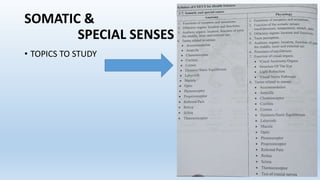
- 1. SOMATIC & SPECIAL SENSES • TOPICS TO STUDY
- 2. • Somatic sense means of the body or bodily ("soma" means body) detect touch, pain pressure, temperature, and tension on the skin and in internal organs. • distributed throughout the body • Special senses detect the sensations of taste, smell, hearing, equilibrium, and sight, only in special sense organs in the head region (a phenomenon known as “cephalization").
- 3. Types of Receptors • 1. Chemoreceptors a) detect chemical concentrations. b)in somatic senses, chemoreceptors can detect ionic, glucose, oxygen, and carbon dioxide concentrations in the blood and tissue fluids. c)in special senses chemoreceptors are responsible for the senses of taste ( in oral cavity) and smell (in nasal cavity). • 2. Nociceptors a)detect pain or tissue damage due to excessive mechanical, electrical, thermal, or chemical forces. b)in somatic senses, nociceptors can be found in the skin and many visceral organs detecting tissue damages. • 3. Thermoreceptors a) detect temperature changes. b)in somatic senses, certain thermoreceptors detect cold temperatures in the skin and some visceral organs, while other thermoreceptors detect high temperatures only.
- 4. • 4. Mechanoreceptors a) detect mechanical forces. b)in somatic senses, proprioceptors detect muscle tension, pressoreceptors detect blood pressure, and stretch receptors detect lung inflation, stomach distention, and urinary bladder expansion. c)in special senses, mechanoreceptors in the inner ear detect the senses of hearing (in cochlea)and equilibrium (in semicircular canals). • 5. photoreceptors a) detect light intensity. b)in special senses, photoreceptors in the eye detect the tones (by rods photoreceptors) and colors (by cones photoreceptors) of visual images
- 5. Functions of Receptors 1. Rod and cone cells of the eye’s retina – respond to electromagnetic radiation of light. 2. Hair cells as receptor in ear – respond to vibration of sound 3. Olfactory neurons – respond to odorant chemicals that binds to them 4. Taste receptor – respond to chemical substances that binds to them 5. Meissner’s corpuscles – respond to touch and its free ending bring sensation of pain
- 6. Function of sensations 1. Detection of sensations allow the human body to be aware of changes (or stimuli) that occur in the environment or inside the body. 2. These senses permit the central nervous system to produce reactions for the stimuli and maintain body homeostasis. 3. All senses are detected by sensory receptors, and after integration and processing being dong done in the central nervous system, motor nerves produce a response. 4. Somatic senses ("soma" means body) detect touch, pain pressure, temperature, and tension on the skin and in internal organs. 5. Special senses detect the sensations of taste, smell, hearing, equilibrium, and sight, only in special sense organs in the head region (a phenomenon known as “cephalization").
- 7. Functions of somatic senses: 1.sensory receptors in the skin, muscles, joints, and visceral organs detect external and internal stimuli of the body. 2. Three types of somatic senses: a)Exteroceptive senses detect changes that occur at body surface, such as touch, pressure and temperature. b)Proprioceptive senses detect changes that occur in muscles, tendons, ligaments and joint tissues. c)Visceroceptive senses detect changes that occur in internal organs.
- 8. • The Nose: -prominent and protruding structure of the face between eyes, serves the sense of smell. • Entrance to the respiratory tract and contains the olfactory organs. • Consists of nasal cavity, shaped by Ethmoid bone and nasal septum associated with cartilage. • Roof of nasal cavity contains olfactory epithelium which is the organ of olfaction, • keep the nose moist. Olfactory organs:
- 9. a) olfactory receptors located in "olfactory organs " are specialized chemoreceptors in the nasal cavity. b) olfactory receptors are bipolar neurons surrounded by ciliated columnar cells. These sensory cells, after being stimulated by olfactory sensations, send nerve impulses along the olfactory (cranial nerve I) to the cerebrum. c) gases in the air entering the nasal cavity are dissolved by nasal mucus (secreted by goblet cells in the columnar epithelium ), and the resulting solution stimulates various olfactory receptors.
- 10. Auditory organs: Ear a) The gross anatomy of the human ear includes the outer ear (consists of auricle and external auditory meatus), middle ear (consists of the tympanic membrane and auditory ossicles, and inner ear (consists of cochlea, 3 semicircular canals, and the vestibulocochlear nerves). https://youtu.be/3G5jiXl2LSM
- 11. b)The outer ear is responsible for transferring sound waves from the environment to the middle ear. c)The middle ear is responsible for amplifying sound waves into strong signals for the hearing receptors to detect. d)The inner ear is responsible for using mechanoreceptors to detect stimuli for hearing (in cochlea) and equilibrium (in semicircular canals) and send the nerve impulses through the vestibulocochlear nerve (nerve VIII) to the brain. e)auditory ossicles include the malleus, incus, and stapes, which are articulated to one another, but not to the skeleton. The malleus is attached to the tympanic membrane, stapes is attached to the cochlea. f)the middle ear also contains the eustachian tube (auditory tube) that connects with the pharynx for equalizing air pressure in the skull.
- 12. h) when the ossicles vibrate waves in the endolymph fluid inside the cochlea are generated. i) endolymph waves bend the strerocilia (modified dendrites) of hearing receptors called hair cells, which are located in the organ of corti on the basilar membrane. Bending of hair cells result in generation of nerve impulses which reach the cerebrum via the cochlear nerve.
- 13. Taste perception: • a) about 10,000 taste buds occur in the oral cavity, mostly on the tongue. Each taste bud contains 40-60 taste receptors. These sensory cells, are stimulated by taste sensations. • b) chemical substances from the food or beverage enter the mouth and are dissolved in the saliva ( secreted by salivary glands and mucous membrane of oral cavity). • c) dissolved chemical substances stimulate various taste buds in different regions of the tongue, and combinations of taste sensations produce hundreds of different taste senses. • d) These sensory cells, after being stimulated by taste sensations, send nerve impulses through the facial nerve (Nerve VII), glossopharyngeal nerve (Nerve IX), or vagus nerve (Nerve X ) to the cerebrum.
- 14. e) four primary taste sensations: -sweet- sensation caused by organic substances such as sugars and amino acids; most sensitive at the tip of tongue. -sour – sensation caused by hydrogen ions (H+) from acidic substances; most sensitive at both sides of the tongue. - salty-sensation caused by metal ions (e.g. Mg++) or inorganic salts (e.g. NACL); most sensitive at the sides and the tip of tongue. - bitter – caused by alkaloids (e.g. nicotine, caffeine) and nonalkaloids (e.g. aspirin) ; most sensitive at the back of tongue
- 15. Process of equilibrium: a)detected by mechanoreceptors in the semicircular canals to help maintain body posture and body stability. b)nerve impulses generated by the receptors in semicircular canals are transmitted by the vestibular nerve to Nerve VIII, and the signals will be processed by the brain. https://youtu.be/YMIMvBa8XGs
- 16. C) two types of equilibrium: Static equilibrium: senses the position of the head and vertical and horizontal movement of the body; helps to maintain body posture. Dynamic equilibrium: senses the motions of the head and body (mainly rotational); helps to maintain overall body stability. D) static equilibrium: receptors called macula located in the semicircular canals detect the position of the head. When the head changes position, fluid in the semicircular canals moves and generates waves that bend the stereocilia on these macula cells. E) dynamic equilibrium: receptors called crista ampullaris located the semicircular canals detect the position of the body. When body moves, similar physiology with equilibrium occurs in the semicircular canals, resulting in nerve impulses being sent to the brain for interpretation.
- 17. Sense of Vision a) Accessory structures of the eye are those that are not directly related the sense of vision, but facilitate the physiology of the eyeballs. • Eyebrows – to shade the eyes from sunlight and to prevent perspiration from reaching the eyes. • Eyelids- to protect the eyes from foreign objects (e.g. dust particles) ,and to prevent desiccation (drying) of the eyes by lubricating fluid. • Conjunctiva- a mucous membrane on the inner lining of eyelids, which produces lubricating and cleansing fluid for the surface of eye. • Lacrimal gland- exocrine gland that secretes a dilute saline solution called tears for moistening the eyes. [Tears contain mucus, antibodies ,and antibacterial enzymes that protect the eye from infections. Emotional tears also contain enzymes that seem to help reduce stress levels].
- 19. Anatomy of The Eye at • The wall of the eyeball consists of 3 layers of tissue: I)Fibrous Tunic: outermost layer, made of fibrous connective tissue with minimal blood vessels. • Contains 2 regions: sclera (a white area th extends from the back of the eye toward the front) and cornea (a transparent tissue in the front for allowing light to enter the eyeball). https://youtu.be/nbwPPcwknPU
- 20. II) Vascular Tunic (Uvea): middle layer, made of thin fibrous connective tissue that contains numerous blood vessels (capillaries). • contains choroids (a pigmented membrane in the back to provide nutrition and to absorb light) and iris (to regulate the amount of light entering the eye by constriction or dilation) • includes specialized structures such as ciliary body (which regulates the shape of lens), suspensory ligaments (which attach the ciliary body to the lens), lens (another transparent tissue that bends the light entering the eye), and pupil (an opening created by the actions of the iris where a large pupil is caused by a dilated iris, while a small pupil is created by a constricted iris) .
- 22. III) sensory tunic (also called retina): • innermost layer, made of specialized nerve tissue. It contains 2 layers of tissue: an outer pigmented layer (which absorbs light and stores vitaminA) and an inner neural layer (that detects light using photoreceptors and sends nerve impulses to the occipital lobe of cerebrum through the optic nerves). • 2 types of photoreceptors are found on the neural layer: rods (detect tones of visual images) and cones (detect colors). These sensory cells, after being stimulated by visual sensations, send nerve impulses through the optic nerve (Nerve II ) to the occipital lobs of cerebrum.
- 26. Chambers and fluids in the eyeball: • Anterior cavity: located between the cornea and lens, contains aqueous humor for supplying oxygen and nutrients to the lens an cornea. • Posterior cavity: located between the lens and retina, contains vitreous humor to transmit light from the lens to the photoreceptors on retina.
- 28. Extrinsic muscles of the Eye Lat. Rectus: innervated by abducens nerve & moves eye lat. Med. Rectus: innervated by oculomotor nerve & moves eye med. Sup. Rectus: innervated by oculomotor nerve & moves eye sup. & med. Inf. Rectus: innervated by oculomotor nerve & moves eye inf. & med. Inf. Oblique: innervated by oculomotor nerve & moves eye sup. & lat. Sup. Oblique: innervated by trochlear nerve & moves eye inf. & lat.
- 30. Human eye Accommodation. Accommodation of the eye refers to the act of physiologically adjusting crystalline lens elements to alter the refractive power and bring objects that are closer to the eye into sharp focus. This tutorial explores changes in the lens structure as objects are relocated with respect to the eye. https://www.olympus-lifescience.com/en/microscope- resource/primer/java/humanvision/accommodation/ humans are concerned accommodation is caused by the increased curvature of anterior area of the eye lens while at the same time its thickness also changes.
- 32. Ampulla • The ampulla contains the sensory area called the ampullary crest. These ampullary crests sense the movement of head and play a role in maintaining the balance of the body. • Electroreception, the ability to detect weak naturally occurring electrostatic fields in the environment. Electroreception is found in a number of vertebrate species, including the members of two distinct lineages of teleosts (a group of ray-finned fishes) and monotremes (egg-laying mammals). Bumblebees also are able to detect weak electric fields. In vertebrates electroreception is made possible through the existence of sensitive electroreceptor organs in the skin. • Electroreception facilitates the detection of prey or other food sources and objects and is used by some species as a means of social communication. In general, terrestrial animals have little use for electroreception, because the high resistance of air limits the flow of electric current. Thus, humans lack electroreceptors; however, through the indiscriminant stimulation of sensory and motor nerve fibres, humans are able to detect strong electric currents (e.g., from batteries or static generators) resulting from either direct contact with an electric source or indirect contact with a conducting medium such as water.
- 33. 12 pairs Cranial nerves:
- 34. https://www.msdmanuals.com/professional/neurologic-disorders/neurologic-examination/how-to-assess-the-cranial- nerves# Test of Cranial Nerves: The cranial nerve exam is a type of neurological examination. It is used to identify problems with the cranial nerves by physical examination. It has nine components. Each test is designed to assess the status of one or more of the twelve cranial nerves (I-XII). These components correspond to testing the sense of smell (I), visual fields and acuity (II), eye movements (III, IV, VI) and pupils (III, sympathetic and parasympathetic), sensory function of face (V), strength of facial (VII) and shoulder girdle muscles (XI), hearing (VII, VIII), taste (VII, IX, X), pharyngeal movement and reflex (IX, X), tongue movements (XII).