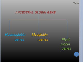
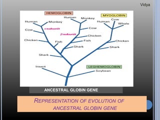
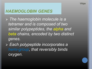
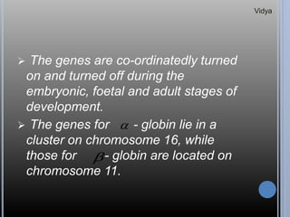
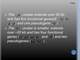
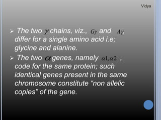
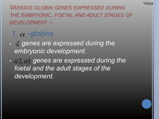
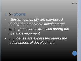
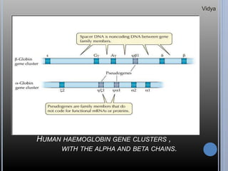
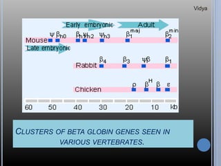
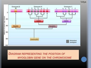
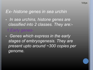
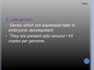
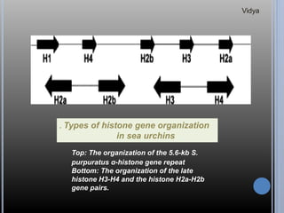
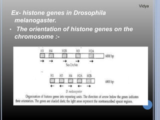
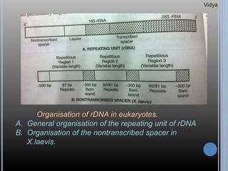
GENE FAMILIES AND
CLUSTERS
D. V. Vidya Lakshmi.
GENE FAMILY
 A gene family is a set of several
similar genes, formed by duplication of
a single original gene, and generally
with similar biochemical functions.
 A gene family is a set of homologous
genes within one organism.
Vidya
 When a gene is present in two or
more copies per genome, the
condition is known as “ redundancy”.
 The members of a gene family may
be either clustered together ,
dispersed on different chromosomes
or present in a combination of both.
Vidya
 If the genes of a gene family encode
proteins, the term “protein family” is
often used in an analogous manner to
gene family.
 One example for such family are the
genes for Human haemoglobin
subunits.
Vidya
GENE CLUSTER
 A gene cluster is part of a gene family.
 A gene cluster is a group of two or
more genes found within an
organism’s DNA that encode for
similar polypeptides or proteins, which
collectively share a generalised
function and are located within a few
thousand base pairs of each other.
Vidya
 The size of gene clusters can vary
significantly, from a few genes to
several hundred genes.
 Genes found in a gene cluster may be
observed near one another on the
same chromosome or on different, but
homologous chromosomes.
Vidya
 Extensive tanden repetition of a gene
normally occurs when the gene
product is needed in unusually large
amounts. E.g., genes for rRNA,
histone genes, etc.
 Sometimes all the members of a gene
family are functional, but often some
members are nonfunctional
pseudogenes.
Vidya
TANDEM REPEAT
 In a tandem repeat, the nucleotide
sequence is repeated in the same
orientation.
 For example, the trinucleotide
sequence GAA is repeated two times
in the DNA segment –GAAGAA-.
GAA GAA
CTT CTT
tandem repeat
Vidya
GLOBIN GENES
 Genes encoding the various globin
proteins evolved from one common
ancestral globin gene, which
duplicated and diverged about 450-
500 million years ago.
 After the duplication events,
differences between the genes in
globin family arose from the
accumulation of mutations.
Vidya
ANCESTRAL GLOBIN GENE
Haemoglobin Myoglobin
genes genes Plant
globin
genes
Vidya
REPRESENTATION OF EVOLUTION OF
ANCESTRAL GLOBIN GENE
Vidya
HAEMOGLOBIN GENES
 The haemoglobin molecule is a
tetramer and is composed of two
similar polypeptides, the alpha and
beta chains, encoded by two distinct
genes.
 Each polypeptide incorporates a
hemi-group, that reversibly binds
oxygen.
Vidya
 The genes are co-ordinatedly turned
on and turned off during the
embryonic, foetal and adult stages of
development.
 The genes for - globin lie in a
cluster on chromosome 16, while
those for - globin are located on
chromosome 11.


Vidya
 The - cluster extends over 50 kb
and has five functional genes(E , ,
, , ) and one pseudogene ( ) .
 The - cluster is smaller, extends
over ~20 kb and has four functional
genes ( , , , , and ) and two
pseudogenes ( , ) .

G A
 

2 1 2 1 
 

Vidya
 The two chains, viz., and ,
differ for a single amino acid i.e;
glycine and alanine.
 The two genes, namely ,
code for the same protein; such
identical genes present in the same
chromosome constitute “non allelic
copies” of the gene.
 G A
 2,1 
Vidya
VARIOUS GLOBIN GENES EXPRESSED DURING
THE EMBRYONIC, FOETAL AND ADULT STAGES OF
DEVELOPMENT :-
1. -globins
• genes are expressed during the
embryonic development.
• genes are expressed during the
foetal and the adult stages of the
development.


1,2 
Vidya
2. - globins
• Epsilon genes (E) are expressed
during the embryonic development.
• genes are expressed during the
foetal development.
• genes are expressed during the
adult stages of development.

 AG ,
 ,
Vidya
HUMAN HAEMOGLOBIN GENE CLUSTERS ,
WITH THE ALPHA AND BETA CHAINS.
Vidya
CLUSTERS OF BETA GLOBIN GENES SEEN IN
VARIOUS VERTEBRATES.
Vidya
MYOGLOBIN GENES
 Myoglobin is an iron- and oxygen-
binding protein found in the muscle
tissue of vertebrates in general and in
almost all mammals.
 It is related to haemoglobin, which is
the iron- and oxygen- binding protein
in blood, specifically in the red blood
cells.
Vidya
 In humans, myoglobin is only found in
the bloodstream after muscle injury.
 Myoglobin is the primary oxygen-
carrying pigment of muscle tissues.
 High concentrations of myoglobin in
muscle cells allow organisms to hold
their breath for a longer period of time.
Vidya
 Diving mammals such as whales and
seals have muscles with particularly
high abundance of myoglobin.
 In humans, myoglobin is encoded by
the MB gene.
Vidya
MEAT COLOUR
 Myoglobin contains hemes, pigments
responsible for the colour of red meat.
 The colour that meat takes is partly
determined by the degree of oxidation
of the myoglobin.
 The higher the degree of oxidation of
myoglobin, the richer the meat colour.
Vidya
ROLE IN DISEASE
 Myoglobin is released from damaged
muscle tissue, which has very high
concentrations of myoglobin.
 Myoglobin is a sensitive marker for
muscle injury, making it a potential
marker for heart attack in patients with
chest pain.
Vidya
DIAGRAM REPRESENTING THE POSITION OF
MYOGLOBIN GENE ON THE CHROMOSOME.
Vidya
HISTONE GENES
 Histones are highly alkaline proteins
found in eukaryotic cell nuclei that
package and order the DNA into
structural units called “nucleosomes”.
 They are the chief protein
components of chromatin, acting as
spools around which DNA winds, and
play a role in gene regulation.
Vidya
 Without histones, the unwound DNA
in chromosomes would be very long.
 Histones are found in the nuclei of the
eukaryotic cells and in certain
Archaea, but not in bacteria.
 The unicellular algae known as
dinoflagellates are the only eukaryotes
that are known to completely lack
histones.
Vidya
 Histone proteins are among the highly
conserved proteins in eukaryotes,
emphasizing their important role in the
biology of the nucleus.
Histone genes are of five types
namely, H1, H2A, H2B,H3 and H4.
Vidya
 Large quantities of histones are
required during the S phase of cell
cycle to package the newly
synthesised DNA.
 To meet this demand multiple copies
of each of the five histone genes are
present per genome in clusters of
tandemly repeated units.
Vidya
 A repeat unit contains one copy each
of five histone genes seperated from
each other by non transcribed spacer
regions.
 The histone genes don’t have intron
sequences. All the exon sequences
(coding sequences) are highly
conserved.
Vidya
 The variation in the length of repeated
units in different organisms is due to
the variation in the lengths of their non
transcribed spacer regions.
 The arrangement of histone genes is
same in all species, but variation is
observed in the orientation of genes
on the chromosome.
Vidya
TYPES OF HISTONE GENES
1.Histone H1:-
• H1 is one of the five main histone
protein families which are components
of chromatin in eukaryotic cells.
• Unlike other histones, H1 doesn’t
make up the nucleosome bead. It sits
on top of the stucture, keeping in place
the DNA that has wrapped around the
nucleosome.
Vidya
2. HISTONE H2A:-
• Histone H2A is one of the five main
histone proteins involved in the
structure of chromatin in eukaryotic
cells.
• H2A is important for packaging DNA
into chromatin.
• H2A plays a major role in determining
the overall structure of chromatin.
Vidya
3. HISTONE H2B:-
• Histone H2B is one of the five main
histone proteins involved in the
structure of chromatin in eukaryotic
cells.
• H2B is involved with the structure of
the nucleosomes of the ‘beads on a
string’ structure.
Vidya
4. HISTONE H3:-
• Histone H3 is one of the five main
histone proteins involved in the
structure of chromatin in eukaryotic
cells.
• H3 is involved with the structure of the
nucleosomes of the ‘beads on a string’
stucture.
Vidya
5. HISTONE H4:-
• Histone H4 is one of the five main
histone proteins involved in the structure
of chromatin in eukaryotic cells.
• H4 is a structural component of the
nucleosome, and is subjected to some
modifications including acetylation and
methylation, which may alter expression
of genes located on DNA associated with
its parent histone octamer.
Vidya
Ex- histone genes in sea urchin
• In sea urchins, histone genes are
classified into 2 classes. They are:-
1.Early genes-
• Genes which express in the early
stages of embryogenesis. They are
present upto around ~300 copies per
genome.
Vidya
2. Late genes:-
• Genes which are expressed later in
embryonic development.
• They are present upto around ~10
copies per genome.
Vidya
. Types of histone gene organization
in sea urchins
Top: The organization of the 5.6-kb S.
purpuratus α-histone gene repeat
Bottom: The organization of the late
histone H3-H4 and the histone H2a-H2b
gene pairs.
Vidya
Ex- histone genes in Drosophila
melanogaster.
• The orientation of histone genes on the
chromosome :-
Vidya
• The variations in length are due to the no.
of repeated units, the non-transcribed
space.
• The ends of the histone genes have
attachment regions seen in the non-
transcribed space.
• These attachment sites may help in the
packing of DNA, in form of nuclear
scaffolds.
Vidya
GENES FOR RIBOSOMAL RNA’S
• Ribosomal RNA (rRNA) constitutes 80-90%
of the total cellular RNA in both prokaryotes
and eukaryotes.
• Both rRNA’s and tRNA’s are present in
multiple copies per genome.
• In eukaryotes, these copies are present in
clusters either confined to a single
chromosome, eg., in X. laevis, or they
spread on more than one chromosome, eg.,
in humans and mouse.
Vidya
• In D. melanogaster, rRNA genes are
present in X and Y chromosomes, the
cluster in X being much larger than that in Y
chromosome.
• The region containing rRNA genes is often
called rDNA (ribosomal DNA).
Prokaryotes vs Eukaryotes
• Both prokaryotic and eukaryotic ribosomes
can be broken down into two subunits.
Vidya
• In prokaryotes, a small 30S ribosomal
subunit contains the 5S ribosomal RNA.
• The large 50S ribosomal subunit contains
two rRNA species (the 16S and 23S
ribosomal RNAs).
• Bacterial 16S rRNA, 23S rRNA and 5S
rRNA genes are typically organised as a co-
transcribed operon.
• There may be one or more copies of the
operon dispersed in the genome. Eg.,
Escherichia coli has seven).
Vidya
• Archaea contains either a single rDNA
operon or multiple copies of the operon.
• The 3’ end of the 16S rRNA (in a ribosome)
binds to a sequence on the 5’ end of mRNA
called the Shine-Dalgarno sequence.
• In eukaryotes, many copies of the rRNA
genes are organised in tandem repeats.
• In humans, ~300-400 repeats are present
in five clusters (on chromosomes 13, 14, 15,
21 and 22).
Vidya
• The 5S rRNA in most eukaryotes is in the
small ribosomal subunit, and the large
subunit contains three rRNA species (the
5.8S, 18S and 28S in mammals, 25S in
plants).
• In eukaryotes, the 5S RNA is transcribed
independently.
• In higher eukaryotes, 5S RNA genes are
organised in separate clusters and their
numbers are far in excess of those coding
18S-28S rRNA.
Vidya
• The number of copies of tRNA genes is
difficult to estimate and most likely the
numbers given in the following table.
ORGANISM rRNA genes tRNA
genes
18S/23S
rRNA
(16S/23S
rRNA)*
5S rRNA
E.Coli 7 7 60
Yeast 140 140 250
Drosophila
X chromosome 250
Y chromosome 150
Human 280 2,000 1,300
Xenopus laevis 450 24,000 1,150
Vidya
• The repeating unit of rRNA gene clusters in
eukaryotes consists of the following:
1. a transcribed region containing,
a) a 5’ leader, followed by b) 18S rRNA ,
c) a transcribed spacer and d) 28S rRNA &
2. Non transcribed spacer.
• After trancription, the leader and the
transcribed spacer are cleaved to generate
the 18S and 28S rRNA sequences within
the same transcription unit, while the non
transcribed spacer seperates different
transcription units.
Vidya
• The transcribed spacer contains some
shorter and useful sequences that are
released by cleavage.
• In bacteria, it contains tRNA sequences, in
mammals and amphibians one short
sequence cleaved from this region forms
5.8S RNA, which forms H-bonds with 28S
rRNA in ribosomes.
• The remaining transcribed spacer and the
leader are presumably degraded.
Vidya
Organisation of rDNA in eukaryotes.
A. General organisation of the repeating unit of rDNA
B. Organisation of the nontranscribed spacer in
X.laevis.
Vidya
• The non transcribed spacer varies widely.
The nontranscribed spacer in yeast is short
(1.75 kb) and is constant in length.
• In drosophila and X. laevis, this region is
much larger and shows a variation in length
between different copies of the repeating
unit.
• Mammals represent the extreme case,
where the non transcribed spacer is ~30 kb
long.
Vidya
• Each nontranscribed spacer is internally
repetitious, variation in length arises from
changes in number of repeats of some of
the sub units.
• In case of Xenopus , it contains 3
repetitious regions each comprised of a
variable number of repeats of a rather short
sequence.
• At the start of each nontranscribed region is
a ~500bp long unique sequence. In
addition, there are 3 sequences called Bam
islands.
Vidya
• They are termed as bam islands because
they are isolated following the digestion of
spacer with BamH1 restriction enzyme.
• Two of the Bam islands separate the (each
~300 bp) 3 repetitious regions, while the
third one lies at the end of the spacer.
• The 60/81 repeats of the NTS are involved
in transcription initiation in a manner
comparable with the enhancer sequences in
relation to RNA polymeraseII.
Vidya
Vidya

Gene families and clusters

  • 1.
  • 2.
    GENE FAMILY  Agene family is a set of several similar genes, formed by duplication of a single original gene, and generally with similar biochemical functions.  A gene family is a set of homologous genes within one organism. Vidya
  • 3.
     When agene is present in two or more copies per genome, the condition is known as “ redundancy”.  The members of a gene family may be either clustered together , dispersed on different chromosomes or present in a combination of both. Vidya
  • 4.
     If thegenes of a gene family encode proteins, the term “protein family” is often used in an analogous manner to gene family.  One example for such family are the genes for Human haemoglobin subunits. Vidya
  • 5.
    GENE CLUSTER  Agene cluster is part of a gene family.  A gene cluster is a group of two or more genes found within an organism’s DNA that encode for similar polypeptides or proteins, which collectively share a generalised function and are located within a few thousand base pairs of each other. Vidya
  • 6.
     The sizeof gene clusters can vary significantly, from a few genes to several hundred genes.  Genes found in a gene cluster may be observed near one another on the same chromosome or on different, but homologous chromosomes. Vidya
  • 7.
     Extensive tandenrepetition of a gene normally occurs when the gene product is needed in unusually large amounts. E.g., genes for rRNA, histone genes, etc.  Sometimes all the members of a gene family are functional, but often some members are nonfunctional pseudogenes. Vidya
  • 8.
    TANDEM REPEAT  Ina tandem repeat, the nucleotide sequence is repeated in the same orientation.  For example, the trinucleotide sequence GAA is repeated two times in the DNA segment –GAAGAA-. GAA GAA CTT CTT tandem repeat Vidya
  • 9.
    GLOBIN GENES  Genesencoding the various globin proteins evolved from one common ancestral globin gene, which duplicated and diverged about 450- 500 million years ago.  After the duplication events, differences between the genes in globin family arose from the accumulation of mutations. Vidya
  • 10.
    ANCESTRAL GLOBIN GENE HaemoglobinMyoglobin genes genes Plant globin genes Vidya
  • 11.
    REPRESENTATION OF EVOLUTIONOF ANCESTRAL GLOBIN GENE ANCESTRAL GLOBIN GENE Vidya
  • 12.
    HAEMOGLOBIN GENES  Thehaemoglobin molecule is a tetramer and is composed of two similar polypeptides, the alpha and beta chains, encoded by two distinct genes.  Each polypeptide incorporates a hemi-group, that reversibly binds oxygen. Vidya
  • 13.
     The genesare co-ordinatedly turned on and turned off during the embryonic, foetal and adult stages of development.  The genes for - globin lie in a cluster on chromosome 16, while those for - globin are located on chromosome 11.   Vidya
  • 14.
     The -cluster extends over 50 kb and has five functional genes(E , , , , ) and one pseudogene ( ) .  The - cluster is smaller, extends over ~20 kb and has four functional genes ( , , , , and ) and two pseudogenes ( , ) .  G A    2 1 2 1     Vidya
  • 15.
     The twochains, viz., and , differ for a single amino acid i.e; glycine and alanine.  The two genes, namely , code for the same protein; such identical genes present in the same chromosome constitute “non allelic copies” of the gene.  G A  2,1  Vidya
  • 16.
    VARIOUS GLOBIN GENESEXPRESSED DURING THE EMBRYONIC, FOETAL AND ADULT STAGES OF DEVELOPMENT :- 1. -globins • genes are expressed during the embryonic development. • genes are expressed during the foetal and the adult stages of the development.   1,2  Vidya
  • 17.
    2. - globins •Epsilon genes (E) are expressed during the embryonic development. • genes are expressed during the foetal development. • genes are expressed during the adult stages of development.   AG ,  , Vidya
  • 18.
    HUMAN HAEMOGLOBIN GENECLUSTERS , WITH THE ALPHA AND BETA CHAINS. Vidya
  • 19.
    CLUSTERS OF BETAGLOBIN GENES SEEN IN VARIOUS VERTEBRATES. Vidya
  • 20.
    MYOGLOBIN GENES  Myoglobinis an iron- and oxygen- binding protein found in the muscle tissue of vertebrates in general and in almost all mammals.  It is related to haemoglobin, which is the iron- and oxygen- binding protein in blood, specifically in the red blood cells. Vidya
  • 21.
     In humans,myoglobin is only found in the bloodstream after muscle injury.  Myoglobin is the primary oxygen- carrying pigment of muscle tissues.  High concentrations of myoglobin in muscle cells allow organisms to hold their breath for a longer period of time. Vidya
  • 22.
     Diving mammalssuch as whales and seals have muscles with particularly high abundance of myoglobin.  In humans, myoglobin is encoded by the MB gene. Vidya
  • 23.
    MEAT COLOUR  Myoglobincontains hemes, pigments responsible for the colour of red meat.  The colour that meat takes is partly determined by the degree of oxidation of the myoglobin.  The higher the degree of oxidation of myoglobin, the richer the meat colour. Vidya
  • 24.
    ROLE IN DISEASE Myoglobin is released from damaged muscle tissue, which has very high concentrations of myoglobin.  Myoglobin is a sensitive marker for muscle injury, making it a potential marker for heart attack in patients with chest pain. Vidya
  • 25.
    DIAGRAM REPRESENTING THEPOSITION OF MYOGLOBIN GENE ON THE CHROMOSOME. Vidya
  • 26.
    HISTONE GENES  Histonesare highly alkaline proteins found in eukaryotic cell nuclei that package and order the DNA into structural units called “nucleosomes”.  They are the chief protein components of chromatin, acting as spools around which DNA winds, and play a role in gene regulation. Vidya
  • 27.
     Without histones,the unwound DNA in chromosomes would be very long.  Histones are found in the nuclei of the eukaryotic cells and in certain Archaea, but not in bacteria.  The unicellular algae known as dinoflagellates are the only eukaryotes that are known to completely lack histones. Vidya
  • 28.
     Histone proteinsare among the highly conserved proteins in eukaryotes, emphasizing their important role in the biology of the nucleus. Histone genes are of five types namely, H1, H2A, H2B,H3 and H4. Vidya
  • 29.
     Large quantitiesof histones are required during the S phase of cell cycle to package the newly synthesised DNA.  To meet this demand multiple copies of each of the five histone genes are present per genome in clusters of tandemly repeated units. Vidya
  • 30.
     A repeatunit contains one copy each of five histone genes seperated from each other by non transcribed spacer regions.  The histone genes don’t have intron sequences. All the exon sequences (coding sequences) are highly conserved. Vidya
  • 31.
     The variationin the length of repeated units in different organisms is due to the variation in the lengths of their non transcribed spacer regions.  The arrangement of histone genes is same in all species, but variation is observed in the orientation of genes on the chromosome. Vidya
  • 32.
    TYPES OF HISTONEGENES 1.Histone H1:- • H1 is one of the five main histone protein families which are components of chromatin in eukaryotic cells. • Unlike other histones, H1 doesn’t make up the nucleosome bead. It sits on top of the stucture, keeping in place the DNA that has wrapped around the nucleosome. Vidya
  • 33.
    2. HISTONE H2A:- •Histone H2A is one of the five main histone proteins involved in the structure of chromatin in eukaryotic cells. • H2A is important for packaging DNA into chromatin. • H2A plays a major role in determining the overall structure of chromatin. Vidya
  • 34.
    3. HISTONE H2B:- •Histone H2B is one of the five main histone proteins involved in the structure of chromatin in eukaryotic cells. • H2B is involved with the structure of the nucleosomes of the ‘beads on a string’ structure. Vidya
  • 35.
    4. HISTONE H3:- •Histone H3 is one of the five main histone proteins involved in the structure of chromatin in eukaryotic cells. • H3 is involved with the structure of the nucleosomes of the ‘beads on a string’ stucture. Vidya
  • 36.
    5. HISTONE H4:- •Histone H4 is one of the five main histone proteins involved in the structure of chromatin in eukaryotic cells. • H4 is a structural component of the nucleosome, and is subjected to some modifications including acetylation and methylation, which may alter expression of genes located on DNA associated with its parent histone octamer. Vidya
  • 37.
    Ex- histone genesin sea urchin • In sea urchins, histone genes are classified into 2 classes. They are:- 1.Early genes- • Genes which express in the early stages of embryogenesis. They are present upto around ~300 copies per genome. Vidya
  • 38.
    2. Late genes:- •Genes which are expressed later in embryonic development. • They are present upto around ~10 copies per genome. Vidya
  • 39.
    . Types ofhistone gene organization in sea urchins Top: The organization of the 5.6-kb S. purpuratus α-histone gene repeat Bottom: The organization of the late histone H3-H4 and the histone H2a-H2b gene pairs. Vidya
  • 40.
    Ex- histone genesin Drosophila melanogaster. • The orientation of histone genes on the chromosome :- Vidya
  • 41.
    • The variationsin length are due to the no. of repeated units, the non-transcribed space. • The ends of the histone genes have attachment regions seen in the non- transcribed space. • These attachment sites may help in the packing of DNA, in form of nuclear scaffolds. Vidya
  • 42.
    GENES FOR RIBOSOMALRNA’S • Ribosomal RNA (rRNA) constitutes 80-90% of the total cellular RNA in both prokaryotes and eukaryotes. • Both rRNA’s and tRNA’s are present in multiple copies per genome. • In eukaryotes, these copies are present in clusters either confined to a single chromosome, eg., in X. laevis, or they spread on more than one chromosome, eg., in humans and mouse. Vidya
  • 43.
    • In D.melanogaster, rRNA genes are present in X and Y chromosomes, the cluster in X being much larger than that in Y chromosome. • The region containing rRNA genes is often called rDNA (ribosomal DNA). Prokaryotes vs Eukaryotes • Both prokaryotic and eukaryotic ribosomes can be broken down into two subunits. Vidya
  • 44.
    • In prokaryotes,a small 30S ribosomal subunit contains the 5S ribosomal RNA. • The large 50S ribosomal subunit contains two rRNA species (the 16S and 23S ribosomal RNAs). • Bacterial 16S rRNA, 23S rRNA and 5S rRNA genes are typically organised as a co- transcribed operon. • There may be one or more copies of the operon dispersed in the genome. Eg., Escherichia coli has seven). Vidya
  • 45.
    • Archaea containseither a single rDNA operon or multiple copies of the operon. • The 3’ end of the 16S rRNA (in a ribosome) binds to a sequence on the 5’ end of mRNA called the Shine-Dalgarno sequence. • In eukaryotes, many copies of the rRNA genes are organised in tandem repeats. • In humans, ~300-400 repeats are present in five clusters (on chromosomes 13, 14, 15, 21 and 22). Vidya
  • 46.
    • The 5SrRNA in most eukaryotes is in the small ribosomal subunit, and the large subunit contains three rRNA species (the 5.8S, 18S and 28S in mammals, 25S in plants). • In eukaryotes, the 5S RNA is transcribed independently. • In higher eukaryotes, 5S RNA genes are organised in separate clusters and their numbers are far in excess of those coding 18S-28S rRNA. Vidya
  • 47.
    • The numberof copies of tRNA genes is difficult to estimate and most likely the numbers given in the following table. ORGANISM rRNA genes tRNA genes 18S/23S rRNA (16S/23S rRNA)* 5S rRNA E.Coli 7 7 60 Yeast 140 140 250 Drosophila X chromosome 250 Y chromosome 150 Human 280 2,000 1,300 Xenopus laevis 450 24,000 1,150 Vidya
  • 48.
    • The repeatingunit of rRNA gene clusters in eukaryotes consists of the following: 1. a transcribed region containing, a) a 5’ leader, followed by b) 18S rRNA , c) a transcribed spacer and d) 28S rRNA & 2. Non transcribed spacer. • After trancription, the leader and the transcribed spacer are cleaved to generate the 18S and 28S rRNA sequences within the same transcription unit, while the non transcribed spacer seperates different transcription units. Vidya
  • 49.
    • The transcribedspacer contains some shorter and useful sequences that are released by cleavage. • In bacteria, it contains tRNA sequences, in mammals and amphibians one short sequence cleaved from this region forms 5.8S RNA, which forms H-bonds with 28S rRNA in ribosomes. • The remaining transcribed spacer and the leader are presumably degraded. Vidya
  • 50.
    Organisation of rDNAin eukaryotes. A. General organisation of the repeating unit of rDNA B. Organisation of the nontranscribed spacer in X.laevis. Vidya
  • 51.
    • The nontranscribed spacer varies widely. The nontranscribed spacer in yeast is short (1.75 kb) and is constant in length. • In drosophila and X. laevis, this region is much larger and shows a variation in length between different copies of the repeating unit. • Mammals represent the extreme case, where the non transcribed spacer is ~30 kb long. Vidya
  • 52.
    • Each nontranscribedspacer is internally repetitious, variation in length arises from changes in number of repeats of some of the sub units. • In case of Xenopus , it contains 3 repetitious regions each comprised of a variable number of repeats of a rather short sequence. • At the start of each nontranscribed region is a ~500bp long unique sequence. In addition, there are 3 sequences called Bam islands. Vidya
  • 53.
    • They aretermed as bam islands because they are isolated following the digestion of spacer with BamH1 restriction enzyme. • Two of the Bam islands separate the (each ~300 bp) 3 repetitious regions, while the third one lies at the end of the spacer. • The 60/81 repeats of the NTS are involved in transcription initiation in a manner comparable with the enhancer sequences in relation to RNA polymeraseII. Vidya
  • 54.