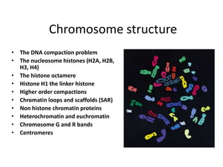
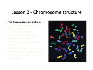
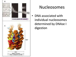
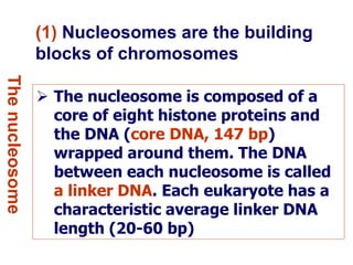
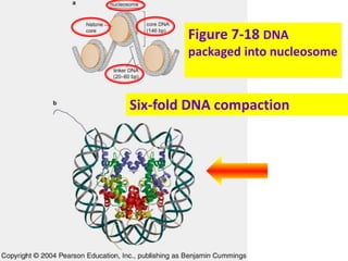
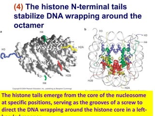
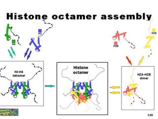
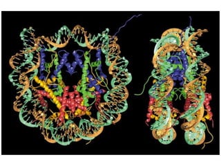
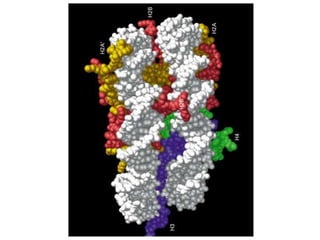
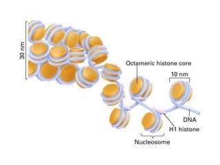
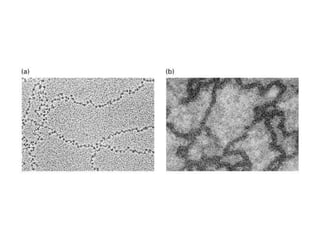
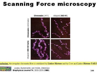
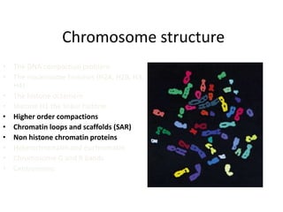
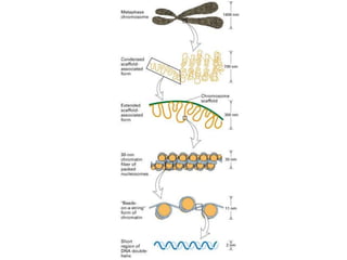
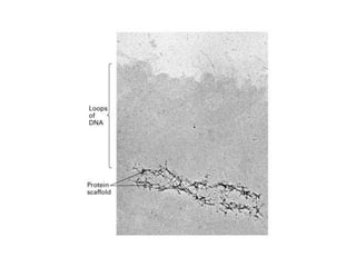
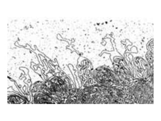
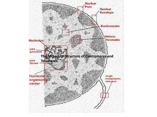
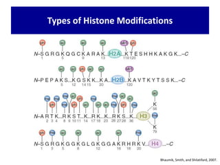
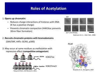
Chromatin Structure & Genome
Organization
What is so special about chromosomes ?
1.They are huge:
One bp = 600 dalton, an average chromosome is 107 bp
long = 109- 1010 dalton !
(for comparison a protein of 3x10⁵ is considered very big.
What is so special about chromosomes ?
1.They are huge:
One bp = 600 dalton, an average chromosome is 107 bp
long = 109- 1010 dalton !
(for comparison a protein of 3x10⁵ is considered very big.
2. They contain a huge amount of non-
redundant information (it is not just a big repetitive
polymer but it has a unique sequence) .
• Philosophically - the cell is there to serve,
protect and propagate the chromosomes.
• Practically - the chromosome must be
protected at the ends - telomers
and it must have “something” that will enable
it to be moved to daughter cells - centromers
Genome Complexity
Chromosome structure
• The DNA compaction problem
• The nucleosome histones (H2A, H2B,
H3, H4)
• The histone octamere
• Histone H1 the linker histone
• Higher order compactions
• Chromatin loops and scaffolds (SAR)
• Non histone chromatin proteins
• Heterochromatin and euchromatin
• Chromosome G and R bands
• Centromeres
Lesson 2 - Chromosome structure
• The DNA compaction problem
• The nucleosome histones (H2A,
H2B, H3, H4)
• The histone octamere
• Histone H1 the linker histone
• Higher order compactions
• Chromatin loops and scaffolds
(SAR)
• Non histone chromatin proteins
• Heterochromatin and euchromatin
• Chromosome G and R bands
• Centromeres
Chromosome is a compact form of the
DNA that readily fits inside the cell
To protect DNA from damage
DNA in a chromosome can be
transmitted efficiently to both daughter
cells during cell division
Chromosome confers an overall
organization to each molecule of DNA,
which facilitates gene expression as
well as recombination
Importance
• Take 4 meters of DNA (string) and compact them into
a ball of 10M. Now 10M are 1/100 of a mm and a
bit small to imagine – so now walk from here to the
main entrance let say 400 meters and try to compact
it all into 1 mm.
• This compaction is very complex and the DNA
isn’t just crammed into the nucleus but is
organized in a very orderly fashion from the
smallest unit - the nucleosome, via loops,
chromosomal domains and bands to the entire
chromosome which has a fixed space in the
nucleus.
Chromatin
 The complexes between eukaryotic DNA and proteins are
called chromatin, which typically contains about twice as much
protein as DNA.
 The major proteins of chromatin are the histones—small proteins
containing a high proportion of basic amino acids (arginine and
lysine) that facilitate binding to the negatively charged DNA
molecule.
 There are five major types of histones—called H1, H2A, H2B, H3,
and H4—which are very similar among different species of
eukaryotes .
 The histones are extremely abundant proteins in eukaryotic cells;
together, their mass is approximately equal to that of the cell's DNA.
chromatin
• In addition, chromatin contains an approximately equal
mass of a wide variety of nonhistone chromosomal
proteins. There are more than a thousand different types
of these proteins, which are involved in a range of
activities, including DNA replication
and gene expression.
• Histones are not found in eubacteria (e.g., E. coli),
although the DNA of these bacteria is associated with
other proteins that presumably function like histones to
package the DNA within the bacterial cell.
Archaebacteria, however, do contain histones that
package their DNAs in structures similar to eukaryotic
chromatin
The nucleosome
Nucleosome & histone
structures
The basic structural unit of chromatin, the nucleosome, was described by Roger
Kornberg in 1974 .
Two types of experiments led to Kornberg's proposal of the nucleosome model.
First, partial digestion of chromatin with micrococcal nuclease (an enzyme that
degrades DNA) was found to yield DNA fragments approximately 200 base pairs
long.
In contrast, a similar digestion of naked DNA (not associated with proteins)
yielded a continuous smear of randomly sized fragments. These results
suggested that the binding of proteins to DNA in chromatin protects regions of
the DNA from nuclease digestion, so that the enzyme can attack DNA only at
sites separated by approximately 200 base pairs.
Consistent with this notion, electron microscopy revealed that chromatin fibers
have a beaded appearance, with the beads spaced at intervals of approximately
200 base pairs. Thus, both the nuclease digestion and the electron microscopic
studies suggested that chromatin is composed of repeating 200-base-pair units,
which were called nucleosomes.
Discovery of nucleosomes
Nucleosomes
• DNA associated with
individual nucleosomes
determined by DNAse I
digestion
(1) Nucleosomes are the building
blocks of chromosomes
Thenucleosome
 The nucleosome is composed of a
core of eight histone proteins and
the DNA (core DNA, 147 bp)
wrapped around them. The DNA
between each nucleosome is called
a linker DNA. Each eukaryote has a
characteristic average linker DNA
length (20-60 bp)
Figure 7-18 DNA
packaged into nucleosome
Six-fold DNA compaction
 Five abundant histones are H1
(linker histone, 20 kd), H2A, H2B,
H3 and H4 (core histones, 11-15 kd).
 The core histones share a common
structural fold, called histone-fold
domain
 The core histones each have an N-
terminal “tail”, the sites of
extensive modifications
(2) Histones are small, positively
charged (basic) proteins
Thenucleosome
Histone
a
Molecular
weight
Number
of amino acids
Percentage
Lysine +
Arginine
H1 22,500 244 30.8
H2A 13,960 129 20.2
H2B 13,774 125 22.4
H3 15,273 135 22.9
H4 11,236 102 24.5
The Major Histone Proteins
The core histones share a common structural fold
(1)(2)
(3) Many DNA sequence-
independent contacts (?) mediate
interaction between between the
core histones and DNA
Thenucleosome
(4) The histone N-terminal tails
stabilize DNA wrapping around the
octamer
The histone tails emerge from the core of the nucleosome
at specific positions, serving as the grooves of a screw to
direct the DNA wrapping around the histone core in a left-
Lesson 2 - Chromosome structure
• The DNA compaction problem
• The nucleosome histones (H2A, H2B,
H3, H4)
• The histone octamere
• Histone H1 the linker histone
• Higher order compactions
• Chromatin loops and scaffolds (SAR)
• Non histone chromatin proteins
• Heterochromatin and euchromatin
• Chromosome G and R bands
• Telomeres
• Centromeres
Chromosome structure
• The DNA compaction problem
• The nucleosome histones (H2A, H2B, H3,
H4)
• The histone octamere
• Histone H1 the linker histone
• Higher order compactions
• Chromatin loops and scaffolds (SAR)
• Non histone chromatin proteins
• Heterochromatin and euchromatin
• Chromosome G and R bands
• Centromeres
SARs are very AT-rich fragments several hundredbase pairs in length that were first
identified as DNA fragments that are retained by nuclear scaffold/matrix
preparations.
They define the bases of the DNA loops that become visible as a halo around extracted
nuclei and that can be traced in suitable electron micro-graphs of histone-depleted
metaphase chromosomes.
They are possibly best described as being composed of numerous clustered, irregularly
spaced runs of As and Ts
Scaffold attachment regions
 Compaction level of interphase chromosomes is
not uniform
 Euchromatin
 Less condensed regions of chromosomes
 Transcriptionally active
 Regions where 30 nm fiber forms radial loop domains
 Heterochromatin
 Tightly compacted regions of chromosomes
 Transcriptionally inactive (in general)
 Radial loop domains compacted even further
Heterochromatin vs Euchromatin
Types of Heterochromatin
Figure 10.20
• Constitutive heterochromatin
– Always heterochromatic
– Permanently inactive with regard to transcription
• Facultative heterochromatin
– Regions that can interconvert between euchromatin
and heterochromatin
– Example: Barr body
Most of the euchromatin in interphase nuclei appears to be in
the form of 30-nm fibers, organized into large loops containing
approximately 50 to 100 kb of DNA.
About 10% of the euchromatin, containing the genes that are
actively transcribed, is in a more decondensed state (the 10-nm
conformation) that allows transcription.
Chromatin structure is thus intimately linked to the control
of gene expression in eukaryotes.
Dynamics of chromatin
The extent of chromatin condensation varies during the life
cycle of the cell.
In interphase (nondividing) cells, most of the chromatin
(called euchromatin) is relatively decondensed and distributed
throughout the nucleus .
 During this period of the cell cycle, genes are transcribed
and the DNA is replicated in preparation for cell division.
Dynamics of chromatin
R-bands are known to replicate early, to contain most
housekeeping genes and are enriched in hyperacetylated
histone H4 and DNase I-sensitive chromatin.
This suggests they have a more open chromatin conformation,
consistent with a central AT-queue with longer loops that reach
the nuclear periphery.
In contrast, Q-bands contain fewer genes and are proposed to have
loops that are shorter and more tightly folded, resulting in an AT-
queue path resembling a coiled spring.
The Molecular Structure of Centromeres and
Telomeres
The Molecular Structure of Centromeres and
Telomeres
Bhaumik, Smith, and Shilatifard, 2007.
Types of Histone Modifications
• Covalently attached groups (usually to histone tails)
• Reversible and Dynamic
– Enzymes that add/remove modification
– Signals
• Have diverse biological functions
Cell, 111:285-91, Nov. 1, 2002
Methyl Acetyl Phospho Ubiquitin SUMO
Features of Histone Modifications
• Small vs. Large groups
• One or up to three groups per residue
Features of Histone Modifications
Jason L J M et al. J. Biol. Chem. 2005
Ub = ~8.5 kDa
H4 = 14 kDa
• Others: Sumoylation (Lysine), ADP Ribosylation (Glutamate)
Histone Modifications and Modifers
Residue Modification Modiying Enzyme
Lysine
Acetylation
Deacetylation
HAT
HDAC
Lysine
Methylation
Demethylation
HMT
HDM
Lysine
Ubiquitylation
Deubiquitylation
Ub ligase
Ub protease
Serine/Threonine
Phosphorylation
Dephosphorylation
Kinase
Phosphatase
Arginine
Methylation
Demethylation
PRMT
Deiminase/Demethylas
e
General Roles of Histone Modifications
• Intrinsic
– Single nucleosome changes
– Example: histone variant specific modifications (H2AX)
• Extrinsic
– Chromatin organization: nucleosome/nucleosome interactions
– Alter chromatin packaging, electrostatic charge
– Example: H4 acetylation
• Effector-mediated
– Recruitment of other proteins to the chromatin via
• Bromodomains bind acetylation
• Chromo-like royal domains (chromo, tudor, MBT) and PHD bind methylation
• 14-3-3 proteins bind phosphorylation
Kouzarides, Cell 2007
- Prevent binding: H3S10P prevents Heterochromatin
Protein 1 binding
Acetylation
• Many lysine residues can be acetylated
• mainly on histone tails (sometimes in core)
• Can be part of large acetylation domains
• Modifying enzymes:
• often multi-enzyme complexes
• can modify multiple residues
• Well correlated with transcriptional activation
• Other roles (chromatin assembly, DNA repair, etc.)
• Two general types:
• Type B: cytoplasmic (newly synthesized histones)
• HAT1
Kouzarides, Cell, 2007.
Histone Acetyl Transferases (HATs/KATs)
Type B
1. Opens up chromatin:
– Reduces charge interactions of histones with DNA
(K has a positive charge)
– Prevents chromatin compaction (H4K16ac prevents
30nm fiber formation)
2. Recruits chromatin proteins with bromodomains
(SWI/SNF, HATs: GCN5, p300)
3. May occur at same residues as methylation with
repressive effect (competitive antagonism)
Roles of Acetylation
Robinson et al., J. Mol. Biol., 2008.
Mujtaba et al., Oncogene, 2007.
PCAF
Yang and Chen, Cell Research, 2011.
H3K27ac
H3K27me3
4. Highly correlated with active transcription
i.e. enriched at TSS of actively transcribed genes
Roles of Acetylation -- Continued
Expression: P1<P2<P3<P4
Heintzman N et al., Nature Genetics, 2007.
Roles of Acetylation
H4Ac H3Ac H3 RNAPII
208 TSS investigated
5. Correlated with binding of activating transcription factors
i.e. enriched at promoters and enhancers
Roles of Acetylation
Heintzman N et al., Nature Genetics, 2007.
H4Ac H3Ac p300
Expression: E1<E2<E3
74 enhancers
(distal p300 binding sites)

Chromatin Structure & Genome Organization by Shivendra Kumar

  • 1.
    Chromatin Structure &Genome Organization
  • 2.
    What is sospecial about chromosomes ? 1.They are huge: One bp = 600 dalton, an average chromosome is 107 bp long = 109- 1010 dalton ! (for comparison a protein of 3x10⁵ is considered very big.
  • 3.
    What is sospecial about chromosomes ? 1.They are huge: One bp = 600 dalton, an average chromosome is 107 bp long = 109- 1010 dalton ! (for comparison a protein of 3x10⁵ is considered very big. 2. They contain a huge amount of non- redundant information (it is not just a big repetitive polymer but it has a unique sequence) .
  • 4.
    • Philosophically -the cell is there to serve, protect and propagate the chromosomes. • Practically - the chromosome must be protected at the ends - telomers and it must have “something” that will enable it to be moved to daughter cells - centromers
  • 5.
  • 6.
    Chromosome structure • TheDNA compaction problem • The nucleosome histones (H2A, H2B, H3, H4) • The histone octamere • Histone H1 the linker histone • Higher order compactions • Chromatin loops and scaffolds (SAR) • Non histone chromatin proteins • Heterochromatin and euchromatin • Chromosome G and R bands • Centromeres
  • 7.
    Lesson 2 -Chromosome structure • The DNA compaction problem • The nucleosome histones (H2A, H2B, H3, H4) • The histone octamere • Histone H1 the linker histone • Higher order compactions • Chromatin loops and scaffolds (SAR) • Non histone chromatin proteins • Heterochromatin and euchromatin • Chromosome G and R bands • Centromeres
  • 8.
    Chromosome is acompact form of the DNA that readily fits inside the cell To protect DNA from damage DNA in a chromosome can be transmitted efficiently to both daughter cells during cell division Chromosome confers an overall organization to each molecule of DNA, which facilitates gene expression as well as recombination Importance
  • 9.
    • Take 4meters of DNA (string) and compact them into a ball of 10M. Now 10M are 1/100 of a mm and a bit small to imagine – so now walk from here to the main entrance let say 400 meters and try to compact it all into 1 mm.
  • 10.
    • This compactionis very complex and the DNA isn’t just crammed into the nucleus but is organized in a very orderly fashion from the smallest unit - the nucleosome, via loops, chromosomal domains and bands to the entire chromosome which has a fixed space in the nucleus.
  • 11.
    Chromatin  The complexesbetween eukaryotic DNA and proteins are called chromatin, which typically contains about twice as much protein as DNA.  The major proteins of chromatin are the histones—small proteins containing a high proportion of basic amino acids (arginine and lysine) that facilitate binding to the negatively charged DNA molecule.  There are five major types of histones—called H1, H2A, H2B, H3, and H4—which are very similar among different species of eukaryotes .  The histones are extremely abundant proteins in eukaryotic cells; together, their mass is approximately equal to that of the cell's DNA.
  • 12.
    chromatin • In addition,chromatin contains an approximately equal mass of a wide variety of nonhistone chromosomal proteins. There are more than a thousand different types of these proteins, which are involved in a range of activities, including DNA replication and gene expression. • Histones are not found in eubacteria (e.g., E. coli), although the DNA of these bacteria is associated with other proteins that presumably function like histones to package the DNA within the bacterial cell. Archaebacteria, however, do contain histones that package their DNAs in structures similar to eukaryotic chromatin
  • 13.
    The nucleosome Nucleosome &histone structures
  • 14.
    The basic structuralunit of chromatin, the nucleosome, was described by Roger Kornberg in 1974 . Two types of experiments led to Kornberg's proposal of the nucleosome model. First, partial digestion of chromatin with micrococcal nuclease (an enzyme that degrades DNA) was found to yield DNA fragments approximately 200 base pairs long. In contrast, a similar digestion of naked DNA (not associated with proteins) yielded a continuous smear of randomly sized fragments. These results suggested that the binding of proteins to DNA in chromatin protects regions of the DNA from nuclease digestion, so that the enzyme can attack DNA only at sites separated by approximately 200 base pairs. Consistent with this notion, electron microscopy revealed that chromatin fibers have a beaded appearance, with the beads spaced at intervals of approximately 200 base pairs. Thus, both the nuclease digestion and the electron microscopic studies suggested that chromatin is composed of repeating 200-base-pair units, which were called nucleosomes. Discovery of nucleosomes
  • 16.
    Nucleosomes • DNA associatedwith individual nucleosomes determined by DNAse I digestion
  • 17.
    (1) Nucleosomes arethe building blocks of chromosomes Thenucleosome  The nucleosome is composed of a core of eight histone proteins and the DNA (core DNA, 147 bp) wrapped around them. The DNA between each nucleosome is called a linker DNA. Each eukaryote has a characteristic average linker DNA length (20-60 bp)
  • 18.
    Figure 7-18 DNA packagedinto nucleosome Six-fold DNA compaction
  • 19.
     Five abundanthistones are H1 (linker histone, 20 kd), H2A, H2B, H3 and H4 (core histones, 11-15 kd).  The core histones share a common structural fold, called histone-fold domain  The core histones each have an N- terminal “tail”, the sites of extensive modifications (2) Histones are small, positively charged (basic) proteins Thenucleosome
  • 20.
    Histone a Molecular weight Number of amino acids Percentage Lysine+ Arginine H1 22,500 244 30.8 H2A 13,960 129 20.2 H2B 13,774 125 22.4 H3 15,273 135 22.9 H4 11,236 102 24.5 The Major Histone Proteins
  • 21.
    The core histonesshare a common structural fold (1)(2)
  • 22.
    (3) Many DNAsequence- independent contacts (?) mediate interaction between between the core histones and DNA Thenucleosome
  • 23.
    (4) The histoneN-terminal tails stabilize DNA wrapping around the octamer The histone tails emerge from the core of the nucleosome at specific positions, serving as the grooves of a screw to direct the DNA wrapping around the histone core in a left-
  • 27.
    Lesson 2 -Chromosome structure • The DNA compaction problem • The nucleosome histones (H2A, H2B, H3, H4) • The histone octamere • Histone H1 the linker histone • Higher order compactions • Chromatin loops and scaffolds (SAR) • Non histone chromatin proteins • Heterochromatin and euchromatin • Chromosome G and R bands • Telomeres • Centromeres
  • 31.
    Chromosome structure • TheDNA compaction problem • The nucleosome histones (H2A, H2B, H3, H4) • The histone octamere • Histone H1 the linker histone • Higher order compactions • Chromatin loops and scaffolds (SAR) • Non histone chromatin proteins • Heterochromatin and euchromatin • Chromosome G and R bands • Centromeres
  • 33.
    SARs are veryAT-rich fragments several hundredbase pairs in length that were first identified as DNA fragments that are retained by nuclear scaffold/matrix preparations. They define the bases of the DNA loops that become visible as a halo around extracted nuclei and that can be traced in suitable electron micro-graphs of histone-depleted metaphase chromosomes. They are possibly best described as being composed of numerous clustered, irregularly spaced runs of As and Ts Scaffold attachment regions
  • 37.
     Compaction levelof interphase chromosomes is not uniform  Euchromatin  Less condensed regions of chromosomes  Transcriptionally active  Regions where 30 nm fiber forms radial loop domains  Heterochromatin  Tightly compacted regions of chromosomes  Transcriptionally inactive (in general)  Radial loop domains compacted even further Heterochromatin vs Euchromatin
  • 38.
    Types of Heterochromatin Figure10.20 • Constitutive heterochromatin – Always heterochromatic – Permanently inactive with regard to transcription • Facultative heterochromatin – Regions that can interconvert between euchromatin and heterochromatin – Example: Barr body
  • 39.
    Most of theeuchromatin in interphase nuclei appears to be in the form of 30-nm fibers, organized into large loops containing approximately 50 to 100 kb of DNA. About 10% of the euchromatin, containing the genes that are actively transcribed, is in a more decondensed state (the 10-nm conformation) that allows transcription. Chromatin structure is thus intimately linked to the control of gene expression in eukaryotes. Dynamics of chromatin
  • 40.
    The extent ofchromatin condensation varies during the life cycle of the cell. In interphase (nondividing) cells, most of the chromatin (called euchromatin) is relatively decondensed and distributed throughout the nucleus .  During this period of the cell cycle, genes are transcribed and the DNA is replicated in preparation for cell division. Dynamics of chromatin
  • 42.
    R-bands are knownto replicate early, to contain most housekeeping genes and are enriched in hyperacetylated histone H4 and DNase I-sensitive chromatin. This suggests they have a more open chromatin conformation, consistent with a central AT-queue with longer loops that reach the nuclear periphery. In contrast, Q-bands contain fewer genes and are proposed to have loops that are shorter and more tightly folded, resulting in an AT- queue path resembling a coiled spring.
  • 43.
    The Molecular Structureof Centromeres and Telomeres The Molecular Structure of Centromeres and Telomeres
  • 44.
    Bhaumik, Smith, andShilatifard, 2007. Types of Histone Modifications
  • 45.
    • Covalently attachedgroups (usually to histone tails) • Reversible and Dynamic – Enzymes that add/remove modification – Signals • Have diverse biological functions Cell, 111:285-91, Nov. 1, 2002 Methyl Acetyl Phospho Ubiquitin SUMO Features of Histone Modifications
  • 46.
    • Small vs.Large groups • One or up to three groups per residue Features of Histone Modifications Jason L J M et al. J. Biol. Chem. 2005 Ub = ~8.5 kDa H4 = 14 kDa
  • 47.
    • Others: Sumoylation(Lysine), ADP Ribosylation (Glutamate) Histone Modifications and Modifers Residue Modification Modiying Enzyme Lysine Acetylation Deacetylation HAT HDAC Lysine Methylation Demethylation HMT HDM Lysine Ubiquitylation Deubiquitylation Ub ligase Ub protease Serine/Threonine Phosphorylation Dephosphorylation Kinase Phosphatase Arginine Methylation Demethylation PRMT Deiminase/Demethylas e
  • 48.
    General Roles ofHistone Modifications • Intrinsic – Single nucleosome changes – Example: histone variant specific modifications (H2AX) • Extrinsic – Chromatin organization: nucleosome/nucleosome interactions – Alter chromatin packaging, electrostatic charge – Example: H4 acetylation • Effector-mediated – Recruitment of other proteins to the chromatin via • Bromodomains bind acetylation • Chromo-like royal domains (chromo, tudor, MBT) and PHD bind methylation • 14-3-3 proteins bind phosphorylation Kouzarides, Cell 2007 - Prevent binding: H3S10P prevents Heterochromatin Protein 1 binding
  • 49.
    Acetylation • Many lysineresidues can be acetylated • mainly on histone tails (sometimes in core) • Can be part of large acetylation domains • Modifying enzymes: • often multi-enzyme complexes • can modify multiple residues • Well correlated with transcriptional activation • Other roles (chromatin assembly, DNA repair, etc.)
  • 50.
    • Two generaltypes: • Type B: cytoplasmic (newly synthesized histones) • HAT1 Kouzarides, Cell, 2007. Histone Acetyl Transferases (HATs/KATs) Type B
  • 51.
    1. Opens upchromatin: – Reduces charge interactions of histones with DNA (K has a positive charge) – Prevents chromatin compaction (H4K16ac prevents 30nm fiber formation) 2. Recruits chromatin proteins with bromodomains (SWI/SNF, HATs: GCN5, p300) 3. May occur at same residues as methylation with repressive effect (competitive antagonism) Roles of Acetylation Robinson et al., J. Mol. Biol., 2008. Mujtaba et al., Oncogene, 2007. PCAF Yang and Chen, Cell Research, 2011. H3K27ac H3K27me3
  • 52.
    4. Highly correlatedwith active transcription i.e. enriched at TSS of actively transcribed genes Roles of Acetylation -- Continued Expression: P1<P2<P3<P4 Heintzman N et al., Nature Genetics, 2007. Roles of Acetylation H4Ac H3Ac H3 RNAPII 208 TSS investigated
  • 53.
    5. Correlated withbinding of activating transcription factors i.e. enriched at promoters and enhancers Roles of Acetylation Heintzman N et al., Nature Genetics, 2007. H4Ac H3Ac p300 Expression: E1<E2<E3 74 enhancers (distal p300 binding sites)