SlideShare a Scribd company logo
Cell Transplantation, Vol. 17, pp. 1277–1293, 2008 0963-6897/08 $90.00 + .00
Printed in the USA. All rights reserved. E-ISSN 1555-3892
Copyright  2008 Cognizant Comm. Corp. www.cognizantcommunication.com
Administration of Autologous Bone Marrow Stem Cells
Into Spinal Cord Injury Patients Via Multiple Routes Is Safe
and Improves Their Quality of Life: Comprehensive Case Studies
L. F. Geffner,* P. Santacruz,* M. Izurieta,* L. Flor,* B. Maldonado,†
A. H. Auad,* X. Montenegro,* R. Gonzalez,‡ and F. Silva‡
*Hospital Luis Vernaza, JBGYE, Guayaquil, Ecuador
†SOLCA, Guayaquil, Ecuador
‡DaVinci Biosciences, LLC, Costa Mesa, CA, USA
Presently, there is no cure or effective treatment for spinal cord injury (SCI). Studies in SCI patients have
shown that for a treatment to be effective it must primarily improve their quality of life. Numerous studies
have shown that stem cells represent an alternative treatment for various disorders and have shown promise
in several disease/trauma states. For instance, the use of autologous CD34+
stem cells has been shown to
ameliorate symptoms of several disorders such as leukemia, cardiomyopathy, diabetes, and several autoim-
mune diseases, including multiple sclerosis. For the first time, we report eight case studies of SCI (four
acute, four chronic) with approximately 2 years of follow-up that were administered bone marrow stem cells
(BMSCs) via multiple routes: directly into the spinal cord, directly into the spinal canal, and intravenous.
Magnetic resonance imaging illustrated morphological changes in the spinal cord of some of the patients
following BMSCs administration. Comprehensive evaluations demonstrate improvements in ASIA, Barthel
(quality of life), Frankel, and Ashworth scoring. Moreover, in order to assess bladder function, we designed
a simple numerical clinical scoring system that demonstrates significant changes in bladder function follow-
ing BMSCs administration. To date, we have administration BMSCs into 52 patients with SCI and have had
no tumor formations, no cases of infection or increased pain, and few instances of minor adverse events.
These studies demonstrate that BMSCs administration via multiple routes is feasible, safe, and may improve
the quality of life for patients living with SCI.
Key words: Spinal cord injury; Bone marrow stem cells (BMSCs); Quality of life
INTRODUCTION strategies (23,35,46). Scientists are compelled to find a
cure or effective treatment for SCI; however, the hetero-
geneity of human SCI represents an enormous challengeSpinal cord injury (SCI) is a devastating disorder af-
flicting millions across the world (18). Presently, there for finding a standard of care. With our present technol-
ogy, realistically the SCI community would greatly ben-is no cure or effective standard of care for SCI. Many
treatments have been tested in clinical trials, including efit with a treatment that promotes partial functional re-
covery leading to an improved quality of life (2,3).the use of methylprednisolone (9,10), GM1 ganglioside
(24), decompression (51), and 4-aminopyridine (14), all Stem cells have been identified in various adult or-
gans where they are thought to contribute to tissue re-of which have only produced marginal benefits with ad-
verse side effects. The present standard of care for SCI pair. The expanding field of adult stem cell research has
demonstrated numerous results concerning the broadis methylprednisolone and/or decompression. However,
neither of these treatments has prevented the pathologi- differentiation potential of adult stem cells. Compared
to embryonic stem cells, adult tissue-specific stem cellscal cascade triggered by SCI and the efficacy of methyl-
prednisolone is questionable (29). have a limited self-renewal ability and plasticity, but yet
they have been proven to be multipotent. For example,Tissue loss from primary trauma to the spinal cord
and the complexity of cell types required for functional neural stem cells were found to repopulate the hemato-
poietic system (5), as well as differentiate into all threerecovery exemplifies a need for cellular replacement
Received May 16, 2008; final acceptance July 22, 2008.
Address correspondence to Francisco Silva, DaVinci Biosciences, 1239 Victoria St., Suite 302, Costa Mesa, CA 92627, USA. Tel (949) 515-2828;
Fax: (949) 515-2929; E-mail: fsilva@dvbiosciences.com
1277
1278 GEFFNER ET AL.
germ layers (16). Furthermore, bone marrow cells (MAPCs) nosuppressants within the last month; no multiple acute
injuries; no active pressure ulcers of the skin, especiallywere shown to generate neuronal phenotypes as well as
endoderm (32), while muscle-derived stem cells have in the iliac crest region; no patients that cannot follow a
strict physical therapy regimen; no obesity; and no pa-been shown to express neural markers (1). These results
demonstrated the feasibility of using different adult stem tients with a life expectancy of less than 2 years. Follow-
ing admittance into the study patients underwent an ex-cell populations for autologous therapeutic applications
in animal models. Many cell-based strategies have been tensive medical evaluation, including magnetic resonance
imaging, psychological examination, and neurologicalsuccessful in SCI animal models, but when clinically
translated to the human condition they have all produced examination by physicians trained with the Frankel scale
and American Spinal Injury Association (ASIA) impair-little or no effect (49).
Numerous studies have shown that CD34+
stem cells ment scale. Patients were also evaluated using the Ash-
worth scale (spasticity), Barthel Index (quality of life),(HSCs) represent an alternative treatment for various
disorders in humans. For instance, the use of autologous and a newly developed bladder function scale designated
the Geffner, Gonzalez, Santacruz, and Flor (GGSF)CD34+
stem cells has been shown to ameliorate symp-
toms of several disorders, such as leukemia (4), cardio- Bladder Function Scale. All patients underwent standard
physical therapy prior to and after transplantation. Pa-myopathy (37), diabetes (45,52), and several autoimmune
diseases, including multiple sclerosis (20). This demon- tients were classified into acute or chronic injury accord-
ing to the International Campaign for Cures of spinalstrates the heterogeneous potential that CD34+
stem cells
have for clinical applications. cord injury Paralysis (ICCP) guidelines (chronic injuries
are defined as patients who have been injured longerBased on the inadequate standard of care for SCI,
we sought to develop a rigorous clinical transplantation than 1 year where the preceding 6 months there were no
changes in functional capacity) (21). All acute patientsprogram. The objective of this study was to demonstrate
that multiple route administration of bone marrow stem were not treated with any other medications prior to
BMSCs transplantation.cells (BMSCs) for SCI is safe and feasible. In addition,
administration with BMSCs via multiple routes: directly
Isolation of Autologous Bone Marrow Cellsinto the spinal cord, directly into the spinal canal, and
for Administrationintravenous improves the quality of life for SCI patients.
This novel multiroute technique of BMSCs administra- Bone marrow was harvested by aspiration at a mini-
mal number of sites under intrathecal or no anesthesiation may be an ideal method to assure that the cells
reach their necessary target in order to promote repair. depending on the individual case. Bone marrow (100
ml) was harvested using only one skin puncture site onUsing this technique, we have had no cases of tumor
formation, infection, or increased pain, and few instances the right and left sides. A multiholed needle was intro-
duced into the iliac bone between both posterior iliacof minor adverse events.
spines; 5-ml aspirations were collected at a time for a
MATERIALS AND METHODS
total of 10 aspirations on the left and 10 aspirations on
Patients Guidelines the right. The bone marrow was placed in a blood col-
lecting bag with 15,000 units of sodium heparin andAll studies were approved in accordance with the eth-
ical committee of Luis Vernaza Hospital, Guayaquil, Ec- kept on ice. Using a satellite bag system and centrifuga-
tion at 1500 rpm for 20 min, we obtained the buffy coat.uador. Acute and chronic patients with spinal cord injur-
ies were enrolled in this study and an informed consent The buffy coat was transferred into a bag containing 75
ml of ficol-hypaque with 5000 units of heparin and cen-was obtained from each patient. All patients were evalu-
ated prior to enrollment in this study. Some of the in- trifuged at 1000 rpm for 30 min. The supernatant, which
contains the mononuclear cells, was then washed withclusion criteria were: have a spinal cord injury with
paraplegia or paraparesia; not have any impediments to sterile saline solution and placed into a blood collecting
bag and a sample was processed for FACS analysis toeffect the analysis of the spinal cord with magnetic reso-
nance imaging; be over the age of 13 with consent of obtain CD34+
/CD45−
cell counts. The mononuclear cells
were resuspended in saline and autologous plasma for athe responsible adult if under 18; have a desire and be
motivated to participate in the study; and have an abso- total volume of 80 ml. The average total of mononuclear
cells obtained for transplantation was 4 × 10⁸
cells.lute understanding of the informed consent. Some of the
exclusion criteria were: doubting that you can follow the Within that administration population there was an aver-
age of 90 × 106
CD34+
cells. The administration quantityspecific study outlined; depression, psychosis, or any
other mental disorders; no alcohol or drug abuse; other was based upon preclinical work on SCI animal models
(35) and human autologous stem cell transplantation intodiseases, especially those with blood-related disorders;
no active infections; no patients who have taken immu- cardiac myopathies (6,41,42).
TRANSPLANT OF STEM CELLS INTO SPINAL CORD INJURY 1279
Identification of Cells by Fluorescence Activated tion with definitions as follows: 0 = no increase in tone,
1 = slight increase in muscle tone, manifested by a catchCell Sorting (FACS)
and release or minimal resistance at the end of the ROMFACS analysis was performed using the FACS Cali-
when the affected part(s) is moved in flexion or extension,bur from Becton, Dickinson (BD) (Franklin Lakes, NJ).
1+ = slight increase in muscle tone, manifested by aThe ISHAGE method was used for obtaining CD34+
cell
catch, followed by minimal resistance throughout the re-counts as previously described (25). Briefly, samples
mainder (less than half) of the ROM, 2 = more markedwere incubated with CD34 and CD45 monoclonal anti-
increase in muscle tone through most of the ROM, butbodies followed by Pharm Lyse Lysing solution,
affected part(s) easily moved, 3 = considerable increase inTruCount Tubes, and Via Probe (7-AAD) (all from BD).
muscle tone, passive movement difficult, and 4 = affectedThe acquisition and analysis of data was composed us-
part(s) rigid in flexion or extension. We evaluated theseing Cell Quest software (BD). Prior to data acquisition,
three clinical parameters because they are widely accepteda gate was established to exclude CD34−
, CD45−
events
assessment methods used in SCI and we are not equippedin order to eliminate any debris that may contaminate
to perform somatosensory evoked potentials or motorthe sample.
evoked potentials.
Administration of Bone Marrow Stem Cells
Quality of Life Evaluation and BladderAll patients were administered with BMSCs using the
Function Evaluationsame paradigm. Under general anesthesia we perform a
The Barthel Index (www.strokecenter.org) was usedradioscopic assessment of the vertebral injury area.
in order to document changes in quality of life followingAfter careful evaluation of the injury site, a laminecto-
administration of BMSCs. Briefly, there are 10 catego-my(s) was performed in order to expose the spinal cord.
ries [feeding (0, 5, 10), bathing (0, 5), grooming (0, 5),Following clear visibility of the spinal cord, we carefully
dressing 0, 5, 10), bowels (0, 5, 10), bladder (0, 5, 10),removed the scar tissue and detethered the cord. Using
toilet use (0, 5, 10), transfers—bed to chair and back (0,a 21-gauge needle attached to a syringe, multiple micro-
5, 10, 15), mobility—on level surfaces (0, 5, 10, 15),punctures were then performed and 1 ml of cell suspen-
and stairs (0, 5, 10)] for a maximum score of 100. Aftersion was injected into multiple locations in and around
carefully evaluating bladder function with ultrasoundsthe injury epicenter and into any intraspinal cavities for
of the kidneys, ureters, and bladder (we were unable toa total of 20 ml. The dura was then sutured shut and
perform urodynamic studies) we noticed a need for aanother 30 ml of the cell suspension was administered
simplified in-depth scoring system that takes into ac-into the spinal canal. The remaining 30 ml was adminis-
count the method of voiding following SCI. We designedtered intravenous for a total of 80 ml of cell suspension.
the Geffner, Gonzalez, Santacruz, and Flor (GGSF) scale,We used a multiple route administration delivery system
which is a bladder function scoring system from 0 to 6.in order to assure that the BMSCs reach their appro-
A score of 0 = no urinary bladder sensation or function,priate target. It has been previously shown that intrave-
1 = patients with cystostomies that when are closed maynous administration of BMSCs had little to no effect on
involuntarily void through the urethra, 2 = bladder sen-SCI patients (48).
sation or autonomic symptoms and inability to void, 3 =
Neurological Evaluation bladder sensation or autonomic symptoms and passive
voiding (spontaneous release of urine), 3.5 = patientsFollowing acceptance into the study all patients under-
went an initial evaluation that consisted of the use of the with open cystostomies that have bladder sensation or
autonomic symptoms and passively void through theASIA, Frankel, and Ashworth scales (Tables 1 and 2).
Follow-up testing was done at approximately 6 months, 1 urethra (spontaneous release of urine), 4 = bladder sen-
sation with incomplete voiding (needs catheterization toand 2 years after administration except in a few cases
indicated so (Table 3). All neurologists involved did not complete voiding), 5 = bladder sensation with active
ability to void; however no control while voiding, 6 =participate in any recruitment for the study. The ASIA
scale was used to evaluate motor and sensory function as complete bladder control. Definitions are listed in Table
5, and the scoring sheet is listed in Table 4.previously described (48). The Frankel score was also
used to classify each SCI patient with definitions as fol-
Magnetic Resonance Imaging (MRI)lows: A = complete paralysis, B = sensory function only
below the injury level, C = incomplete motor function be- For all MRI studies a General Electric 1.0 Tesla
closed system was used. In order to determine the sitelow injury level, D = fair to good motor function below
injury level, and E = normal function. The widely used of injury, we analyzed several T1 and T2-weighted im-
ages as previously described (22). Prior to acceptanceand accepted modified Ashworth score (34) was used in
order to measure spasticity changes following transplanta- into the study, patients underwent MRI and only patients
1280 GEFFNER ET AL.
Table 1. Neurological Evaluations: Asia Impairment Grade/Frankel Grade/
Ashworth Score
Prior to 6 Months After 1 Year After 2 Years After
Case Study Administration Administration Administration Administration
Acute
Case 1 A/B/0 C/C/2 C/C/3 C/C/1
Case 2 A/A/3 A/C/1 A/C/1 C/C/2*
Case 3 A/A/0 ND A/C/1 A/C/1
Case 4 A/A/0 C/C/1 C/C/1 C/C/1
Chronic
Case 5 B/C/1 B/C/0 C/D/1 C/D/1
Case 6 C/D/3.5 C/D/3 D/D/3.5 D/D/ND
Case 7 A/A/0 C/C/1 C/C/1 C/C/1+†
Case 8 C/C/2 C/C/2 C/D/1 C/D/0
A summary of neurological evaluations for eight case studies. ND indicates not done. Case 4,
5, 6, 7, and 8 can walk with the aid of a walker or other medical device. There were few
changes in spasticity (Ashworth score) for all cases described. The criteria of scoring systems
are described below.
*1 year 6 months.
†1 year 3 months.
ASIA Impairment Scale
A complete: no preservation of function below level of injury, and no sacral sparing (S4–S5)
B incomplete: sensory but no motor function is preserved below the neurological level and
includes the sacral segments S4–S5
C incomplete: motor function is preserved below the neurological level, and more than half of
key muscles below the neurological level have a muscle grade less than 3
D incomplete: motor function is preserved below the neurological level, and at least half of
key muscles below the neurological level have a grade of 3 or more
E normal: motor and sensory function are normal
Frankel Scale
A complete paralysis
B sensory function only below the injury level
C incomplete motor function below injury level
D fair to good motor function below injury level
E normal function
Modified Ashworth
0 no increase in tone
1 slight increase in muscle tone, manifested by a catch and release or minimal resistance at
the end of the ROM when the affected part(s) is moved in flexion or extension
1+ slight increase in muscle tone, manifested by a catch, followed by minimal resistance
throughout the remainder (less than half) of the ROM
2 more marked increase in muscle tone through most of the ROM, but affected part(s) easily
moved
3 considerable increase in muscle tone, passive movement difficult
4 affected part(s) rigid in flexion or extension
with clear midsagittal T2-weighted images of the lesion an average total of 90.0 × 10⁶
CD34+
cells per adminis-
tration (Table 3). Prior to BMSCs administration eachsite were allow into the study. The lesion site was quan-
tified according to the midsagittal T2-weighted image patient underwent an MRI and neurological examina-
tion. At approximately 6 months, 1 year, and 2 yearsand the vertebral segments were identified in order to
isolate the area of interest for administration of BMSCs. following BMSCs administration the patients all under-
went follow-up MRIs and neurological exams. Follow-
ing administration of BMSCs there are noticeable mor-
RESULTS
phological changes within the spinal cord as illustrated
by sequential MRIs of an acute patient (Fig. 2a–d) andTable 3 illustrates the demographics of each case
(Cases 1–4 acute; Cases 5–8 chronic). Bone marrow chronic patient (Fig. 2e–h) taken prior to administration,
6 months after BMSCs administration, 1 year afterisolated from each patient was evaluated by FACS anal-
ysis for the presence of CD34+
stem cells (Fig. 1, Table BMSCs administration, and approximately 2 years after
BMSCs administration (Fig. 2). These studies illustrate3). The patients were administered with an average of
1.2 × 10⁶
CD34+
cells per kilogram of body weight for that administration of BMSCs directly into the spinal
TRANSPLANT OF STEM CELLS INTO SPINAL CORD INJURY 1281
Table 2. ASIA Motor Score/Sensory Light Touch Score/Sensory Pin Prick Score
Prior to 6 Months After 1 Year After 2 Years After
Case Study Administration Administration Administration Administration
Acute
Case 1 50/64/64 54/72/72 54/83/80 56/90/90
Case 2 50/49/49 52/50/50 52/54/54 52/54/57*
Case 3 50/42/42 ND 51/58/58 51/60/64
Case 4 50/76/76 52/78/78 56/80/82 58/80/84
Chronic
Case 5 52/88/88 54/88/88 64/100/101 68/100/101
Case 6 62/66/66 62/80/80 78/94/94 85/94/94
Case 7 50/70/70 54/70/70 54/70/73 61/72/73†
Case 8 58/86/86 58/88/88 66/98/98 70/98/98
Detailed ASIA motor and sensory scores for all eight cases. Motor scoring: 0 = total paralysis,
1 = palpable or visible contraction, 2 = active movement, gravity eliminated, 3 = active move-
ment against gravity, 4 = active movement against some resistance, and 5 = active movement
against full resistance. Scores are accumulated from right and left sides and are based upon
evaluating a total of five arm and five leg muscle groups (total of 100 points maximum). The
ASIA sensory scoring is for light touch and pin prick: 0 = absent, 1 = impaired, and 2 = normal.
There are 28 dermatomes assessed for a total of 112 possible points. ND indicated not done.
*1 year 6 months.
†1 year 3 months.
cord, directly into the spinal canal, and intravenously kel grade B) with no motor functions preserved below
the level of injury. Moreover, there was no sensationmay cause morphological changes to the spinal cord.
(light touch or pinprick; score of 64, 64, respectively)
Case 1 below the S2 dermatome (Table 1, Table 2, Fig. 3a–c).
Comprehensive data analysis indicates an initial BarthelA 28-year-old male sustained a gunshot wound to the
T9 vertebral body resulting in a lesion and contusion scoring of 20 (Fig. 4a), Ashworth 0 (Table 1), and blad-
der function (GGSF) score of 0 (Fig. 5a). Forty daysinjury with metallic fragment in the spinal canal. Initial
evaluation of the patient’s MRI illustrated a lateral hemi- postinjury the patient had the bullet removed and was
administered BMSCs. MRI of the vertebral column ap-section of the spinal cord at T9.1–T9.2 resulting from
the bullet. In addition there was a contusion at the T8.1– proximately 2 years following administration illustrated
the continual existence of the lateral hemisection withT10.1 levels with edema and spinal cord thickening
(Fig. 2a). The patient’s evaluation prior to BMSCs ad- residual cavities formed from T7.1 to T9.1 and spinal
cord thickening (Fig. 2d). In addition, there appeared toministration demonstrates that he sustained a complete
injury (ASIA impairment grade A, motor score 50, Fran- be the formation of a small bridge of tissue with the
Table 3. Case Studies Demographics
Time of BMSCs
CD34/kg Viability Administration
Case No. Gender Age Weight (kg) Injury Level Injury Type Cell/10e6 CD34+
(%) After SCI
1 M 28 80 T9 gunshot 1.43 89.62 1.5 months
2 F 33 75 T4 gunshot 1.1 82.22 7 months
3 M 28 79 T5–6 fall 1.5 77.62 13 days
4 M 31 67 T12–L1 fall 0.94 96.27 5 days
5 M 37 86 T12 car accident 1.2 91.22 6 years 3 months
6 M 42 72 T4 gunshot 1.3 91.93 21 years 10 months
7 M 27 80 T11 gunshot 0.88 91.15 5 years 10 months
8 M 44 68 T12 fall 1.43 89.62 6 years 9 months
Demographics of each patient are described including the type of injury, number, and viability of CD34+
cells that were administered in
the cell suspension, and time of administration following SCI. Each patient had an average of 1.2 million CD34+
cells/kg of body weight
administered (average of 90 million CD34+
cells total transplanted). Cases 1–4 are acute injuries while cases 4–8 are chronic.
1282 GEFFNER ET AL.
Figure 1. Fluorescence activated cell sorting (FACS) results. Following mononuclear cell isolation each case had an aliquot of
cells sent for identification of CD34+ cells. A representative plot of all cases demonstrates the identification of CD34+ cells in the
bone marrow of patients with SCI.
same MRI intensity of normal tissue and dorsal recuper- though she underwent a strict rehabilitation regimen;
therefore, administration of BMSCs was performed 7ation of normal spinal cord signal below the injury level.
Approximately 2 years post-BMSCs administration the months after spinal cord injury. At 6 months following
BMSCs administration the patient progressively startedpatient has progressed significantly with an ASIA im-
pairment grade of C, motor score 56, and Frankel grade to improve (Tables 1 and 2, Figs. 3a–c, 4a, 5a). At 6
months postadministration her ASIA motor and sensoryof C. In addition, there was sensation through the S4–5
dermatome (light touch or pinprick; score of 90, 90, re- scoring had improved slightly (52, 50, and 50 for motor,
sensory light touch, and sensory pin prick, respectively)spectively) (Tables 1 and 2, Fig. 3a-c). Quality of life
(Barthel score) increased from a score of 20 to 90 (Fig. (Table 2, Fig. 3a–c) while her quality of life (Barthel
Score) improved 35 points (Fig. 4a). Her most recent4a) and bladder function improved from no function to
complete bladder control (Fig. 5a). The patient can stand MRI (18 months) illustrated the persistence of a com-
plete transection at T5.2–T6.1 with scar tissue at T5.2–on parallel bars and takes small strides with the use of
a walker or crutches. Interestingly, the patient now T6.1. In addition, there was retraction of supraposterior
cord fibers. There was no evidence of edema. At 18comes to physical therapy riding a quad motor bike.
months following BMSCs administration, her bladder
Case 2 function has improved from none to having sensation
(Fig. 5a). Also, she improved from an ASIA impairmentA 33-year-old female sustained a gunshot wound to
the thoracic area resulting in an injury to the spinal cord grade A to C (52, 54, and 57 for motor, sensory light
touch, and sensory pinprick, respectively), Frankel A toat the T4 vertebral level. Initial evaluation of the pa-
tient’s MRI illustrated a transection at T5.2–T6.1 and a C, and her spasticity had decreased from a 3 to 2 (Tables
1 and 2, Fig. 3a–c). There was no sensation below thesevere contusion injury with edema from T4.3 to T5.1.
The patient’s evaluation prior to BMSCs administration T9 dermatome. Overall, we have noticed a qualitative
increase in her life. She can now stand on the parallelsdemonstrated that she sustained a complete injury (ASIA
impairment grade A, motor score 50, Frankel grade A) with braces on.
with no motor functions preserved below the level of
Case 3injury. In addition, there was no sensation (light touch
or pinprick; score of 47, 47, respectively) below the T7 A 28-year-old male fell from a tree approximately 8
m and sustained an injury to his spinal cord at the T5–6dermatome (Tables 1 and 2, Fig. 3a–c). Eleven days fol-
lowing her SCI, the patient underwent a first surgery in vertebral spinal level. Initial MRI illustrates an oblique
hemisection on the left side at T5.1–T6.1 and contusionorder to decompress the spinal cord and remove the bul-
let fragment from the spinal canal. Following decom- with edema cranial at T4.3–T3.1 and caudal at T6.2–
T7.3. There is dilation of the ependyma below the injurypression, the patient had no functional changes even
TRANSPLANT OF STEM CELLS INTO SPINAL CORD INJURY 1283
and posterior vertebral displacement narrowing the ca- below the injury area (motor score 50) (Tables 1 and 2,
Fig. 3a–c). In addition, there was no sensation (lightnal. Initial neurological evaluation demonstrates that the
patient sustained a complete injury (ASIA impairment touch or pinprick; score of 42, 42, respectively) below
the T5 dermatome (Table 2, Fig. 3b, c). Patient was ad-grade A, Frankel A) with no motor function preserved
Figure 2. MRI evaluations were performed for each case at several intervals. MRI images of an acute patient (Case 1) prior to
administration (a), at 6 months (b), at 1 year (c), and approximately 2 years (d) after administration of BMSCs demonstrates
structural changes of the spinal cord as time progresses following administration of BMSCs. The images illustrate a lesion of the
spinal cord at T9 from a bullet (a). As time progresses there is the formation of a syringomyelic cavity with spinal cord thickening
and the recuperation of normal signal below the injury site (d). MRI images of a chronic patient (Case 5) prior to administration
(e), at 6 months (f), at 1 year (g), and at approximately 2 years (h) after administration demonstrates structural changes of the
spinal cord as time progresses following administration of BMSCs. The images illustrate a lateral hemisection of the spinal cord at
T11 with residual cavities at T12.1–T12.2 (e). At approximately 2 years following BMSCs administration the MRI illustrates a
decrease in the residual cavity at T12.1 (h). Arrows denote injury area. (a–d) Acute injury; (e–h) chronic injury.
1284 GEFFNER ET AL.
Table 4. Geffner, Gonzalez, Santacruz, and Flor (GGSF) Bladder Function Scale
0. No urinary bladder sensation or function*†‡§
1. Patients with cystostomies that when are closed may involuntarily void through the urethra*
2. Bladder sensation or autonomic symptoms and inability to void*†‡§
3. Bladder sensation or autonomic symptoms and passive voiding (spontaneous release of urine)*†‡
3.5. Patients with open cystostomies that have bladder sensation or autonomic symptoms and passively
void through the urethra (spontaneous release of urine)*
4. Bladder sensation with incomplete voiding (needs catheterization to complete voiding)†‡§
5. Bladder sensation with active ability to void; however no control while voiding
6. Complete bladder control
*Patients use suprapubic cystostomies in order to void their urinary bladder.
†Patients use a catheter in order to void their urinary bladder.
‡Patients with a urine collector.
§Patients that manually compress (massage) the hypogastric region in order to void their urinary bladder.
ministered BMSCs 13 days following SCI and metal and passive voiding (spontaneous release of urine) (Fig.
5a). We were unable to perform additional MRIs afterrods were placed in order to stabilize his vertebral col-
umn. The patient never showed up for his rehabilitation placement of the metal rods. Considering the challenges
presented with his bed sore and the fact that he has notregimen and was given inadequate care at home immo-
bile for 32 days. The patient acquired a giant bed sore performed any rehabilitation regimen, this patient has
improved his quality of life following administration ofthat had to be surgically removed. The bed sore caused
an infection, leading to a definitive colostomy and re- BMSCs.
moval of the left femoral head. Due to the severe bed
Case 4sore the patient had many autologous skin graft surger-
ies in order to repair the damaged area. The patient had A 31-year-old male fell from a ladder approximately
3 m and sustained an injury to his spinal cord at thean initial Barthel score of 5 (Fig. 4a), bladder function
score of 0 (Fig. 5a), and Ashworth (spasticity) score of T12–L1 vertebral level. The patient was evaluated im-
mediately after injury. MRI of the vertebral column il-0 (Table 1). At approximately 2 years post-BMSCs ad-
ministration the patient has an improved Frankel grade lustrated posterior vertebral displacement of L1 over
T12 grade II and severe narrowing of the spinal canal.of C, an ASIA impairment grade of A with some im-
provements in his sensory score (60, 64 for light touch There was a contusion with edema at T12.2–L1.3 with
a hematoma. The initial neurological evaluation demon-and pin prick, respectively), and no sensation below the
T11 dermatome (Tables 1 and 2, Fig. 3b, c). His quality strates that the patient sustained a complete injury
(ASIA impairment grade A, Frankel A) with no motorof life score (Barthel Index) has increased from 5 to 40
(Fig. 4a) and bladder function increased from no bladder function preserved below the injury level (motor score
50) (Tables 1 and 2, Fig. 3a). There was no sensationfunction to bladder sensation or autonomic symptoms
Table 5. Definitions and Terms
Bladder sensation: A feeling or awareness of conditions within the urinary bladder resulting from the
stimulation of sensory receptors
Bladder function: The action performed by the urinary bladder (i.e., the ability to void urine)
Suprapubic cystostomies: A connection between the urinary bladder and the skin to drain urine from
the bladder
Void: The ability to evacuate (empty) the urinary bladder
Involuntary: Independent of or contrary to volition
Dysesthesia: Abnormal sensations on the skin, such as a feeling of numbness, tingling, burning, itching,
electric shock, pins and needles
Autonomic symptoms: Dysesthesia, pyloric erection, pain, burning, sudden hypertension, sweating, and
bradycardia normally triggered by distention of the urinary bladder
Passive voiding: Evacuation or partial evacuation of the urinary bladder without any control
Manuel bladder (hypogastric) compression for voiding: Through active compression (massage) of the
hypogastric region, urine is released through the urethra
TRANSPLANT OF STEM CELLS INTO SPINAL CORD INJURY 1285
Figure 3. Graphical representations of the ASIA motor, ASIA sensory light touch, and ASIA sensory pin prick. There are motor
improvements in all cases but most notable are acute cases 1 and 4, and chronic cases 5, 6, 7, and 8 (a). ASIA sensory light touch
and pin prick scores demonstrate that there are improvements in all cases but most notable for acute cases 1 and 3 and chronic
cases 5, 6, and 8 (b, c). *The last follow-up for Case 2 was at 1 year 6 months. **The last follow-up for Case 7 was at 1 year 3
months. ND, not done.
1286 GEFFNER ET AL.
Figure 4. Quality of life evaluation performed using the Barthel Index Score for eight cases.
Barthel scoring indicates an improvement in quality of life for all four acute spinal cord injury
cases (a) and all four chronic spinal cord injury cases (b) following administration of BMSCs. The
greatest improvements occurred from prior to administration to 6 months after administration of
BMSCs (a, b). ND, not done. **The last follow-up for Case 2 was at 1 year 6 months. *The last
follow-up for Case 7 was at 1 year 3 months.
(light touch or pinprick; score of 76 and 76, respec- kel score C) with active movement against gravity at the
hip flexors and sensation through the L1 dermatome andtively) below the T12 dermatome (Table 2, Fig. 3b, c)
and he had an initial Ashworth score of 0 (Table 1). at the S2–S3 dermatome (light touch and pin prick; 84
and 80, respectively) (Tables 1 and 2, Fig. 3a–c). Bar-Quality of life scoring demonstrates an initial Barthel
score of 30 (Fig. 4a) and no bladder function (Fig. 5a). thel scoring demonstrates an improvement from an ini-
tial score of 30 to 90 (Fig. 4a) while bladder functionFive days following initial trauma posterior rods were
placed in order to stabilize the vertebral column and ad- went from absolutely no function to bladder sensation
with active ability to void; however, no control whileministration of BMSCs was done. At 2 years following
BMSCs administration the patient has improved signifi- voiding (Fig. 5a). We were unable to do any more MRI
due to the placement of metal rods. This patient cancantly (ASIA impairment grade C, motor score 58, Fran-
TRANSPLANT OF STEM CELLS INTO SPINAL CORD INJURY 1287
walk a couple of blocks with the use of a walker and lowing injury. MRI prior to BMSCs administration illus-
trated an anterior disc herniation at T11–12 and a lateralankle braces.
hemisection of the spinal cord at T11.3. In addition,
Case 5 there was a residual cavity from T12.1 to T12.2 (Fig.
2e). The initial neurological evaluation demonstratedA 37-year-old male sustained an injury to the spinal
cord at the T12 vertebral spinal level from a car acci- that the patient sustained an incomplete injury (ASIA
impairment grade B, Frankel C) with only palpable ordent. The patient was evaluated 6 years 2 months fol-
Figure 5. Evaluation of bladder function using a newly designed bladder function assessment
scoring system entitled Geffner, Gonzalez, Santacruz, and Flor (GGSF) Bladder Function Score.
There were significant improvements in bladder function for acute SCI Cases 1 and 4 (a) and
chronic SCI Case 7 (b) after administration of BMSCs. In addition, chronic SCI Cases 5 and 6
improved to complete bladder control. Overall, all SCI cases evaluated had an improvement in
bladder function as assessed using the GGSF scale. Full scale descriptions are attached as an
appendix. ND. not done. **The last follow-up for Case 2 was at 1 year 6 months. The last follow-
up for Case 7 was at 1 year 3 months.
1288 GEFFNER ET AL.
visible contractions at the L2 level (ASIA motor score 94, and Frankel D grade (Tables 1 and 2, Fig. 3a). More-
over, his sensory scoring has improved from a 66 light52, 1 point on each side at the L2 level) (Tables 1 and
2, Fig. 3a). In addition, there was impaired sensation touch/66 pin prick to a 94 light touch/94 pin prick (Ta-
ble 2, Fig. 3b, c). Barthel score has improved dramati-(light touch or pinprick; score of 88 and 88, respec-
tively) through the S4–5 dermatome (Table 2, Fig. 3b, cally to a maximum score of 100 and he has complete
bladder function (Figs. 4b and 5b, respectively). He nowc) and an Ashworth score of 1 (Table 1). Initial Barthel
score was a 65 (Fig. 4b) and he had bladder sensation has the ability to walk using forearm crutches and is
able to walk up and down stairs.with active ability to void; however, no control while
voiding (GGSF score of 5) (Fig. 5b). Six years and 3
Case 7months following SCI the patient underwent a partial
discectomy and administration of BMSCs. At 2 years A 27-year-old male sustained a gunshot wound to the
scapula region that passed through his spine and exitedfollowing administration the patient has improved sig-
nificantly with the MRI illustrating a disc herniation at his chest, causing a spinal cord injury at the T11 verte-
bral level. The patient was evaluated for BMSCs admin-T11–12, a lesion at T11.3, and a small cavity at T12.1
(Fig. 2h). His neurological evaluation demonstrates an istration 5 years 9 months follow SCI. Initial MRI illus-
trated a fracture at T11.1 with a cavity from theASIA impairment grade of C (improved motor score of
68, scores of 1–3 from the L2–S1 level), and Frankel D projectile in the vertebral body and an oblique hemisec-
tion of the spinal cord at T11.1–T11.2 His neurological(Tables 1 and 2, Fig. 3a). His ASIA sensory score has
increased from 88 light touch and 88 pin prick to 100 evaluation prior to BMSCs administration demonstrated
that he has an complete injury (ASIA impairment gradelight touch and 101 pin prick (Table 2, Fig. 3c, b). Bar-
thel score is now 100 (maximum value) and he has full A, Frankel A) with a motor score of 50, meaning the
absence of all key muscles below the injury level (Ta-control of his bladder (Figs. 4b and 5b, respectively).
This patient can now walk with braces and crutches for bles 1 and 2, Fig. 3a). There was no sensation below the
T11 dermatome (70 and 70 for light touch and pin prick,more than 1 h.
respectively) and his Ashworth score was 0 (Tables 1
Case 6 and 2, Fig. 3b, c). His Barthel score was 35 and he had
no bladder function (Figs. 4b and 5b, respectively). MRIA 42-year-old male sustained a gunshot wound to the
chest in 1984 that penetrated his vertebral column and evaluation 1 year 3 months following BMSCs adminis-
tration illustrated the existence of the fracture at T11.1was lodged in his dura at the T4 vertebral spinal level,
causing an injury to his spinal cord. The patient immedi- and the oblique hemisection injury has expanded from
T11.1–T12.1. There was a small residual cavity aboveately underwent a laminectomy at T3–4 and surgical re-
moval of the bullet. MRI prior to BMSCs administration the injury from T10.1–T10.3. Although his MRI demon-
strates an expanded injury, his neurological evaluationillustrated that he has an oblique lesion at T3.2–T4.1
and hypoplasia. There is a residual cavity at T3.2–T4.2. indicated a significant functional improvement (ASIA
impairment grade C, improved motor score of 61) (Ta-His neurological evaluation prior to administration (21
years 10 months after initial trauma) demonstrated that bles 1 and 2, Fig. 3a). In addition, his sensory score has
increased slightly with impaired sensation at the S2–S3he is categorized as an incomplete injury (ASIA impair-
ment grade C, Frankel D) with active movement, gravity dermatome level (light touch 72, pin prick 73) (Table 2,
Fig. 3b, c). There are little changes in his spasticity. Bar-eliminated through the L4 level on the left/L3 level on
the right and palpable or visible contractions at the thel scoring indicates a significantly improvement to a
score of 85 (Fig. 4b) and his bladder function has im-L5–S1 level on the left side only (ASIA motor score
62) (Tables 1 and 2, Fig. 3a). In addition, there was proved from 0 to a 3 (sensation or autonomic symptoms
and passive voiding) (Fig. 5b). One year 3 months fol-no sensation below the T7 dermatome on the right and
impaired through the S4–5 dermatome from T6 (light lowing BMSCs administration with the aid of a walker
and braces, this patient has gained the ability to walk.touch or pinprick; score of 66 and 66, respectively) (Ta-
ble 2, Fig. 3b, c). Initial Barthel score was 55 (Fig. 4b)
Case 8and he had bladder sensation with active ability to void;
however, no control while voiding (score of 5) (Fig. 5b). In 1999, a 44-year-old male fell 8 m and sustained
an injury to the spinal cord at the T12 vertebral spinalHis 2-year follow-up MRI illustrated minor changes
with an oblique lesion at T3.3–T4.3 and hypoplasia. level. Five days following SCI the patient had metal
rods placed in order to stabilize his vertebral column,There remains the residual cavity at T3.2–T4.2. Neuro-
logical evaluation 2 years after BMSCs administration which were subsequently removed 4.5 years later. The
patient was evaluated at the hospital for BMSCs admin-demonstrated a significant improvement of motor func-
tion with an ASIA impairment grade of D, motor score istration in early 2006. His MRI prior to BMSCs admin-
TRANSPLANT OF STEM CELLS INTO SPINAL CORD INJURY 1289
istration illustrated a compression of the vertebral body BMSCs via multiple routes is feasible, safe, and most
importantly improves the quality of life for both acuteat T12 with a 10% posterior displacement. There was
compression of the spinal cord at T11.2–T12.1 with a and chronic patients suffering from SCI. Moreover,
there has been no incidence of infection or severe ad-residual cavity at T11.2–T12.3. His initial neurological
examination with us indicated that he had an incomplete verse reactions for any cases documented to date. This
study clearly documents the feasibility of cell replace-injury (ASIA impairment grade C, Frankel C) with ac-
tive movement, gravity eliminated at L2 and L3 (motor ment strategies as an alternative therapy for SCI. In ad-
dition, we have a unique method of delivering the cells;score 58) (Tables 1 and 2, Fig. 3a). There was no sensa-
tion below the L3 dermatome (86 and 86, light touch all patients get cells directly into the spinal cord, directly
into the spinal canal, and an intravenous injection ofand pin prick, respectively) and his Ashworth score was
2 (Tables 1 and 2, Fig. 3b, c). His Barthel score was a 55 cells. Although multiple route administration limits the
mechanistic understanding for cell-based applications,(Fig. 4b) and he had bladder sensation with incomplete
voiding (score of 4) (Fig. 5b). Two years following this assures us that the BMSCs will reach the necessary
target in order to fulfill their niche and possibly promoteBMSCs administration his MRI demonstrated a lesion
at T11.3–T12.1 with a residual cavity that has shrunk at host endogenous repair within the SCI area. Shi et al.
demonstrated that bone marrow-derived CD34+
EPCsT11.2–T11.3. There was a small edema below the lesion
at T12.2–T12.3. Neurological evaluation indicated func- from canines migrated to the vascular injury site (44).
In another study, autologous bone marrow CD34+
cellstional improvement (ASIA C, improved motor score of
70, and Frankel D) and a reduction in spasticity (Tables labeled with magnetic nanoparticles that were delivered
into the spinal cord via lumbar puncture migrated into1 and 2, Fig. 3a). There was impaired sensation through
the S4–5 dermatome with an improved sensory score of the injured site in patients with chronic SCI (13).
Following primary trauma to the adult spinal cord98 and 98 for light touch and pin prick, respectively
(Table 2, Fig 3b, c). Barthel scoring indicates he has there is evidence of hemorrhage and blood flow is atten-
uated within the spinal cord. This disruption in bloodimproved to the maximum score of 100 (Fig. 4b) and he
has the ability to actively void his bladder (GGSF score flow leads to spinal cord infarction, the disruption of the
blood–spinal cord injury barrier, edema, the release of5) (Fig. 5b). Interestingly, even though he has the ability
to walk with crutches and can go up and down stairs; vasoactive molecules influencing spinal cord perfusion,
and ischemia. This primary disruption ultimately leadsbecause he has active movement against full resistance
(score 5) in only 40% of his key muscles he continues to the death of millions of cells and the activation of
secondary degeneration, which is a cascade of eventsto be considered ASIA impairment grade C.
that exacerbate primary trauma (18,36,38). Secondary
DISCUSSION degeneration is the present target for pharmaceutical in-
tervention in acute SCI; however, any interventionThe standard of care for acute SCI is methylpredniso-
lone and/or decompression. Clinical studies using surgi- would have to be applied rapidly as within 72 h the
white matter in the spinal cord is thought to be irrevers-cal intervention for acute SCI (decompression) have
shown no difference in neurological recovery between ibly damaged (8) and cell death progresses for months
due to the complicated cascade of secondary degenera-operated and nonoperated patients (50,51). In three Na-
tional Acute Spinal Cord Injury Studies (NASCIS I, II, tion (19).
Autologous stem cells represent an alternative ther-and III), the use of methylprednisolone demonstrated
minimal overall benefits (10–12). These benefits did not apy when conventional methods have not worked for
various devastating disorders. Specifically, CD34+
stemcome without much skepticism of the post hoc analysis
of the data (17,30,40). Indeed, the use of methylprednis- cells (HSCs) and/or endothelial progenitor cells (EPCs),
which are easily attainable from bone marrow or periph-olone or any steroid does not come without risk. In all
three NASCIS studies the incidence of sepsis and pneu- eral blood. HSCs and EPCs are well known for their
ability to promote angiogenesis (43,54). Angiogenesis ismonia was higher in the methylprednisolone groups
compared to other treatment groups. Moreover, gastroin- necessary for wound healing and likely plays a critical
role in establishing a growth-permissive environment.testinal complications were also reported following
high-dose methylprednisolone (10–12). According to a Results have shown that EPCs play an essential role in
repairing injured blood vessels and transplantation ofcommittee of Canadian physicians, the use of high-dose
methylprednisolone is not an evidence-based standard of EPCs into ischemic limbs of rats induced collateral ves-
sel formation (31). Moreover, it was noted that trans-care for SCI (29).
It has been demonstrated that intravenous and intra- planted EPCs secrete proangiogenic growth factors such
as VEGF, HGF, and IGF-1, which lead to new vesselarterial transplantation of mononuclear cells into SCI is
safe (48). Here we demonstrate that administration of development (39,53). Studies indicate that progenitor
1290 GEFFNER ET AL.
cells contribute not only to the restoration of injured tis- ors and knee extensors. This individual can walk with
crutches and can go up and down stairs, which is a vastsues, but also to the pathological remodeling (26,33,53).
Interestingly, in SCI revascularization occurs prior to improvement in his quality of life. Other challenges are
whether to separate out the ASIA upper and lower ex-nerve regeneration (55). Because BMSCs possess angio-
genic properties, we hypothesize that improved blood tremity scores. In almost all of our cases lower extremity
scores were nonexistent (total motor score of 50) and aflow and oxygen supply within the injury area may have
contributed to the functional improvements seen in SCI 5–10 point increase may make a large difference if one
separates the upper and lower extremity scores. In agree-patients transplanted with autologous BMSCs. Alterna-
tively, it is well documented that BMSCs promote host ment with the ICCP panel, our personal observation is
that separation of lower and upper extremities in theendogenous repair (15,27,28). For instance, transplanta-
tion of marrow stromal cells into SCI rats promotes axo- ASIA motor scale can better indicate functional im-
provement compared to combining the data, which maynal regeneration (28), and transplantation of BMSCs
into a mouse model of chemically induced pancreatic illustrate a nominal effect (47).
A survey of the SCI community illustrates that onedamage causes endogenous pancreatic regeneration (27).
Clearly, administration of autologous BMSCs is a prom- of the most important aspects in improving their quality
of life would be the ability to control bladder functionising cell replacement strategy for several human disor-
ders. (2). This study indicated that presently we would greatly
benefit from the development of treatments that lead toOne of the greatest challenges in finding a treatment
or improving the quality of life for the SCI community partial functional recovery in order to improve the qual-
ity of life for the SCI community. Our data clearly dem-is the vast heterogeneity of human SCI; no two injuries
are alike. It is well understood that tremendous chal- onstrate that administration of autologous BMSCs im-
proves the quality of life for SCI patients (Figs. 4 andlenges exist in gathering data in order to validate a spe-
cific treatment type for SCI (47,49). Presently, we have 5). Because bladder function is of great importance, we
designed the GGSF bladder function score to facilitateperformed administration of autologous BMSCs using
our unique multiple route delivery system into 52 SCI bladder function assessment (Tables 4 and 5). It is more
in depth compared to questionnaires in quality of lifepatients. However, it has been challenging to gather
comprehensive long-term data. Therefore, in this study scoring systems, such as the Barthel index, which are a
crude method of assessing bladder function (0 = inconti-we present the eight cases that we have with comprehen-
sive long-term data. We presently have 25 cases of SCI nent, or catheterized and unable to manage alone; 5 =
occasional accident; 10 = continent) yet measure overallwith slightly greater than 3 months comprehensive fol-
low-up. There have been no cases of tumor formation, quality of life changes. It is well understood that sponta-
neous recovery in SCI can occur for as long as 1 yearincreased pain, and/or deterioration of function follow-
ing administration of BMSCs. There were very few ad- following SCI (21). Indeed, this may have occurred
within our acute SCI patients; however, it is importantverse reactions observed following administration of
BMSCs. We observed a transient lack of erection and/ to note that all of our acute patients improved signifi-
cantly with no signs of deterioration or impediment ofor ejaculation in four patients, sweating on one half of
the body on two patients, and spinal cord canal fistula presumed spontaneous recovery. Moreover, Case 2 had
a laminectomy followed by physical therapy for 6in one patient whose metal rods had to be withdrawn.
Nonetheless, there is an overall improvement in the months with no improvements. Following administra-
tion of BMSCs, her quality of life improved.quality of life for patients with SCI following adminis-
tration of BMSCs. In conclusion, these studies document an alternative
cell-based method of treating SCI. Interestingly, it docu-One of the challenges in this study was with patients
that had clear improvements that cannot be reflected ments that BMSCs administration via multiple routes
may be an effective treatment for patients that sufferedcorrectly by the ASIA impairment grade. There are sev-
eral individuals that have large improvements in only a SCI from a gunshot wound. To the best of our knowl-
edge this is the first documentation of SCI patients beingone to two major muscle groups yielding a definite im-
provement in quality of life; however, ASIA impairment administered BMSCs directly into the spinal cord fol-
lowing gunshot wounds. Presently, we are investigatinggrade does not reflect these beneficial changes (7). For
instance, Case 8 has gone from an initial ASIA motor long-term effects of BMSCs administration in quadri-
plegic injuries. However, the difficulties of tracking,score of 58 (motor score of 2 on each side at the L2 and
L3 levels) to an ASIA motor score of 70 (motor score monitoring, and assessing several parameters in patients
that have been administered BMSCs remains our largestof 5 on each side at the L2 and L3 levels), meaning
active movement against full resistance for the hip flex- challenge. Nevertheless, we report here that administra-
TRANSPLANT OF STEM CELLS INTO SPINAL CORD INJURY 1291
11. Bracken, M. B.; Shepard, M. J.; Hellenbrand, K. G.;tion of BMSCs via multiple routes is safe and feasible
Collins, W. F.; Leo, L. S.; Freeman, D. F.; Wagner, F. C.;for several SCI types. Most importantly, it may improve
Flamm, E. S.; Eisenburg, H. M.; Goodman, J. H.; Perot,
the quality of life for many suffering from SCI. P. L.; Green, B. A.; Grossman, R. G.; Meagher, J. N.;
Young, W.; Fischer, B.; Clifton, G. L.; Hunt, W. E.; Rif-ACKNOWLEDGMENTS: We thank the Junta de Beneficencia
kinson, N. Methylprednisolone and neurological functionde Guayaquil, specifically Oscar Orrantia, Lautaro Aspiazu,
1 year after spinal cord injury. Results of the Nationaland Estefano Isaias, for commencing and expanding the stem
Acute Spinal Cord Injury Study. J. Neurosurg. 63:704–cell research programs at Luis Vernaza Hospital (LVH). We
713; 1985.thank the LVH principles Werner Mo¨eller, Jorge Tola, and
12. Bracken, M. B.; Shepard, M. J.; Holford, T. R.; Leo-Antonio Ortega for their support. We also thank the Physio-
Summers, L.; Aldrich, E. F.; Fazl, M.; Fehlings, M.; Herr,therapy, Rehabilitation, and Medical staff of LVH for their
D. L.; Hitchon, P. W.; Marshall, L. F.; Nockels, R. P.;hard work and praise, specifically Bolivar Cardenas, Erin Sa-
Pascale, V.; Perot, Jr., P. L.; Piepmeier, J. M.; Sonntag,lazar, Angel Auad Saab, Eduardo Landivar, and Margarita
V. K.; Wagner, F.; Wilberger, J. E.; Winn, H. R.; Young,Villavicencio. We thank SOLCA Guayaquil hospital for their
W. Administration of methylprednisolone for 24 or 48collaborative effort and their Hematology and Cytometry staff
hours or tirilazad mesylate for 48 hours in the treatmentfor their diligence.
of acute spinal cord injury. Results of the Third National
Acute Spinal Cord Injury Randomized Controlled Trial.
REFERENCES National Acute Spinal Cord Injury Study. J. Am. Med.
Assoc. 277:1597–1604; 1997.1. Alessandri, G.; Pagano, S.; Bez, A.; Benetti, A.; Pozzi,
S.; Iannolo, G.; Baronio, M.; Invernici, G.; Caruso, A.; 13. Callera, F.; de Melo, C. M. Magnetic resonance tracking
of magnetically labeled autologous bone marrow CD34+Muneretto, C.; Bisleri, G.; Parati, E. Isolation and culture
of human muscle-derived stem cells able to differentiate cells transplanted into the spinal cord via lumbar puncture
technique in patients with chronic spinal cord injury:into myogenic and neurogenic cell lineages. Lancet 364:
1872–1883; 2004. CD34+ cells’ migration into the injured site. Stem Cells
Dev. 16:461–466; 2007.2. Anderson, K. D. Targeting recovery: Priorities of the spi-
nal cord-injured population. J. Neurotrauma 21:1371– 14. Cardenas, D. D.; Ditunno, J.; Graziani, V.; Jackson, A. B.;
Lammertse, D.; Potter, P.; Sipski, M.; Cohen, R.; Blight,1383; 2004.
3. Anderson, K. D.; Borisoff, J. F.; Johnson, R. D.; Stiens, A. R. Phase 2 trial of sustained-release fampridine in
chronic spinal cord injury. Spinal Cord 45:158–168; 2007.S. A.; Elliott, S. L. The impact of spinal cord injury on
sexual function: Concerns of the general population. Spi- 15. Chopp, M.; Zhang, X. H.; Li, Y.; Wang, L.; Chen, J.; Lu,
D.; Lu, M.; Rosenblum, M. Spinal cord injury in rat:nal Cord 45:328–337; 2007.
4. Aversa, F.; Martelli, M. F. Transplantation of haploidenti- Treatment with bone marrow stromal cell transplantation.
Neuroreport 11:3001–3005; 2000.cally mismatched stem cells for the treatment of malignant
diseases. Springer Semin. Immunopathol. 26:155–168; 16. Clarke, D. L.; Johansson, C. B.; Wilbertz, J.; Veress, B.;
Nilsson, E.; Karlstro¨m, H.; Lendahl, U.; Frise´n, J. Gener-2004.
5. Bjornson, C. R.; Rietze, R. L.; Reynolds, B. A.; Magli, alized potential of adult neural stem cells. Science 288:
1660–1663; 2000.M. C.; Vescovi, A. L. Turning brain into blood: A hema-
topoietic fate adopted by adult neural stem cells in vivo. 17. Coleman, W. P.; Benzel, D.; Cahill, D. W.; Ducker, T.;
Geisler, F.; Green, B.; Gropper, M. R.; Goffin, J.; Madsen,Science 283:534–537; 1999.
6. Blatt, A.; Cotter, G.; Leitman, M.; Krakover, R.; Kaluski, P. W.; Maiman, D. J.; Ondra, S. L.; Rosner, M.; Sasso,
R. C.; Trost, G. R.; Zeidman, S. A critical appraisal of theE.; Milo-Cotter, O.; Resnick, I. B.; Samuel, S.; Gozal, D.;
Vered, Z.; Slavin, S.; Shapira, M. Y. Intracoronary admin- reporting of the National Acute Spinal Cord Injury Studies
(II and III) of methylprednisolone in acute spinal cord in-istration of autologous mononuclear cells after induction
of short ischemia is safe and may improve hibernation and jury. J. Spinal Disord. 13:185–199; 2000.
18. Dumont, R. J.; Okonkwo, D. O.; Verma, S.; Hurlbert,ischemia in patients with ischemic cardiomyopathy. Am.
Heart J. 150:986.e1–986.e7; 2005. R. J.; Boulos, P. T.; Ellegala, D. B.; Dumont, A. S. Acute
spinal cord injury, part I: Pathophysiological mechanisms,7. Blight, A. R.; Tuszynski, M. H. Clinical trials in spinal
cord injury. J. Neurotrauma 23:586–593; 2006. & part II: Contemporary pharmacotherapy. Clin. Neuro-
pharmacol. 24:254–279; 2001.8. Blight, A. R.; Young, W. Central axons in injured cat spi-
nal cord recover electrophysiological function following 19. Emery, E.; Aldana, P.; Bunge, M. B.; Puckett, W.; Srini-
vasan, A.; Keane, R. W.; Bethea, J.; Levi, A. D. Apoptosisremyelination by Schwann cells. J. Neurol. Sci. 91:15–34;
1989. after traumatic human spinal cord injury. J. Neurosurg. 89:
911–920 1998.9. Bracken, M. B. Methylprednisolone in the management of
acute spinal cord injuries. Med. J. Aust. 153:368; 1990. 20. Fassas, A. Autologous stem cell transplants in treatment
of multiple sclerosis: Where we stand and future pros-10. Bracken, M. B.; Shepard, M. J.; Collins, W. F.; Holford,
T. R.; Young, W.; Baskin, D. S.; Eisenberg, H. M.; pects. Int. J. Hematol. 76(Suppl. 1):223–225; 2002.
21. Fawcett, J. W.; Curt, A.; Steeves, J. D.; Coleman, W. P.;Flamm, E.; Leo-Summers, L.; Maroon, J.; Marshall, L. F.;
Perot, P. L.; Piepmeier, J. M.; Sonntag, V. K.; Wagner, Tuszynski, M. H.; Lammertse, D.; Bartlett, P. F.; Blight,
A. R.; Dietz, V.; Ditunno, J.; Dobkin, B. H.; Havton,F. C.; Wilberger, J. E.; Winn, H. R. A randomized, con-
trolled trial of methylprednisolone or naloxone in the L. A.; Ellaway, P. H.; Fehlings, M. G.; Privat, A.; Gross-
man, R.; Guest, J. D.; Kleitman, N.; Nakamura, M.; Gavi-treatment of acute spinal-cord injury. Results of the Sec-
ond National Acute Spinal Cord Injury Study. N. Engl. J. ria, M.; Short, D. Guidelines for the conduct of clinical
trials for spinal cord injury as developed by the ICCPMed. 322:1405–1411; 1990.
1292 GEFFNER ET AL.
panel: spontaneous recovery after spinal cord injury and 37. Manginas, A.; Goussetis, E.; Koutelou, M.; Karatasakis,
G.; Peristeri, I.; Theodorakos, A.; Leontiadis, E.; Plessas,statistical power needed for therapeutic clinical trials. Spi-
nal Cord 45:190–205; 2006. N.; Theodosaki, M.; Graphakos, S.; Cokkinos, D. V. Pilot
study to evaluate the safety and feasibility of intracoro-22. Flanders, A. E.; Spettell, C. M.; Freidman, D. P.; Marino,
R. J.; Herbison, G. J. The relationship between the func- nary CD133(+) and CD133(−) CD34(+) cell therapy in
patients with nonviable anterior myocardial infarction.tional abilities of patients with cervical spinal cord injury
and the severity of damage revealed by MR imaging. Am. Catheter Cardiovasc. Interv. 69:773–781; 2007.
38. Mautes, A. E. M.; Weinzierl, M. R.; Donovan, F.; Noble,J. Neuroradiol. 20:926–934; 1999.
23. Garbossa, D.; Fontanella, M.; Fronda, C.; Benevello, C.; L. J. Vascular events after spinal cord injury: Contribution
to secondary pathogenesis. Phys. Ther. 80:673–687; 2000.Muraca, G.; Vercelli, A. New strategies for repairing the
injured spinal cord: The role of stem cells. Neurol. Res. 39. Murasawa, S.; Kawamoto, A.; Horii, M.; Nakamori, S.;
Asahara, T. Niche-dependent translineage commitment of28:500–504; 2006.
24. Geisler, F. H.; Coleman, W. P.; Grieco, G.; Poonian, D. endothelial progenitor cells, not cell fusion in general, into
myocardial lineage cells. Arterioscler. Thromb. Vasc.Sysgen Study Group 2001. The Sygen multicenter acute
spinal cord injury study. Spine 26(Suppl. 24):S87–S98; Biol. 25:1388–1394; 2005.
40. Nesathurai, S. Steroids and spinal cord injury: Revisiting2001.
25. Gratama, J. W.; Keeney, M.; Sutherland, D. R. Enumera- the NASCIS 2 and NASCIS 3 trials. J. Trauma 45:1088–
1093; 1998.tion of CD34 hematopoietic stem and progenitor cells.
Curr. Prot. Cytometry 6.4.1–6.4.22; 1999. 41. Ozbaran, M.; Omay, S. B.; Nalbantgil, S.; Kultursay, H.;
Kumanlioglu, K.; Nart, D.; Pektok, E. Autologous periph-26. He, T.; Smith, L. A.; Harrington, S.; Nath, K. A.; Caplice,
N. M.; Katusic, Z. S. Transplantation of circulating endo- eral stem cell transplantation in patients with congestive
heart failure due to ischemic heart disease. Eur. J. Cardio-thelial progenitor cells restores endothelial function of de-
nuded rabbit carotid arteries. Stroke 10:2378–2384; 2004. thorac. Surg. 25:342–350; 2004.
42. Patel, A. N.; Geffner, L.; Vina, R. F.; Saslavsky, J.;27. Hess, D.; Li, L.; Martin, M.; Sakano, S.; Hill, D.; Strutt,
B.; Thyssen, S.; Gray, D. A.; Bhatia, M. Bone marrow- Urschel, H. C.; Kormos, R.; Benetti, F. Surgical treatment
for congestive heart failure with autologous adult stemderived stem cells initiate pancreatic regeneration. Nat.
Biotech. 21:763–770; 2003. cell transplantation: A prospective randomized study. J.
Thorac. Cardiovasc. Surg. 130:1631–1638; 2005.28. Hofstetter, C. P.; Schwarz, E. J.; Hess, D.; Widenfalk, J.;
El Manira, A.; Prockop, D. J.; Olson, L. Marrow stromal 43. Popa, E. R.; Harmsen, M. C.; Tio, R. A.; van der Strate,
B. W. A.; Brouwer, L. A.; Schipper, M.; Koerts, J.; Decells form guiding strands in the injured spinal cord and
promote recovery. Proc. Natl. Acad. Sci. USA 99:2199– Jongste, M. J. L.; Hazenberg, A.; Hendriks, M.; van Luyn,
M. J. A. Circulating CD34+
progenitor cells modulate host2204; 2002.
29. Hugenholtz, H. Methylprednisolone for acute spinal cord angiogensis and inflammation in vivo. J. Mol. Cell.
Cardiol. 41:86–96; 2006.injury: Not a standard of care. Can. Med. Assoc. J. 168:
1145–1146; 2003. 44. Shi, Q.; Rafii, S.; Wu, M. H.; Wijelath, E. S.; Yu, C.;
Ishida, A.; Fujita, Y.; Kothari, S.; Mohle, R.; Sauvage,30. Hurlbert, R. J. Methylprednisolone for acute spinal cord
injury: An inappropriate standard of care. J. Neurosurg. L. R.; Moore, M. A.; Storb, R. F.; Hammond, W. P. Evi-
dence for circulating bone marrow-derived endothelial93(Suppl. 1):1–7; 2000.
31. Iba, O.; Matsubara, H.; Nozawa, Y.; Fujiyama, S.; cells. Blood 92:362–367; 1998.
45. Skyler, J. S. Cellular therapy for type 1 diabetes: Has theAmano, K.; Mori, Y.; Kojima, H.; Iwasaka, T. Angiogen-
esis by implantation of peripheral blood mononuclear cells time come? J. Am. Med. Assoc. 297:1599–1600; 2007.
46. Steeves, J. D.; Fawcett, J.; Tuszynski, M. Report of inter-and platelets into ischemic limbs. Circulation 106:2019–
2025; 2002. national clinical trials workshop on spinal cord injury;
February 20–21, 2004, Vancouver, Canada. Spinal Cord32. Jiang, Y.; Henderson, D.; Blackstad, M.; Chen, A.; Miller,
R. F.; Verfaillie, C. M. Neuroectodermal differentiation 42:591–597; 2004.
47. Steeves, J. D.; Lammertse, D.; Curt, A.; Fawcett, J. W.;from mouse multipotent adult progenitor cells. Proc. Natl.
Acad. Sci. USA 100(Suppl. 1):11854–11860; 2003. Tuszynski, M. H.; Ditunno, J. F.; Ellaway, P. H.; Fehlings,
M. G.; Guest, J. D.; Kleitman, N.; Bartlett, P. F.; Blight,33. Kalka, C.; Masuda, H.; Takahashi, T.; Kalka-Moll, W. M.;
Silver, M.; Kearney, M.; Li, T.; Isner, J. M.; Asahara, T. A. R.; Dietz, V.; Dobkin, B. H.; Grossman, R.; Short, D.;
Nakamura, M.; Coleman, W. P.; Gaviria, M.; Privat, A.Transplantation of ex vivo expanded endothelial progeni-
tor cells for therapeutic neovascularization. Proc. Natl. Guidelines for the conduct of clinical trials for spinal cord
injury as developed by the ICCP panel: Clinical trial out-Acad. Sci. USA 97:3422–3427; 2000.
34. Katz, R. T. Spasticity. In: O’Young, B.; Young, M. A.; come measures. Spinal Cord 45:206–221; 2006.
48. Sykova´, E.; Homola, A.; Mazanec, R.; Lachmann, H.;Stiens, S. A., eds. PM&R secrets. Philadelphia: Hanley &
Belfus; 1997:487. Konra´dova´, S. L.; Kobylka, P.; Pa´dr, R.; Neuwirth, J.;
Komrska, V.; Va´vra, V.; Stulı´k, J.; Bojar, M. Autologous35. Keirstead, H. S.; Nistor, G.; Bernal, G.; Totoiu, M.; Clout-
ier, F.; Sharp, K.; Steward, O. Human embryonic stem bone marrow transplantation in patients with subacute and
chronic spinal cord injury. Cell Transplant. 15:675–687;cell-derived oligodendrocyte progenitor cell transplants
remyelinate and restore locomotion after spinal cord in- 2006.
49. Tator, C. H. Review of treatment trials in human spinaljury. J. Neurosci. 25:4694–4705; 2005.
36. Kwon, B. K.; Tetzlaff, W.; Grauer, J. N.; Beiner, J.; cord injury: Issues, difficulties, and recommendations.
Neurosurgery 59:957–987; 2006.Vaccaro, A. R. Pathophysiology and pharmacologic treat-
ment of acute spinal cord injury. Spine J. 4:451–464; 50. Tator, C. H.; Duncan, E. G.; Edmonds, V. E.; Lapczak,
L. I.; Andrews, D. F. Comparison of surgical and conser-2004.
TRANSPLANT OF STEM CELLS INTO SPINAL CORD INJURY 1293
vative management in 208 patients with acute spinal cord 53. Werner, N.; Junk, S.; Laufs, U.; Link, A.; Walenta, K.;
Bohm, M.; Nickenig, G. Intravenous transfusion of endo-injury. Can. J. Neurol. Sci. 14:60–69; 1987.
51. Vaccaro, A. R.; Daugherty, R. J.; Sheehan, T. P.; Dante, thelial progenitor cells reduces neointima formation after
vascular injury. Circ. Res. 93:e17–24; 2003.S. J.; Cotler, J. M.; Balderston, R. A.; Herbison, G. J.;
Northrup, B. E. Neurologic outcome of early versus late 54. Zhang, Q.; She, M. Biological behaviour and role of endo-
thelial progenitor cells in vascular diseases. Chin. Med. J.surgery for cervical spinal cord injury. Spine 22:2609–
2613; 1997. (Engl.) 120:2297–2303; 2007.
55. Zhang, Z.; Guth, L. Experimental spinal cord injury: Wal-52. Voltarelli, J. C.; Couri, C. E.; Stracieri, A. B.; Oliveira,
M. C.; Moraes, D. A.; Pieroni, F.; Coutinho, M.; Malme- lerian degeneration in the dorsal column is followed by
revascularization, glial proliferation, and nerve regenera-grim, K. C.; Foss-Freitas, M. C.; Simo˜es, B. P.; Foss, M.
C.; Squiers, E.; Burt, R. K. Autologous nonmyeloablative tion. Exp. Neurol. 147:159–171; 1997.
hematopoietic stem cell transplantation in newly diag-
nosed type 1 diabetes mellitus. J. Am. Med. Assoc. 297:
1568–1576; 2007.
Administration of Autologous Bone Marrow Stem Cells Into Spinal Cord Injury Patients Via Multiple Routes Is Safe and Improves Their Quality of Life: Comprehensive Case Studies

More Related Content

What's hot

Role of Mesenchymal Stem Cells in Orthopedic Treatment of Tendon and Ligament...
Role of Mesenchymal Stem Cells in Orthopedic Treatment of Tendon and Ligament...Role of Mesenchymal Stem Cells in Orthopedic Treatment of Tendon and Ligament...
Role of Mesenchymal Stem Cells in Orthopedic Treatment of Tendon and Ligament...
Daniel Janisz
 
Study of cell therapy assisted regeneration of cartilage
Study of cell therapy assisted regeneration of cartilageStudy of cell therapy assisted regeneration of cartilage
Study of cell therapy assisted regeneration of cartilage
Sourabh Harankhedkar
 
Biological and molecular Characterization of a canine hemangiosarcoma-derived...
Biological and molecular Characterization of a canine hemangiosarcoma-derived...Biological and molecular Characterization of a canine hemangiosarcoma-derived...
Biological and molecular Characterization of a canine hemangiosarcoma-derived...
Rodrigo Shamed Cedillo Flores
 

What's hot (20)

Stemcells in Orthopaedic suergery.
Stemcells  in Orthopaedic suergery.Stemcells  in Orthopaedic suergery.
Stemcells in Orthopaedic suergery.
 
8
88
8
 
Vascular_Poster
Vascular_PosterVascular_Poster
Vascular_Poster
 
Stem Cells for Orthopedic Conditions
Stem Cells for Orthopedic ConditionsStem Cells for Orthopedic Conditions
Stem Cells for Orthopedic Conditions
 
Role of Mesenchymal Stem Cells in Orthopedic Treatment of Tendon and Ligament...
Role of Mesenchymal Stem Cells in Orthopedic Treatment of Tendon and Ligament...Role of Mesenchymal Stem Cells in Orthopedic Treatment of Tendon and Ligament...
Role of Mesenchymal Stem Cells in Orthopedic Treatment of Tendon and Ligament...
 
Stem cell research
Stem cell researchStem cell research
Stem cell research
 
Orthopedic applications of stem cells
Orthopedic applications of stem cells Orthopedic applications of stem cells
Orthopedic applications of stem cells
 
Bone and Cartilage regeneration with cells and tissue engineering products - ...
Bone and Cartilage regeneration with cells and tissue engineering products - ...Bone and Cartilage regeneration with cells and tissue engineering products - ...
Bone and Cartilage regeneration with cells and tissue engineering products - ...
 
Lessons from-geron 2014
Lessons from-geron 2014Lessons from-geron 2014
Lessons from-geron 2014
 
Cell-Replacement Therapy with Stem Cells in Neurodegenerative Diseases
Cell-Replacement Therapy with Stem Cells in Neurodegenerative DiseasesCell-Replacement Therapy with Stem Cells in Neurodegenerative Diseases
Cell-Replacement Therapy with Stem Cells in Neurodegenerative Diseases
 
Orthobiology
OrthobiologyOrthobiology
Orthobiology
 
Study of cell therapy assisted regeneration of cartilage
Study of cell therapy assisted regeneration of cartilageStudy of cell therapy assisted regeneration of cartilage
Study of cell therapy assisted regeneration of cartilage
 
Autologous Mesenchymal Stem Cells in Orthopaedics
Autologous Mesenchymal Stem Cells in OrthopaedicsAutologous Mesenchymal Stem Cells in Orthopaedics
Autologous Mesenchymal Stem Cells in Orthopaedics
 
Sessio formacio bst terapies avancades
Sessio formacio bst terapies avancadesSessio formacio bst terapies avancades
Sessio formacio bst terapies avancades
 
Umbilical cord-derived-whartons-jelly-for-regenerative-medicine-applications
Umbilical cord-derived-whartons-jelly-for-regenerative-medicine-applicationsUmbilical cord-derived-whartons-jelly-for-regenerative-medicine-applications
Umbilical cord-derived-whartons-jelly-for-regenerative-medicine-applications
 
iPSCs in treating Cardiovascular disorders
iPSCs in treating Cardiovascular disordersiPSCs in treating Cardiovascular disorders
iPSCs in treating Cardiovascular disorders
 
Presentatie Prof. dr. Doevendans en Prof. dr. Clevers
Presentatie Prof. dr. Doevendans en Prof. dr. CleversPresentatie Prof. dr. Doevendans en Prof. dr. Clevers
Presentatie Prof. dr. Doevendans en Prof. dr. Clevers
 
Stem Cell Therapy of Spinal Cord Injury
Stem Cell Therapy of Spinal Cord InjuryStem Cell Therapy of Spinal Cord Injury
Stem Cell Therapy of Spinal Cord Injury
 
Biological and molecular Characterization of a canine hemangiosarcoma-derived...
Biological and molecular Characterization of a canine hemangiosarcoma-derived...Biological and molecular Characterization of a canine hemangiosarcoma-derived...
Biological and molecular Characterization of a canine hemangiosarcoma-derived...
 
Nrneph.2014.170
Nrneph.2014.170Nrneph.2014.170
Nrneph.2014.170
 

Viewers also liked

Presentation of stem cell and bone marrow tranplan tation
Presentation of stem cell and bone marrow tranplan tationPresentation of stem cell and bone marrow tranplan tation
Presentation of stem cell and bone marrow tranplan tation
goverment nursing college.
 
The Transformation and Future of Health Care | Mary Tolan
The Transformation and Future of Health Care  |  Mary TolanThe Transformation and Future of Health Care  |  Mary Tolan
The Transformation and Future of Health Care | Mary Tolan
Mary Tolan
 
Victor_Lau_Business Journal Article
Victor_Lau_Business Journal ArticleVictor_Lau_Business Journal Article
Victor_Lau_Business Journal Article
Victor Lau
 
RESUME AS MODIFIED 21 Apr 2014
RESUME AS MODIFIED 21 Apr 2014RESUME AS MODIFIED 21 Apr 2014
RESUME AS MODIFIED 21 Apr 2014
Jim Prendergast
 

Viewers also liked (13)

Stem cell therapy for spinal cord injuries
Stem cell therapy for spinal cord injuriesStem cell therapy for spinal cord injuries
Stem cell therapy for spinal cord injuries
 
Stem cells in cardiac care
Stem cells in cardiac careStem cells in cardiac care
Stem cells in cardiac care
 
Presentation of stem cell and bone marrow tranplan tation
Presentation of stem cell and bone marrow tranplan tationPresentation of stem cell and bone marrow tranplan tation
Presentation of stem cell and bone marrow tranplan tation
 
Stem Cells - Biology ppt slides
Stem Cells - Biology ppt slidesStem Cells - Biology ppt slides
Stem Cells - Biology ppt slides
 
The Transformation and Future of Health Care | Mary Tolan
The Transformation and Future of Health Care  |  Mary TolanThe Transformation and Future of Health Care  |  Mary Tolan
The Transformation and Future of Health Care | Mary Tolan
 
Victor_Lau_Business Journal Article
Victor_Lau_Business Journal ArticleVictor_Lau_Business Journal Article
Victor_Lau_Business Journal Article
 
Florida Supercon 2016 Program Guide
Florida Supercon 2016 Program GuideFlorida Supercon 2016 Program Guide
Florida Supercon 2016 Program Guide
 
RESUME AS MODIFIED 21 Apr 2014
RESUME AS MODIFIED 21 Apr 2014RESUME AS MODIFIED 21 Apr 2014
RESUME AS MODIFIED 21 Apr 2014
 
A putative mesenchymal stem cells population isolated from adult human testes
A putative mesenchymal stem cells population isolated from adult human testesA putative mesenchymal stem cells population isolated from adult human testes
A putative mesenchymal stem cells population isolated from adult human testes
 
Epistaxis
EpistaxisEpistaxis
Epistaxis
 
The Grail Message - Volume 1, Lecture 4 - Morality
The Grail Message - Volume 1, Lecture 4 - Morality The Grail Message - Volume 1, Lecture 4 - Morality
The Grail Message - Volume 1, Lecture 4 - Morality
 
tensil paper
tensil papertensil paper
tensil paper
 
Environmental responsibility
Environmental responsibilityEnvironmental responsibility
Environmental responsibility
 

Similar to Administration of Autologous Bone Marrow Stem Cells Into Spinal Cord Injury Patients Via Multiple Routes Is Safe and Improves Their Quality of Life: Comprehensive Case Studies

Martin Pera stem cells and the future of medicine
Martin Pera stem cells and the future of medicineMartin Pera stem cells and the future of medicine
Martin Pera stem cells and the future of medicine
igorod
 
CellTherapyTrends10
CellTherapyTrends10CellTherapyTrends10
CellTherapyTrends10
nanog
 

Similar to Administration of Autologous Bone Marrow Stem Cells Into Spinal Cord Injury Patients Via Multiple Routes Is Safe and Improves Their Quality of Life: Comprehensive Case Studies (20)

07 Stem Cell Therapy
07 Stem Cell Therapy07 Stem Cell Therapy
07 Stem Cell Therapy
 
Stem Cells: A Dynamic Landscape
Stem Cells: A Dynamic LandscapeStem Cells: A Dynamic Landscape
Stem Cells: A Dynamic Landscape
 
Peddinti, Zeine et al. Clinical Cancer Research 2007
Peddinti, Zeine et al. Clinical Cancer Research 2007Peddinti, Zeine et al. Clinical Cancer Research 2007
Peddinti, Zeine et al. Clinical Cancer Research 2007
 
Martin Pera stem cells and the future of medicine
Martin Pera stem cells and the future of medicineMartin Pera stem cells and the future of medicine
Martin Pera stem cells and the future of medicine
 
Mesenchymal stem cells in Orthopaedics
Mesenchymal stem cells in OrthopaedicsMesenchymal stem cells in Orthopaedics
Mesenchymal stem cells in Orthopaedics
 
Autologous Bone Marrow Cell Therapy for Autism: An Open Label Uncontrolled C...
Autologous Bone Marrow Cell Therapy for Autism: An  Open Label Uncontrolled C...Autologous Bone Marrow Cell Therapy for Autism: An  Open Label Uncontrolled C...
Autologous Bone Marrow Cell Therapy for Autism: An Open Label Uncontrolled C...
 
stemcells treatment on Neurogenic bladder repair using ms cs
 stemcells treatment on Neurogenic bladder repair using ms cs stemcells treatment on Neurogenic bladder repair using ms cs
stemcells treatment on Neurogenic bladder repair using ms cs
 
Stem Cell Therapy in Bladder Dysfunction
Stem Cell Therapy in Bladder DysfunctionStem Cell Therapy in Bladder Dysfunction
Stem Cell Therapy in Bladder Dysfunction
 
Stem cells for Bladder Dysfunction
Stem cells for Bladder DysfunctionStem cells for Bladder Dysfunction
Stem cells for Bladder Dysfunction
 
Радикальный новый метод лечения рассеянного склероза
Радикальный новый метод лечения рассеянного склерозаРадикальный новый метод лечения рассеянного склероза
Радикальный новый метод лечения рассеянного склероза
 
Autologous Bone Marrow Mononuclear Cell Therapy for Autism: An Open Label Pro...
Autologous Bone Marrow Mononuclear Cell Therapy for Autism: An Open Label Pro...Autologous Bone Marrow Mononuclear Cell Therapy for Autism: An Open Label Pro...
Autologous Bone Marrow Mononuclear Cell Therapy for Autism: An Open Label Pro...
 
Stem Cell Therapy in Liver Cirrhosis
Stem Cell Therapy in Liver CirrhosisStem Cell Therapy in Liver Cirrhosis
Stem Cell Therapy in Liver Cirrhosis
 
Regenerative healthcare in South East Asia
Regenerative healthcare in South East AsiaRegenerative healthcare in South East Asia
Regenerative healthcare in South East Asia
 
WJSC-6-432
WJSC-6-432WJSC-6-432
WJSC-6-432
 
Metastatic Breast Cancer and The Tumor Microenvironment
Metastatic Breast Cancer and The Tumor MicroenvironmentMetastatic Breast Cancer and The Tumor Microenvironment
Metastatic Breast Cancer and The Tumor Microenvironment
 
Stem-cell therapy in medicine–how far we came and what we can expect?
Stem-cell therapy in medicine–how far we came and what we can expect?Stem-cell therapy in medicine–how far we came and what we can expect?
Stem-cell therapy in medicine–how far we came and what we can expect?
 
Stem cell therapy neurological disorders
Stem cell therapy neurological disordersStem cell therapy neurological disorders
Stem cell therapy neurological disorders
 
STEM CELLS 2022
STEM CELLS 2022STEM CELLS 2022
STEM CELLS 2022
 
CellTherapyTrends10
CellTherapyTrends10CellTherapyTrends10
CellTherapyTrends10
 
Webinar about stem cell therapies for spinal cord injury_Oct2014
Webinar about stem cell therapies for spinal cord injury_Oct2014Webinar about stem cell therapies for spinal cord injury_Oct2014
Webinar about stem cell therapies for spinal cord injury_Oct2014
 

More from ◂ Justin (M) Gaines ▸

Human Prenatal Small Intestine Cells as a Valuable Source of Stem Cells and E...
Human Prenatal Small Intestine Cells as a Valuable Source of Stem Cells and E...Human Prenatal Small Intestine Cells as a Valuable Source of Stem Cells and E...
Human Prenatal Small Intestine Cells as a Valuable Source of Stem Cells and E...
◂ Justin (M) Gaines ▸
 
Human Primary and Cultured Cells (Toxicology Spotlight)
Human Primary and Cultured Cells (Toxicology Spotlight)Human Primary and Cultured Cells (Toxicology Spotlight)
Human Primary and Cultured Cells (Toxicology Spotlight)
◂ Justin (M) Gaines ▸
 

More from ◂ Justin (M) Gaines ▸ (13)

Thermo Fisher 2015 Innovation Day Semifinalist
Thermo Fisher 2015 Innovation Day SemifinalistThermo Fisher 2015 Innovation Day Semifinalist
Thermo Fisher 2015 Innovation Day Semifinalist
 
DV Biologics at a glance...
DV Biologics at a glance...DV Biologics at a glance...
DV Biologics at a glance...
 
DV Biologics Product Menu
DV Biologics Product MenuDV Biologics Product Menu
DV Biologics Product Menu
 
Chronic Kidney Disease & Human Renal Fibroblasts
Chronic Kidney Disease & Human Renal FibroblastsChronic Kidney Disease & Human Renal Fibroblasts
Chronic Kidney Disease & Human Renal Fibroblasts
 
Cell_Lines_HTS_HCS
Cell_Lines_HTS_HCSCell_Lines_HTS_HCS
Cell_Lines_HTS_HCS
 
Primary_Cells_Recombinant_Cell_Lines
Primary_Cells_Recombinant_Cell_LinesPrimary_Cells_Recombinant_Cell_Lines
Primary_Cells_Recombinant_Cell_Lines
 
Biosensors_multiplexed_screening_890_compounds
Biosensors_multiplexed_screening_890_compoundsBiosensors_multiplexed_screening_890_compounds
Biosensors_multiplexed_screening_890_compounds
 
Human Prenatal Small Intestine Cells as a Valuable Source of Stem Cells and E...
Human Prenatal Small Intestine Cells as a Valuable Source of Stem Cells and E...Human Prenatal Small Intestine Cells as a Valuable Source of Stem Cells and E...
Human Prenatal Small Intestine Cells as a Valuable Source of Stem Cells and E...
 
DV Biologics Overview
DV Biologics OverviewDV Biologics Overview
DV Biologics Overview
 
Keratinocyte selection plan
Keratinocyte selection planKeratinocyte selection plan
Keratinocyte selection plan
 
Human Primary and Cultured Cells (Toxicology Spotlight)
Human Primary and Cultured Cells (Toxicology Spotlight)Human Primary and Cultured Cells (Toxicology Spotlight)
Human Primary and Cultured Cells (Toxicology Spotlight)
 
Nomad Biosensors for HCS
Nomad Biosensors for HCSNomad Biosensors for HCS
Nomad Biosensors for HCS
 
DV Biologics_Brochure
DV Biologics_BrochureDV Biologics_Brochure
DV Biologics_Brochure
 

Recently uploaded

Exomoons & Exorings with the Habitable Worlds Observatory I: On the Detection...
Exomoons & Exorings with the Habitable Worlds Observatory I: On the Detection...Exomoons & Exorings with the Habitable Worlds Observatory I: On the Detection...
Exomoons & Exorings with the Habitable Worlds Observatory I: On the Detection...
Sérgio Sacani
 
Pests of Green Manures_Bionomics_IPM_Dr.UPR.pdf
Pests of Green Manures_Bionomics_IPM_Dr.UPR.pdfPests of Green Manures_Bionomics_IPM_Dr.UPR.pdf
Pests of Green Manures_Bionomics_IPM_Dr.UPR.pdf
PirithiRaju
 
Pests of sugarcane_Binomics_IPM_Dr.UPR.pdf
Pests of sugarcane_Binomics_IPM_Dr.UPR.pdfPests of sugarcane_Binomics_IPM_Dr.UPR.pdf
Pests of sugarcane_Binomics_IPM_Dr.UPR.pdf
PirithiRaju
 
Anemia_ different types_causes_ conditions
Anemia_ different types_causes_ conditionsAnemia_ different types_causes_ conditions
Anemia_ different types_causes_ conditions
muralinath2
 
The solar dynamo begins near the surface
The solar dynamo begins near the surfaceThe solar dynamo begins near the surface
The solar dynamo begins near the surface
Sérgio Sacani
 
Detectability of Solar Panels as a Technosignature
Detectability of Solar Panels as a TechnosignatureDetectability of Solar Panels as a Technosignature
Detectability of Solar Panels as a Technosignature
Sérgio Sacani
 
THYROID-PARATHYROID medical surgical nursing
THYROID-PARATHYROID medical surgical nursingTHYROID-PARATHYROID medical surgical nursing
THYROID-PARATHYROID medical surgical nursing
Jocelyn Atis
 
Mammalian Pineal Body Structure and Also Functions
Mammalian Pineal Body Structure and Also FunctionsMammalian Pineal Body Structure and Also Functions
Mammalian Pineal Body Structure and Also Functions
YOGESH DOGRA
 

Recently uploaded (20)

Exomoons & Exorings with the Habitable Worlds Observatory I: On the Detection...
Exomoons & Exorings with the Habitable Worlds Observatory I: On the Detection...Exomoons & Exorings with the Habitable Worlds Observatory I: On the Detection...
Exomoons & Exorings with the Habitable Worlds Observatory I: On the Detection...
 
Constraints on Neutrino Natal Kicks from Black-Hole Binary VFTS 243
Constraints on Neutrino Natal Kicks from Black-Hole Binary VFTS 243Constraints on Neutrino Natal Kicks from Black-Hole Binary VFTS 243
Constraints on Neutrino Natal Kicks from Black-Hole Binary VFTS 243
 
Pests of Green Manures_Bionomics_IPM_Dr.UPR.pdf
Pests of Green Manures_Bionomics_IPM_Dr.UPR.pdfPests of Green Manures_Bionomics_IPM_Dr.UPR.pdf
Pests of Green Manures_Bionomics_IPM_Dr.UPR.pdf
 
THE IMPORTANCE OF MARTIAN ATMOSPHERE SAMPLE RETURN.
THE IMPORTANCE OF MARTIAN ATMOSPHERE SAMPLE RETURN.THE IMPORTANCE OF MARTIAN ATMOSPHERE SAMPLE RETURN.
THE IMPORTANCE OF MARTIAN ATMOSPHERE SAMPLE RETURN.
 
Pests of sugarcane_Binomics_IPM_Dr.UPR.pdf
Pests of sugarcane_Binomics_IPM_Dr.UPR.pdfPests of sugarcane_Binomics_IPM_Dr.UPR.pdf
Pests of sugarcane_Binomics_IPM_Dr.UPR.pdf
 
Anemia_ different types_causes_ conditions
Anemia_ different types_causes_ conditionsAnemia_ different types_causes_ conditions
Anemia_ different types_causes_ conditions
 
mixotrophy in cyanobacteria: a dual nutritional strategy
mixotrophy in cyanobacteria: a dual nutritional strategymixotrophy in cyanobacteria: a dual nutritional strategy
mixotrophy in cyanobacteria: a dual nutritional strategy
 
Richard's entangled aventures in wonderland
Richard's entangled aventures in wonderlandRichard's entangled aventures in wonderland
Richard's entangled aventures in wonderland
 
GBSN - Biochemistry (Unit 5) Chemistry of Lipids
GBSN - Biochemistry (Unit 5) Chemistry of LipidsGBSN - Biochemistry (Unit 5) Chemistry of Lipids
GBSN - Biochemistry (Unit 5) Chemistry of Lipids
 
The solar dynamo begins near the surface
The solar dynamo begins near the surfaceThe solar dynamo begins near the surface
The solar dynamo begins near the surface
 
INSIGHT Partner Profile: Tampere University
INSIGHT Partner Profile: Tampere UniversityINSIGHT Partner Profile: Tampere University
INSIGHT Partner Profile: Tampere University
 
electrochemical gas sensors and their uses.pptx
electrochemical gas sensors and their uses.pptxelectrochemical gas sensors and their uses.pptx
electrochemical gas sensors and their uses.pptx
 
Erythropoiesis- Dr.E. Muralinath-C Kalyan
Erythropoiesis- Dr.E. Muralinath-C KalyanErythropoiesis- Dr.E. Muralinath-C Kalyan
Erythropoiesis- Dr.E. Muralinath-C Kalyan
 
Gliese 12 b, a temperate Earth-sized planet at 12 parsecs discovered with TES...
Gliese 12 b, a temperate Earth-sized planet at 12 parsecs discovered with TES...Gliese 12 b, a temperate Earth-sized planet at 12 parsecs discovered with TES...
Gliese 12 b, a temperate Earth-sized planet at 12 parsecs discovered with TES...
 
Lab report on liquid viscosity of glycerin
Lab report on liquid viscosity of glycerinLab report on liquid viscosity of glycerin
Lab report on liquid viscosity of glycerin
 
Detectability of Solar Panels as a Technosignature
Detectability of Solar Panels as a TechnosignatureDetectability of Solar Panels as a Technosignature
Detectability of Solar Panels as a Technosignature
 
THYROID-PARATHYROID medical surgical nursing
THYROID-PARATHYROID medical surgical nursingTHYROID-PARATHYROID medical surgical nursing
THYROID-PARATHYROID medical surgical nursing
 
insect taxonomy importance systematics and classification
insect taxonomy importance systematics and classificationinsect taxonomy importance systematics and classification
insect taxonomy importance systematics and classification
 
Mammalian Pineal Body Structure and Also Functions
Mammalian Pineal Body Structure and Also FunctionsMammalian Pineal Body Structure and Also Functions
Mammalian Pineal Body Structure and Also Functions
 
NuGOweek 2024 full programme - hosted by Ghent University
NuGOweek 2024 full programme - hosted by Ghent UniversityNuGOweek 2024 full programme - hosted by Ghent University
NuGOweek 2024 full programme - hosted by Ghent University
 

Administration of Autologous Bone Marrow Stem Cells Into Spinal Cord Injury Patients Via Multiple Routes Is Safe and Improves Their Quality of Life: Comprehensive Case Studies

  • 1. Cell Transplantation, Vol. 17, pp. 1277–1293, 2008 0963-6897/08 $90.00 + .00 Printed in the USA. All rights reserved. E-ISSN 1555-3892 Copyright  2008 Cognizant Comm. Corp. www.cognizantcommunication.com Administration of Autologous Bone Marrow Stem Cells Into Spinal Cord Injury Patients Via Multiple Routes Is Safe and Improves Their Quality of Life: Comprehensive Case Studies L. F. Geffner,* P. Santacruz,* M. Izurieta,* L. Flor,* B. Maldonado,† A. H. Auad,* X. Montenegro,* R. Gonzalez,‡ and F. Silva‡ *Hospital Luis Vernaza, JBGYE, Guayaquil, Ecuador †SOLCA, Guayaquil, Ecuador ‡DaVinci Biosciences, LLC, Costa Mesa, CA, USA Presently, there is no cure or effective treatment for spinal cord injury (SCI). Studies in SCI patients have shown that for a treatment to be effective it must primarily improve their quality of life. Numerous studies have shown that stem cells represent an alternative treatment for various disorders and have shown promise in several disease/trauma states. For instance, the use of autologous CD34+ stem cells has been shown to ameliorate symptoms of several disorders such as leukemia, cardiomyopathy, diabetes, and several autoim- mune diseases, including multiple sclerosis. For the first time, we report eight case studies of SCI (four acute, four chronic) with approximately 2 years of follow-up that were administered bone marrow stem cells (BMSCs) via multiple routes: directly into the spinal cord, directly into the spinal canal, and intravenous. Magnetic resonance imaging illustrated morphological changes in the spinal cord of some of the patients following BMSCs administration. Comprehensive evaluations demonstrate improvements in ASIA, Barthel (quality of life), Frankel, and Ashworth scoring. Moreover, in order to assess bladder function, we designed a simple numerical clinical scoring system that demonstrates significant changes in bladder function follow- ing BMSCs administration. To date, we have administration BMSCs into 52 patients with SCI and have had no tumor formations, no cases of infection or increased pain, and few instances of minor adverse events. These studies demonstrate that BMSCs administration via multiple routes is feasible, safe, and may improve the quality of life for patients living with SCI. Key words: Spinal cord injury; Bone marrow stem cells (BMSCs); Quality of life INTRODUCTION strategies (23,35,46). Scientists are compelled to find a cure or effective treatment for SCI; however, the hetero- geneity of human SCI represents an enormous challengeSpinal cord injury (SCI) is a devastating disorder af- flicting millions across the world (18). Presently, there for finding a standard of care. With our present technol- ogy, realistically the SCI community would greatly ben-is no cure or effective standard of care for SCI. Many treatments have been tested in clinical trials, including efit with a treatment that promotes partial functional re- covery leading to an improved quality of life (2,3).the use of methylprednisolone (9,10), GM1 ganglioside (24), decompression (51), and 4-aminopyridine (14), all Stem cells have been identified in various adult or- gans where they are thought to contribute to tissue re-of which have only produced marginal benefits with ad- verse side effects. The present standard of care for SCI pair. The expanding field of adult stem cell research has demonstrated numerous results concerning the broadis methylprednisolone and/or decompression. However, neither of these treatments has prevented the pathologi- differentiation potential of adult stem cells. Compared to embryonic stem cells, adult tissue-specific stem cellscal cascade triggered by SCI and the efficacy of methyl- prednisolone is questionable (29). have a limited self-renewal ability and plasticity, but yet they have been proven to be multipotent. For example,Tissue loss from primary trauma to the spinal cord and the complexity of cell types required for functional neural stem cells were found to repopulate the hemato- poietic system (5), as well as differentiate into all threerecovery exemplifies a need for cellular replacement Received May 16, 2008; final acceptance July 22, 2008. Address correspondence to Francisco Silva, DaVinci Biosciences, 1239 Victoria St., Suite 302, Costa Mesa, CA 92627, USA. Tel (949) 515-2828; Fax: (949) 515-2929; E-mail: fsilva@dvbiosciences.com 1277
  • 2. 1278 GEFFNER ET AL. germ layers (16). Furthermore, bone marrow cells (MAPCs) nosuppressants within the last month; no multiple acute injuries; no active pressure ulcers of the skin, especiallywere shown to generate neuronal phenotypes as well as endoderm (32), while muscle-derived stem cells have in the iliac crest region; no patients that cannot follow a strict physical therapy regimen; no obesity; and no pa-been shown to express neural markers (1). These results demonstrated the feasibility of using different adult stem tients with a life expectancy of less than 2 years. Follow- ing admittance into the study patients underwent an ex-cell populations for autologous therapeutic applications in animal models. Many cell-based strategies have been tensive medical evaluation, including magnetic resonance imaging, psychological examination, and neurologicalsuccessful in SCI animal models, but when clinically translated to the human condition they have all produced examination by physicians trained with the Frankel scale and American Spinal Injury Association (ASIA) impair-little or no effect (49). Numerous studies have shown that CD34+ stem cells ment scale. Patients were also evaluated using the Ash- worth scale (spasticity), Barthel Index (quality of life),(HSCs) represent an alternative treatment for various disorders in humans. For instance, the use of autologous and a newly developed bladder function scale designated the Geffner, Gonzalez, Santacruz, and Flor (GGSF)CD34+ stem cells has been shown to ameliorate symp- toms of several disorders, such as leukemia (4), cardio- Bladder Function Scale. All patients underwent standard physical therapy prior to and after transplantation. Pa-myopathy (37), diabetes (45,52), and several autoimmune diseases, including multiple sclerosis (20). This demon- tients were classified into acute or chronic injury accord- ing to the International Campaign for Cures of spinalstrates the heterogeneous potential that CD34+ stem cells have for clinical applications. cord injury Paralysis (ICCP) guidelines (chronic injuries are defined as patients who have been injured longerBased on the inadequate standard of care for SCI, we sought to develop a rigorous clinical transplantation than 1 year where the preceding 6 months there were no changes in functional capacity) (21). All acute patientsprogram. The objective of this study was to demonstrate that multiple route administration of bone marrow stem were not treated with any other medications prior to BMSCs transplantation.cells (BMSCs) for SCI is safe and feasible. In addition, administration with BMSCs via multiple routes: directly Isolation of Autologous Bone Marrow Cellsinto the spinal cord, directly into the spinal canal, and for Administrationintravenous improves the quality of life for SCI patients. This novel multiroute technique of BMSCs administra- Bone marrow was harvested by aspiration at a mini- mal number of sites under intrathecal or no anesthesiation may be an ideal method to assure that the cells reach their necessary target in order to promote repair. depending on the individual case. Bone marrow (100 ml) was harvested using only one skin puncture site onUsing this technique, we have had no cases of tumor formation, infection, or increased pain, and few instances the right and left sides. A multiholed needle was intro- duced into the iliac bone between both posterior iliacof minor adverse events. spines; 5-ml aspirations were collected at a time for a MATERIALS AND METHODS total of 10 aspirations on the left and 10 aspirations on Patients Guidelines the right. The bone marrow was placed in a blood col- lecting bag with 15,000 units of sodium heparin andAll studies were approved in accordance with the eth- ical committee of Luis Vernaza Hospital, Guayaquil, Ec- kept on ice. Using a satellite bag system and centrifuga- tion at 1500 rpm for 20 min, we obtained the buffy coat.uador. Acute and chronic patients with spinal cord injur- ies were enrolled in this study and an informed consent The buffy coat was transferred into a bag containing 75 ml of ficol-hypaque with 5000 units of heparin and cen-was obtained from each patient. All patients were evalu- ated prior to enrollment in this study. Some of the in- trifuged at 1000 rpm for 30 min. The supernatant, which contains the mononuclear cells, was then washed withclusion criteria were: have a spinal cord injury with paraplegia or paraparesia; not have any impediments to sterile saline solution and placed into a blood collecting bag and a sample was processed for FACS analysis toeffect the analysis of the spinal cord with magnetic reso- nance imaging; be over the age of 13 with consent of obtain CD34+ /CD45− cell counts. The mononuclear cells were resuspended in saline and autologous plasma for athe responsible adult if under 18; have a desire and be motivated to participate in the study; and have an abso- total volume of 80 ml. The average total of mononuclear cells obtained for transplantation was 4 × 10⁸ cells.lute understanding of the informed consent. Some of the exclusion criteria were: doubting that you can follow the Within that administration population there was an aver- age of 90 × 106 CD34+ cells. The administration quantityspecific study outlined; depression, psychosis, or any other mental disorders; no alcohol or drug abuse; other was based upon preclinical work on SCI animal models (35) and human autologous stem cell transplantation intodiseases, especially those with blood-related disorders; no active infections; no patients who have taken immu- cardiac myopathies (6,41,42).
  • 3. TRANSPLANT OF STEM CELLS INTO SPINAL CORD INJURY 1279 Identification of Cells by Fluorescence Activated tion with definitions as follows: 0 = no increase in tone, 1 = slight increase in muscle tone, manifested by a catchCell Sorting (FACS) and release or minimal resistance at the end of the ROMFACS analysis was performed using the FACS Cali- when the affected part(s) is moved in flexion or extension,bur from Becton, Dickinson (BD) (Franklin Lakes, NJ). 1+ = slight increase in muscle tone, manifested by aThe ISHAGE method was used for obtaining CD34+ cell catch, followed by minimal resistance throughout the re-counts as previously described (25). Briefly, samples mainder (less than half) of the ROM, 2 = more markedwere incubated with CD34 and CD45 monoclonal anti- increase in muscle tone through most of the ROM, butbodies followed by Pharm Lyse Lysing solution, affected part(s) easily moved, 3 = considerable increase inTruCount Tubes, and Via Probe (7-AAD) (all from BD). muscle tone, passive movement difficult, and 4 = affectedThe acquisition and analysis of data was composed us- part(s) rigid in flexion or extension. We evaluated theseing Cell Quest software (BD). Prior to data acquisition, three clinical parameters because they are widely accepteda gate was established to exclude CD34− , CD45− events assessment methods used in SCI and we are not equippedin order to eliminate any debris that may contaminate to perform somatosensory evoked potentials or motorthe sample. evoked potentials. Administration of Bone Marrow Stem Cells Quality of Life Evaluation and BladderAll patients were administered with BMSCs using the Function Evaluationsame paradigm. Under general anesthesia we perform a The Barthel Index (www.strokecenter.org) was usedradioscopic assessment of the vertebral injury area. in order to document changes in quality of life followingAfter careful evaluation of the injury site, a laminecto- administration of BMSCs. Briefly, there are 10 catego-my(s) was performed in order to expose the spinal cord. ries [feeding (0, 5, 10), bathing (0, 5), grooming (0, 5),Following clear visibility of the spinal cord, we carefully dressing 0, 5, 10), bowels (0, 5, 10), bladder (0, 5, 10),removed the scar tissue and detethered the cord. Using toilet use (0, 5, 10), transfers—bed to chair and back (0,a 21-gauge needle attached to a syringe, multiple micro- 5, 10, 15), mobility—on level surfaces (0, 5, 10, 15),punctures were then performed and 1 ml of cell suspen- and stairs (0, 5, 10)] for a maximum score of 100. Aftersion was injected into multiple locations in and around carefully evaluating bladder function with ultrasoundsthe injury epicenter and into any intraspinal cavities for of the kidneys, ureters, and bladder (we were unable toa total of 20 ml. The dura was then sutured shut and perform urodynamic studies) we noticed a need for aanother 30 ml of the cell suspension was administered simplified in-depth scoring system that takes into ac-into the spinal canal. The remaining 30 ml was adminis- count the method of voiding following SCI. We designedtered intravenous for a total of 80 ml of cell suspension. the Geffner, Gonzalez, Santacruz, and Flor (GGSF) scale,We used a multiple route administration delivery system which is a bladder function scoring system from 0 to 6.in order to assure that the BMSCs reach their appro- A score of 0 = no urinary bladder sensation or function,priate target. It has been previously shown that intrave- 1 = patients with cystostomies that when are closed maynous administration of BMSCs had little to no effect on involuntarily void through the urethra, 2 = bladder sen-SCI patients (48). sation or autonomic symptoms and inability to void, 3 = Neurological Evaluation bladder sensation or autonomic symptoms and passive voiding (spontaneous release of urine), 3.5 = patientsFollowing acceptance into the study all patients under- went an initial evaluation that consisted of the use of the with open cystostomies that have bladder sensation or autonomic symptoms and passively void through theASIA, Frankel, and Ashworth scales (Tables 1 and 2). Follow-up testing was done at approximately 6 months, 1 urethra (spontaneous release of urine), 4 = bladder sen- sation with incomplete voiding (needs catheterization toand 2 years after administration except in a few cases indicated so (Table 3). All neurologists involved did not complete voiding), 5 = bladder sensation with active ability to void; however no control while voiding, 6 =participate in any recruitment for the study. The ASIA scale was used to evaluate motor and sensory function as complete bladder control. Definitions are listed in Table 5, and the scoring sheet is listed in Table 4.previously described (48). The Frankel score was also used to classify each SCI patient with definitions as fol- Magnetic Resonance Imaging (MRI)lows: A = complete paralysis, B = sensory function only below the injury level, C = incomplete motor function be- For all MRI studies a General Electric 1.0 Tesla closed system was used. In order to determine the sitelow injury level, D = fair to good motor function below injury level, and E = normal function. The widely used of injury, we analyzed several T1 and T2-weighted im- ages as previously described (22). Prior to acceptanceand accepted modified Ashworth score (34) was used in order to measure spasticity changes following transplanta- into the study, patients underwent MRI and only patients
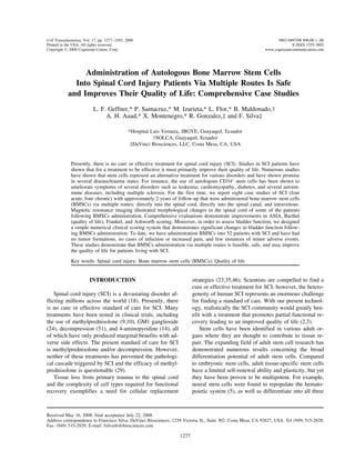
  • 4. 1280 GEFFNER ET AL. Table 1. Neurological Evaluations: Asia Impairment Grade/Frankel Grade/ Ashworth Score Prior to 6 Months After 1 Year After 2 Years After Case Study Administration Administration Administration Administration Acute Case 1 A/B/0 C/C/2 C/C/3 C/C/1 Case 2 A/A/3 A/C/1 A/C/1 C/C/2* Case 3 A/A/0 ND A/C/1 A/C/1 Case 4 A/A/0 C/C/1 C/C/1 C/C/1 Chronic Case 5 B/C/1 B/C/0 C/D/1 C/D/1 Case 6 C/D/3.5 C/D/3 D/D/3.5 D/D/ND Case 7 A/A/0 C/C/1 C/C/1 C/C/1+† Case 8 C/C/2 C/C/2 C/D/1 C/D/0 A summary of neurological evaluations for eight case studies. ND indicates not done. Case 4, 5, 6, 7, and 8 can walk with the aid of a walker or other medical device. There were few changes in spasticity (Ashworth score) for all cases described. The criteria of scoring systems are described below. *1 year 6 months. †1 year 3 months. ASIA Impairment Scale A complete: no preservation of function below level of injury, and no sacral sparing (S4–S5) B incomplete: sensory but no motor function is preserved below the neurological level and includes the sacral segments S4–S5 C incomplete: motor function is preserved below the neurological level, and more than half of key muscles below the neurological level have a muscle grade less than 3 D incomplete: motor function is preserved below the neurological level, and at least half of key muscles below the neurological level have a grade of 3 or more E normal: motor and sensory function are normal Frankel Scale A complete paralysis B sensory function only below the injury level C incomplete motor function below injury level D fair to good motor function below injury level E normal function Modified Ashworth 0 no increase in tone 1 slight increase in muscle tone, manifested by a catch and release or minimal resistance at the end of the ROM when the affected part(s) is moved in flexion or extension 1+ slight increase in muscle tone, manifested by a catch, followed by minimal resistance throughout the remainder (less than half) of the ROM 2 more marked increase in muscle tone through most of the ROM, but affected part(s) easily moved 3 considerable increase in muscle tone, passive movement difficult 4 affected part(s) rigid in flexion or extension with clear midsagittal T2-weighted images of the lesion an average total of 90.0 × 10⁶ CD34+ cells per adminis- tration (Table 3). Prior to BMSCs administration eachsite were allow into the study. The lesion site was quan- tified according to the midsagittal T2-weighted image patient underwent an MRI and neurological examina- tion. At approximately 6 months, 1 year, and 2 yearsand the vertebral segments were identified in order to isolate the area of interest for administration of BMSCs. following BMSCs administration the patients all under- went follow-up MRIs and neurological exams. Follow- ing administration of BMSCs there are noticeable mor- RESULTS phological changes within the spinal cord as illustrated by sequential MRIs of an acute patient (Fig. 2a–d) andTable 3 illustrates the demographics of each case (Cases 1–4 acute; Cases 5–8 chronic). Bone marrow chronic patient (Fig. 2e–h) taken prior to administration, 6 months after BMSCs administration, 1 year afterisolated from each patient was evaluated by FACS anal- ysis for the presence of CD34+ stem cells (Fig. 1, Table BMSCs administration, and approximately 2 years after BMSCs administration (Fig. 2). These studies illustrate3). The patients were administered with an average of 1.2 × 10⁶ CD34+ cells per kilogram of body weight for that administration of BMSCs directly into the spinal
  • 5. TRANSPLANT OF STEM CELLS INTO SPINAL CORD INJURY 1281 Table 2. ASIA Motor Score/Sensory Light Touch Score/Sensory Pin Prick Score Prior to 6 Months After 1 Year After 2 Years After Case Study Administration Administration Administration Administration Acute Case 1 50/64/64 54/72/72 54/83/80 56/90/90 Case 2 50/49/49 52/50/50 52/54/54 52/54/57* Case 3 50/42/42 ND 51/58/58 51/60/64 Case 4 50/76/76 52/78/78 56/80/82 58/80/84 Chronic Case 5 52/88/88 54/88/88 64/100/101 68/100/101 Case 6 62/66/66 62/80/80 78/94/94 85/94/94 Case 7 50/70/70 54/70/70 54/70/73 61/72/73† Case 8 58/86/86 58/88/88 66/98/98 70/98/98 Detailed ASIA motor and sensory scores for all eight cases. Motor scoring: 0 = total paralysis, 1 = palpable or visible contraction, 2 = active movement, gravity eliminated, 3 = active move- ment against gravity, 4 = active movement against some resistance, and 5 = active movement against full resistance. Scores are accumulated from right and left sides and are based upon evaluating a total of five arm and five leg muscle groups (total of 100 points maximum). The ASIA sensory scoring is for light touch and pin prick: 0 = absent, 1 = impaired, and 2 = normal. There are 28 dermatomes assessed for a total of 112 possible points. ND indicated not done. *1 year 6 months. †1 year 3 months. cord, directly into the spinal canal, and intravenously kel grade B) with no motor functions preserved below the level of injury. Moreover, there was no sensationmay cause morphological changes to the spinal cord. (light touch or pinprick; score of 64, 64, respectively) Case 1 below the S2 dermatome (Table 1, Table 2, Fig. 3a–c). Comprehensive data analysis indicates an initial BarthelA 28-year-old male sustained a gunshot wound to the T9 vertebral body resulting in a lesion and contusion scoring of 20 (Fig. 4a), Ashworth 0 (Table 1), and blad- der function (GGSF) score of 0 (Fig. 5a). Forty daysinjury with metallic fragment in the spinal canal. Initial evaluation of the patient’s MRI illustrated a lateral hemi- postinjury the patient had the bullet removed and was administered BMSCs. MRI of the vertebral column ap-section of the spinal cord at T9.1–T9.2 resulting from the bullet. In addition there was a contusion at the T8.1– proximately 2 years following administration illustrated the continual existence of the lateral hemisection withT10.1 levels with edema and spinal cord thickening (Fig. 2a). The patient’s evaluation prior to BMSCs ad- residual cavities formed from T7.1 to T9.1 and spinal cord thickening (Fig. 2d). In addition, there appeared toministration demonstrates that he sustained a complete injury (ASIA impairment grade A, motor score 50, Fran- be the formation of a small bridge of tissue with the Table 3. Case Studies Demographics Time of BMSCs CD34/kg Viability Administration Case No. Gender Age Weight (kg) Injury Level Injury Type Cell/10e6 CD34+ (%) After SCI 1 M 28 80 T9 gunshot 1.43 89.62 1.5 months 2 F 33 75 T4 gunshot 1.1 82.22 7 months 3 M 28 79 T5–6 fall 1.5 77.62 13 days 4 M 31 67 T12–L1 fall 0.94 96.27 5 days 5 M 37 86 T12 car accident 1.2 91.22 6 years 3 months 6 M 42 72 T4 gunshot 1.3 91.93 21 years 10 months 7 M 27 80 T11 gunshot 0.88 91.15 5 years 10 months 8 M 44 68 T12 fall 1.43 89.62 6 years 9 months Demographics of each patient are described including the type of injury, number, and viability of CD34+ cells that were administered in the cell suspension, and time of administration following SCI. Each patient had an average of 1.2 million CD34+ cells/kg of body weight administered (average of 90 million CD34+ cells total transplanted). Cases 1–4 are acute injuries while cases 4–8 are chronic.
  • 6. 1282 GEFFNER ET AL. Figure 1. Fluorescence activated cell sorting (FACS) results. Following mononuclear cell isolation each case had an aliquot of cells sent for identification of CD34+ cells. A representative plot of all cases demonstrates the identification of CD34+ cells in the bone marrow of patients with SCI. same MRI intensity of normal tissue and dorsal recuper- though she underwent a strict rehabilitation regimen; therefore, administration of BMSCs was performed 7ation of normal spinal cord signal below the injury level. Approximately 2 years post-BMSCs administration the months after spinal cord injury. At 6 months following BMSCs administration the patient progressively startedpatient has progressed significantly with an ASIA im- pairment grade of C, motor score 56, and Frankel grade to improve (Tables 1 and 2, Figs. 3a–c, 4a, 5a). At 6 months postadministration her ASIA motor and sensoryof C. In addition, there was sensation through the S4–5 dermatome (light touch or pinprick; score of 90, 90, re- scoring had improved slightly (52, 50, and 50 for motor, sensory light touch, and sensory pin prick, respectively)spectively) (Tables 1 and 2, Fig. 3a-c). Quality of life (Barthel score) increased from a score of 20 to 90 (Fig. (Table 2, Fig. 3a–c) while her quality of life (Barthel Score) improved 35 points (Fig. 4a). Her most recent4a) and bladder function improved from no function to complete bladder control (Fig. 5a). The patient can stand MRI (18 months) illustrated the persistence of a com- plete transection at T5.2–T6.1 with scar tissue at T5.2–on parallel bars and takes small strides with the use of a walker or crutches. Interestingly, the patient now T6.1. In addition, there was retraction of supraposterior cord fibers. There was no evidence of edema. At 18comes to physical therapy riding a quad motor bike. months following BMSCs administration, her bladder Case 2 function has improved from none to having sensation (Fig. 5a). Also, she improved from an ASIA impairmentA 33-year-old female sustained a gunshot wound to the thoracic area resulting in an injury to the spinal cord grade A to C (52, 54, and 57 for motor, sensory light touch, and sensory pinprick, respectively), Frankel A toat the T4 vertebral level. Initial evaluation of the pa- tient’s MRI illustrated a transection at T5.2–T6.1 and a C, and her spasticity had decreased from a 3 to 2 (Tables 1 and 2, Fig. 3a–c). There was no sensation below thesevere contusion injury with edema from T4.3 to T5.1. The patient’s evaluation prior to BMSCs administration T9 dermatome. Overall, we have noticed a qualitative increase in her life. She can now stand on the parallelsdemonstrated that she sustained a complete injury (ASIA impairment grade A, motor score 50, Frankel grade A) with braces on. with no motor functions preserved below the level of Case 3injury. In addition, there was no sensation (light touch or pinprick; score of 47, 47, respectively) below the T7 A 28-year-old male fell from a tree approximately 8 m and sustained an injury to his spinal cord at the T5–6dermatome (Tables 1 and 2, Fig. 3a–c). Eleven days fol- lowing her SCI, the patient underwent a first surgery in vertebral spinal level. Initial MRI illustrates an oblique hemisection on the left side at T5.1–T6.1 and contusionorder to decompress the spinal cord and remove the bul- let fragment from the spinal canal. Following decom- with edema cranial at T4.3–T3.1 and caudal at T6.2– T7.3. There is dilation of the ependyma below the injurypression, the patient had no functional changes even
  • 7. TRANSPLANT OF STEM CELLS INTO SPINAL CORD INJURY 1283 and posterior vertebral displacement narrowing the ca- below the injury area (motor score 50) (Tables 1 and 2, Fig. 3a–c). In addition, there was no sensation (lightnal. Initial neurological evaluation demonstrates that the patient sustained a complete injury (ASIA impairment touch or pinprick; score of 42, 42, respectively) below the T5 dermatome (Table 2, Fig. 3b, c). Patient was ad-grade A, Frankel A) with no motor function preserved Figure 2. MRI evaluations were performed for each case at several intervals. MRI images of an acute patient (Case 1) prior to administration (a), at 6 months (b), at 1 year (c), and approximately 2 years (d) after administration of BMSCs demonstrates structural changes of the spinal cord as time progresses following administration of BMSCs. The images illustrate a lesion of the spinal cord at T9 from a bullet (a). As time progresses there is the formation of a syringomyelic cavity with spinal cord thickening and the recuperation of normal signal below the injury site (d). MRI images of a chronic patient (Case 5) prior to administration (e), at 6 months (f), at 1 year (g), and at approximately 2 years (h) after administration demonstrates structural changes of the spinal cord as time progresses following administration of BMSCs. The images illustrate a lateral hemisection of the spinal cord at T11 with residual cavities at T12.1–T12.2 (e). At approximately 2 years following BMSCs administration the MRI illustrates a decrease in the residual cavity at T12.1 (h). Arrows denote injury area. (a–d) Acute injury; (e–h) chronic injury.
  • 8. 1284 GEFFNER ET AL. Table 4. Geffner, Gonzalez, Santacruz, and Flor (GGSF) Bladder Function Scale 0. No urinary bladder sensation or function*†‡§ 1. Patients with cystostomies that when are closed may involuntarily void through the urethra* 2. Bladder sensation or autonomic symptoms and inability to void*†‡§ 3. Bladder sensation or autonomic symptoms and passive voiding (spontaneous release of urine)*†‡ 3.5. Patients with open cystostomies that have bladder sensation or autonomic symptoms and passively void through the urethra (spontaneous release of urine)* 4. Bladder sensation with incomplete voiding (needs catheterization to complete voiding)†‡§ 5. Bladder sensation with active ability to void; however no control while voiding 6. Complete bladder control *Patients use suprapubic cystostomies in order to void their urinary bladder. †Patients use a catheter in order to void their urinary bladder. ‡Patients with a urine collector. §Patients that manually compress (massage) the hypogastric region in order to void their urinary bladder. ministered BMSCs 13 days following SCI and metal and passive voiding (spontaneous release of urine) (Fig. 5a). We were unable to perform additional MRIs afterrods were placed in order to stabilize his vertebral col- umn. The patient never showed up for his rehabilitation placement of the metal rods. Considering the challenges presented with his bed sore and the fact that he has notregimen and was given inadequate care at home immo- bile for 32 days. The patient acquired a giant bed sore performed any rehabilitation regimen, this patient has improved his quality of life following administration ofthat had to be surgically removed. The bed sore caused an infection, leading to a definitive colostomy and re- BMSCs. moval of the left femoral head. Due to the severe bed Case 4sore the patient had many autologous skin graft surger- ies in order to repair the damaged area. The patient had A 31-year-old male fell from a ladder approximately 3 m and sustained an injury to his spinal cord at thean initial Barthel score of 5 (Fig. 4a), bladder function score of 0 (Fig. 5a), and Ashworth (spasticity) score of T12–L1 vertebral level. The patient was evaluated im- mediately after injury. MRI of the vertebral column il-0 (Table 1). At approximately 2 years post-BMSCs ad- ministration the patient has an improved Frankel grade lustrated posterior vertebral displacement of L1 over T12 grade II and severe narrowing of the spinal canal.of C, an ASIA impairment grade of A with some im- provements in his sensory score (60, 64 for light touch There was a contusion with edema at T12.2–L1.3 with a hematoma. The initial neurological evaluation demon-and pin prick, respectively), and no sensation below the T11 dermatome (Tables 1 and 2, Fig. 3b, c). His quality strates that the patient sustained a complete injury (ASIA impairment grade A, Frankel A) with no motorof life score (Barthel Index) has increased from 5 to 40 (Fig. 4a) and bladder function increased from no bladder function preserved below the injury level (motor score 50) (Tables 1 and 2, Fig. 3a). There was no sensationfunction to bladder sensation or autonomic symptoms Table 5. Definitions and Terms Bladder sensation: A feeling or awareness of conditions within the urinary bladder resulting from the stimulation of sensory receptors Bladder function: The action performed by the urinary bladder (i.e., the ability to void urine) Suprapubic cystostomies: A connection between the urinary bladder and the skin to drain urine from the bladder Void: The ability to evacuate (empty) the urinary bladder Involuntary: Independent of or contrary to volition Dysesthesia: Abnormal sensations on the skin, such as a feeling of numbness, tingling, burning, itching, electric shock, pins and needles Autonomic symptoms: Dysesthesia, pyloric erection, pain, burning, sudden hypertension, sweating, and bradycardia normally triggered by distention of the urinary bladder Passive voiding: Evacuation or partial evacuation of the urinary bladder without any control Manuel bladder (hypogastric) compression for voiding: Through active compression (massage) of the hypogastric region, urine is released through the urethra
  • 9. TRANSPLANT OF STEM CELLS INTO SPINAL CORD INJURY 1285 Figure 3. Graphical representations of the ASIA motor, ASIA sensory light touch, and ASIA sensory pin prick. There are motor improvements in all cases but most notable are acute cases 1 and 4, and chronic cases 5, 6, 7, and 8 (a). ASIA sensory light touch and pin prick scores demonstrate that there are improvements in all cases but most notable for acute cases 1 and 3 and chronic cases 5, 6, and 8 (b, c). *The last follow-up for Case 2 was at 1 year 6 months. **The last follow-up for Case 7 was at 1 year 3 months. ND, not done.
  • 10. 1286 GEFFNER ET AL. Figure 4. Quality of life evaluation performed using the Barthel Index Score for eight cases. Barthel scoring indicates an improvement in quality of life for all four acute spinal cord injury cases (a) and all four chronic spinal cord injury cases (b) following administration of BMSCs. The greatest improvements occurred from prior to administration to 6 months after administration of BMSCs (a, b). ND, not done. **The last follow-up for Case 2 was at 1 year 6 months. *The last follow-up for Case 7 was at 1 year 3 months. (light touch or pinprick; score of 76 and 76, respec- kel score C) with active movement against gravity at the hip flexors and sensation through the L1 dermatome andtively) below the T12 dermatome (Table 2, Fig. 3b, c) and he had an initial Ashworth score of 0 (Table 1). at the S2–S3 dermatome (light touch and pin prick; 84 and 80, respectively) (Tables 1 and 2, Fig. 3a–c). Bar-Quality of life scoring demonstrates an initial Barthel score of 30 (Fig. 4a) and no bladder function (Fig. 5a). thel scoring demonstrates an improvement from an ini- tial score of 30 to 90 (Fig. 4a) while bladder functionFive days following initial trauma posterior rods were placed in order to stabilize the vertebral column and ad- went from absolutely no function to bladder sensation with active ability to void; however, no control whileministration of BMSCs was done. At 2 years following BMSCs administration the patient has improved signifi- voiding (Fig. 5a). We were unable to do any more MRI due to the placement of metal rods. This patient cancantly (ASIA impairment grade C, motor score 58, Fran-
  • 11. TRANSPLANT OF STEM CELLS INTO SPINAL CORD INJURY 1287 walk a couple of blocks with the use of a walker and lowing injury. MRI prior to BMSCs administration illus- trated an anterior disc herniation at T11–12 and a lateralankle braces. hemisection of the spinal cord at T11.3. In addition, Case 5 there was a residual cavity from T12.1 to T12.2 (Fig. 2e). The initial neurological evaluation demonstratedA 37-year-old male sustained an injury to the spinal cord at the T12 vertebral spinal level from a car acci- that the patient sustained an incomplete injury (ASIA impairment grade B, Frankel C) with only palpable ordent. The patient was evaluated 6 years 2 months fol- Figure 5. Evaluation of bladder function using a newly designed bladder function assessment scoring system entitled Geffner, Gonzalez, Santacruz, and Flor (GGSF) Bladder Function Score. There were significant improvements in bladder function for acute SCI Cases 1 and 4 (a) and chronic SCI Case 7 (b) after administration of BMSCs. In addition, chronic SCI Cases 5 and 6 improved to complete bladder control. Overall, all SCI cases evaluated had an improvement in bladder function as assessed using the GGSF scale. Full scale descriptions are attached as an appendix. ND. not done. **The last follow-up for Case 2 was at 1 year 6 months. The last follow- up for Case 7 was at 1 year 3 months.
  • 12. 1288 GEFFNER ET AL. visible contractions at the L2 level (ASIA motor score 94, and Frankel D grade (Tables 1 and 2, Fig. 3a). More- over, his sensory scoring has improved from a 66 light52, 1 point on each side at the L2 level) (Tables 1 and 2, Fig. 3a). In addition, there was impaired sensation touch/66 pin prick to a 94 light touch/94 pin prick (Ta- ble 2, Fig. 3b, c). Barthel score has improved dramati-(light touch or pinprick; score of 88 and 88, respec- tively) through the S4–5 dermatome (Table 2, Fig. 3b, cally to a maximum score of 100 and he has complete bladder function (Figs. 4b and 5b, respectively). He nowc) and an Ashworth score of 1 (Table 1). Initial Barthel score was a 65 (Fig. 4b) and he had bladder sensation has the ability to walk using forearm crutches and is able to walk up and down stairs.with active ability to void; however, no control while voiding (GGSF score of 5) (Fig. 5b). Six years and 3 Case 7months following SCI the patient underwent a partial discectomy and administration of BMSCs. At 2 years A 27-year-old male sustained a gunshot wound to the scapula region that passed through his spine and exitedfollowing administration the patient has improved sig- nificantly with the MRI illustrating a disc herniation at his chest, causing a spinal cord injury at the T11 verte- bral level. The patient was evaluated for BMSCs admin-T11–12, a lesion at T11.3, and a small cavity at T12.1 (Fig. 2h). His neurological evaluation demonstrates an istration 5 years 9 months follow SCI. Initial MRI illus- trated a fracture at T11.1 with a cavity from theASIA impairment grade of C (improved motor score of 68, scores of 1–3 from the L2–S1 level), and Frankel D projectile in the vertebral body and an oblique hemisec- tion of the spinal cord at T11.1–T11.2 His neurological(Tables 1 and 2, Fig. 3a). His ASIA sensory score has increased from 88 light touch and 88 pin prick to 100 evaluation prior to BMSCs administration demonstrated that he has an complete injury (ASIA impairment gradelight touch and 101 pin prick (Table 2, Fig. 3c, b). Bar- thel score is now 100 (maximum value) and he has full A, Frankel A) with a motor score of 50, meaning the absence of all key muscles below the injury level (Ta-control of his bladder (Figs. 4b and 5b, respectively). This patient can now walk with braces and crutches for bles 1 and 2, Fig. 3a). There was no sensation below the T11 dermatome (70 and 70 for light touch and pin prick,more than 1 h. respectively) and his Ashworth score was 0 (Tables 1 Case 6 and 2, Fig. 3b, c). His Barthel score was 35 and he had no bladder function (Figs. 4b and 5b, respectively). MRIA 42-year-old male sustained a gunshot wound to the chest in 1984 that penetrated his vertebral column and evaluation 1 year 3 months following BMSCs adminis- tration illustrated the existence of the fracture at T11.1was lodged in his dura at the T4 vertebral spinal level, causing an injury to his spinal cord. The patient immedi- and the oblique hemisection injury has expanded from T11.1–T12.1. There was a small residual cavity aboveately underwent a laminectomy at T3–4 and surgical re- moval of the bullet. MRI prior to BMSCs administration the injury from T10.1–T10.3. Although his MRI demon- strates an expanded injury, his neurological evaluationillustrated that he has an oblique lesion at T3.2–T4.1 and hypoplasia. There is a residual cavity at T3.2–T4.2. indicated a significant functional improvement (ASIA impairment grade C, improved motor score of 61) (Ta-His neurological evaluation prior to administration (21 years 10 months after initial trauma) demonstrated that bles 1 and 2, Fig. 3a). In addition, his sensory score has increased slightly with impaired sensation at the S2–S3he is categorized as an incomplete injury (ASIA impair- ment grade C, Frankel D) with active movement, gravity dermatome level (light touch 72, pin prick 73) (Table 2, Fig. 3b, c). There are little changes in his spasticity. Bar-eliminated through the L4 level on the left/L3 level on the right and palpable or visible contractions at the thel scoring indicates a significantly improvement to a score of 85 (Fig. 4b) and his bladder function has im-L5–S1 level on the left side only (ASIA motor score 62) (Tables 1 and 2, Fig. 3a). In addition, there was proved from 0 to a 3 (sensation or autonomic symptoms and passive voiding) (Fig. 5b). One year 3 months fol-no sensation below the T7 dermatome on the right and impaired through the S4–5 dermatome from T6 (light lowing BMSCs administration with the aid of a walker and braces, this patient has gained the ability to walk.touch or pinprick; score of 66 and 66, respectively) (Ta- ble 2, Fig. 3b, c). Initial Barthel score was 55 (Fig. 4b) Case 8and he had bladder sensation with active ability to void; however, no control while voiding (score of 5) (Fig. 5b). In 1999, a 44-year-old male fell 8 m and sustained an injury to the spinal cord at the T12 vertebral spinalHis 2-year follow-up MRI illustrated minor changes with an oblique lesion at T3.3–T4.3 and hypoplasia. level. Five days following SCI the patient had metal rods placed in order to stabilize his vertebral column,There remains the residual cavity at T3.2–T4.2. Neuro- logical evaluation 2 years after BMSCs administration which were subsequently removed 4.5 years later. The patient was evaluated at the hospital for BMSCs admin-demonstrated a significant improvement of motor func- tion with an ASIA impairment grade of D, motor score istration in early 2006. His MRI prior to BMSCs admin-
  • 13. TRANSPLANT OF STEM CELLS INTO SPINAL CORD INJURY 1289 istration illustrated a compression of the vertebral body BMSCs via multiple routes is feasible, safe, and most importantly improves the quality of life for both acuteat T12 with a 10% posterior displacement. There was compression of the spinal cord at T11.2–T12.1 with a and chronic patients suffering from SCI. Moreover, there has been no incidence of infection or severe ad-residual cavity at T11.2–T12.3. His initial neurological examination with us indicated that he had an incomplete verse reactions for any cases documented to date. This study clearly documents the feasibility of cell replace-injury (ASIA impairment grade C, Frankel C) with ac- tive movement, gravity eliminated at L2 and L3 (motor ment strategies as an alternative therapy for SCI. In ad- dition, we have a unique method of delivering the cells;score 58) (Tables 1 and 2, Fig. 3a). There was no sensa- tion below the L3 dermatome (86 and 86, light touch all patients get cells directly into the spinal cord, directly into the spinal canal, and an intravenous injection ofand pin prick, respectively) and his Ashworth score was 2 (Tables 1 and 2, Fig. 3b, c). His Barthel score was a 55 cells. Although multiple route administration limits the mechanistic understanding for cell-based applications,(Fig. 4b) and he had bladder sensation with incomplete voiding (score of 4) (Fig. 5b). Two years following this assures us that the BMSCs will reach the necessary target in order to fulfill their niche and possibly promoteBMSCs administration his MRI demonstrated a lesion at T11.3–T12.1 with a residual cavity that has shrunk at host endogenous repair within the SCI area. Shi et al. demonstrated that bone marrow-derived CD34+ EPCsT11.2–T11.3. There was a small edema below the lesion at T12.2–T12.3. Neurological evaluation indicated func- from canines migrated to the vascular injury site (44). In another study, autologous bone marrow CD34+ cellstional improvement (ASIA C, improved motor score of 70, and Frankel D) and a reduction in spasticity (Tables labeled with magnetic nanoparticles that were delivered into the spinal cord via lumbar puncture migrated into1 and 2, Fig. 3a). There was impaired sensation through the S4–5 dermatome with an improved sensory score of the injured site in patients with chronic SCI (13). Following primary trauma to the adult spinal cord98 and 98 for light touch and pin prick, respectively (Table 2, Fig 3b, c). Barthel scoring indicates he has there is evidence of hemorrhage and blood flow is atten- uated within the spinal cord. This disruption in bloodimproved to the maximum score of 100 (Fig. 4b) and he has the ability to actively void his bladder (GGSF score flow leads to spinal cord infarction, the disruption of the blood–spinal cord injury barrier, edema, the release of5) (Fig. 5b). Interestingly, even though he has the ability to walk with crutches and can go up and down stairs; vasoactive molecules influencing spinal cord perfusion, and ischemia. This primary disruption ultimately leadsbecause he has active movement against full resistance (score 5) in only 40% of his key muscles he continues to the death of millions of cells and the activation of secondary degeneration, which is a cascade of eventsto be considered ASIA impairment grade C. that exacerbate primary trauma (18,36,38). Secondary DISCUSSION degeneration is the present target for pharmaceutical in- tervention in acute SCI; however, any interventionThe standard of care for acute SCI is methylpredniso- lone and/or decompression. Clinical studies using surgi- would have to be applied rapidly as within 72 h the white matter in the spinal cord is thought to be irrevers-cal intervention for acute SCI (decompression) have shown no difference in neurological recovery between ibly damaged (8) and cell death progresses for months due to the complicated cascade of secondary degenera-operated and nonoperated patients (50,51). In three Na- tional Acute Spinal Cord Injury Studies (NASCIS I, II, tion (19). Autologous stem cells represent an alternative ther-and III), the use of methylprednisolone demonstrated minimal overall benefits (10–12). These benefits did not apy when conventional methods have not worked for various devastating disorders. Specifically, CD34+ stemcome without much skepticism of the post hoc analysis of the data (17,30,40). Indeed, the use of methylprednis- cells (HSCs) and/or endothelial progenitor cells (EPCs), which are easily attainable from bone marrow or periph-olone or any steroid does not come without risk. In all three NASCIS studies the incidence of sepsis and pneu- eral blood. HSCs and EPCs are well known for their ability to promote angiogenesis (43,54). Angiogenesis ismonia was higher in the methylprednisolone groups compared to other treatment groups. Moreover, gastroin- necessary for wound healing and likely plays a critical role in establishing a growth-permissive environment.testinal complications were also reported following high-dose methylprednisolone (10–12). According to a Results have shown that EPCs play an essential role in repairing injured blood vessels and transplantation ofcommittee of Canadian physicians, the use of high-dose methylprednisolone is not an evidence-based standard of EPCs into ischemic limbs of rats induced collateral ves- sel formation (31). Moreover, it was noted that trans-care for SCI (29). It has been demonstrated that intravenous and intra- planted EPCs secrete proangiogenic growth factors such as VEGF, HGF, and IGF-1, which lead to new vesselarterial transplantation of mononuclear cells into SCI is safe (48). Here we demonstrate that administration of development (39,53). Studies indicate that progenitor
  • 14. 1290 GEFFNER ET AL. cells contribute not only to the restoration of injured tis- ors and knee extensors. This individual can walk with crutches and can go up and down stairs, which is a vastsues, but also to the pathological remodeling (26,33,53). Interestingly, in SCI revascularization occurs prior to improvement in his quality of life. Other challenges are whether to separate out the ASIA upper and lower ex-nerve regeneration (55). Because BMSCs possess angio- genic properties, we hypothesize that improved blood tremity scores. In almost all of our cases lower extremity scores were nonexistent (total motor score of 50) and aflow and oxygen supply within the injury area may have contributed to the functional improvements seen in SCI 5–10 point increase may make a large difference if one separates the upper and lower extremity scores. In agree-patients transplanted with autologous BMSCs. Alterna- tively, it is well documented that BMSCs promote host ment with the ICCP panel, our personal observation is that separation of lower and upper extremities in theendogenous repair (15,27,28). For instance, transplanta- tion of marrow stromal cells into SCI rats promotes axo- ASIA motor scale can better indicate functional im- provement compared to combining the data, which maynal regeneration (28), and transplantation of BMSCs into a mouse model of chemically induced pancreatic illustrate a nominal effect (47). A survey of the SCI community illustrates that onedamage causes endogenous pancreatic regeneration (27). Clearly, administration of autologous BMSCs is a prom- of the most important aspects in improving their quality of life would be the ability to control bladder functionising cell replacement strategy for several human disor- ders. (2). This study indicated that presently we would greatly benefit from the development of treatments that lead toOne of the greatest challenges in finding a treatment or improving the quality of life for the SCI community partial functional recovery in order to improve the qual- ity of life for the SCI community. Our data clearly dem-is the vast heterogeneity of human SCI; no two injuries are alike. It is well understood that tremendous chal- onstrate that administration of autologous BMSCs im- proves the quality of life for SCI patients (Figs. 4 andlenges exist in gathering data in order to validate a spe- cific treatment type for SCI (47,49). Presently, we have 5). Because bladder function is of great importance, we designed the GGSF bladder function score to facilitateperformed administration of autologous BMSCs using our unique multiple route delivery system into 52 SCI bladder function assessment (Tables 4 and 5). It is more in depth compared to questionnaires in quality of lifepatients. However, it has been challenging to gather comprehensive long-term data. Therefore, in this study scoring systems, such as the Barthel index, which are a crude method of assessing bladder function (0 = inconti-we present the eight cases that we have with comprehen- sive long-term data. We presently have 25 cases of SCI nent, or catheterized and unable to manage alone; 5 = occasional accident; 10 = continent) yet measure overallwith slightly greater than 3 months comprehensive fol- low-up. There have been no cases of tumor formation, quality of life changes. It is well understood that sponta- neous recovery in SCI can occur for as long as 1 yearincreased pain, and/or deterioration of function follow- ing administration of BMSCs. There were very few ad- following SCI (21). Indeed, this may have occurred within our acute SCI patients; however, it is importantverse reactions observed following administration of BMSCs. We observed a transient lack of erection and/ to note that all of our acute patients improved signifi- cantly with no signs of deterioration or impediment ofor ejaculation in four patients, sweating on one half of the body on two patients, and spinal cord canal fistula presumed spontaneous recovery. Moreover, Case 2 had a laminectomy followed by physical therapy for 6in one patient whose metal rods had to be withdrawn. Nonetheless, there is an overall improvement in the months with no improvements. Following administra- tion of BMSCs, her quality of life improved.quality of life for patients with SCI following adminis- tration of BMSCs. In conclusion, these studies document an alternative cell-based method of treating SCI. Interestingly, it docu-One of the challenges in this study was with patients that had clear improvements that cannot be reflected ments that BMSCs administration via multiple routes may be an effective treatment for patients that sufferedcorrectly by the ASIA impairment grade. There are sev- eral individuals that have large improvements in only a SCI from a gunshot wound. To the best of our knowl- edge this is the first documentation of SCI patients beingone to two major muscle groups yielding a definite im- provement in quality of life; however, ASIA impairment administered BMSCs directly into the spinal cord fol- lowing gunshot wounds. Presently, we are investigatinggrade does not reflect these beneficial changes (7). For instance, Case 8 has gone from an initial ASIA motor long-term effects of BMSCs administration in quadri- plegic injuries. However, the difficulties of tracking,score of 58 (motor score of 2 on each side at the L2 and L3 levels) to an ASIA motor score of 70 (motor score monitoring, and assessing several parameters in patients that have been administered BMSCs remains our largestof 5 on each side at the L2 and L3 levels), meaning active movement against full resistance for the hip flex- challenge. Nevertheless, we report here that administra-
  • 15. TRANSPLANT OF STEM CELLS INTO SPINAL CORD INJURY 1291 11. Bracken, M. B.; Shepard, M. J.; Hellenbrand, K. G.;tion of BMSCs via multiple routes is safe and feasible Collins, W. F.; Leo, L. S.; Freeman, D. F.; Wagner, F. C.;for several SCI types. Most importantly, it may improve Flamm, E. S.; Eisenburg, H. M.; Goodman, J. H.; Perot, the quality of life for many suffering from SCI. P. L.; Green, B. A.; Grossman, R. G.; Meagher, J. N.; Young, W.; Fischer, B.; Clifton, G. L.; Hunt, W. E.; Rif-ACKNOWLEDGMENTS: We thank the Junta de Beneficencia kinson, N. Methylprednisolone and neurological functionde Guayaquil, specifically Oscar Orrantia, Lautaro Aspiazu, 1 year after spinal cord injury. Results of the Nationaland Estefano Isaias, for commencing and expanding the stem Acute Spinal Cord Injury Study. J. Neurosurg. 63:704–cell research programs at Luis Vernaza Hospital (LVH). We 713; 1985.thank the LVH principles Werner Mo¨eller, Jorge Tola, and 12. Bracken, M. B.; Shepard, M. J.; Holford, T. R.; Leo-Antonio Ortega for their support. We also thank the Physio- Summers, L.; Aldrich, E. F.; Fazl, M.; Fehlings, M.; Herr,therapy, Rehabilitation, and Medical staff of LVH for their D. L.; Hitchon, P. W.; Marshall, L. F.; Nockels, R. P.;hard work and praise, specifically Bolivar Cardenas, Erin Sa- Pascale, V.; Perot, Jr., P. L.; Piepmeier, J. M.; Sonntag,lazar, Angel Auad Saab, Eduardo Landivar, and Margarita V. K.; Wagner, F.; Wilberger, J. E.; Winn, H. R.; Young,Villavicencio. We thank SOLCA Guayaquil hospital for their W. Administration of methylprednisolone for 24 or 48collaborative effort and their Hematology and Cytometry staff hours or tirilazad mesylate for 48 hours in the treatmentfor their diligence. of acute spinal cord injury. Results of the Third National Acute Spinal Cord Injury Randomized Controlled Trial. REFERENCES National Acute Spinal Cord Injury Study. J. Am. Med. Assoc. 277:1597–1604; 1997.1. Alessandri, G.; Pagano, S.; Bez, A.; Benetti, A.; Pozzi, S.; Iannolo, G.; Baronio, M.; Invernici, G.; Caruso, A.; 13. Callera, F.; de Melo, C. M. Magnetic resonance tracking of magnetically labeled autologous bone marrow CD34+Muneretto, C.; Bisleri, G.; Parati, E. Isolation and culture of human muscle-derived stem cells able to differentiate cells transplanted into the spinal cord via lumbar puncture technique in patients with chronic spinal cord injury:into myogenic and neurogenic cell lineages. Lancet 364: 1872–1883; 2004. CD34+ cells’ migration into the injured site. Stem Cells Dev. 16:461–466; 2007.2. Anderson, K. D. Targeting recovery: Priorities of the spi- nal cord-injured population. J. Neurotrauma 21:1371– 14. Cardenas, D. D.; Ditunno, J.; Graziani, V.; Jackson, A. B.; Lammertse, D.; Potter, P.; Sipski, M.; Cohen, R.; Blight,1383; 2004. 3. Anderson, K. D.; Borisoff, J. F.; Johnson, R. D.; Stiens, A. R. Phase 2 trial of sustained-release fampridine in chronic spinal cord injury. Spinal Cord 45:158–168; 2007.S. A.; Elliott, S. L. The impact of spinal cord injury on sexual function: Concerns of the general population. Spi- 15. Chopp, M.; Zhang, X. H.; Li, Y.; Wang, L.; Chen, J.; Lu, D.; Lu, M.; Rosenblum, M. Spinal cord injury in rat:nal Cord 45:328–337; 2007. 4. Aversa, F.; Martelli, M. F. Transplantation of haploidenti- Treatment with bone marrow stromal cell transplantation. Neuroreport 11:3001–3005; 2000.cally mismatched stem cells for the treatment of malignant diseases. Springer Semin. Immunopathol. 26:155–168; 16. Clarke, D. L.; Johansson, C. B.; Wilbertz, J.; Veress, B.; Nilsson, E.; Karlstro¨m, H.; Lendahl, U.; Frise´n, J. Gener-2004. 5. Bjornson, C. R.; Rietze, R. L.; Reynolds, B. A.; Magli, alized potential of adult neural stem cells. Science 288: 1660–1663; 2000.M. C.; Vescovi, A. L. Turning brain into blood: A hema- topoietic fate adopted by adult neural stem cells in vivo. 17. Coleman, W. P.; Benzel, D.; Cahill, D. W.; Ducker, T.; Geisler, F.; Green, B.; Gropper, M. R.; Goffin, J.; Madsen,Science 283:534–537; 1999. 6. Blatt, A.; Cotter, G.; Leitman, M.; Krakover, R.; Kaluski, P. W.; Maiman, D. J.; Ondra, S. L.; Rosner, M.; Sasso, R. C.; Trost, G. R.; Zeidman, S. A critical appraisal of theE.; Milo-Cotter, O.; Resnick, I. B.; Samuel, S.; Gozal, D.; Vered, Z.; Slavin, S.; Shapira, M. Y. Intracoronary admin- reporting of the National Acute Spinal Cord Injury Studies (II and III) of methylprednisolone in acute spinal cord in-istration of autologous mononuclear cells after induction of short ischemia is safe and may improve hibernation and jury. J. Spinal Disord. 13:185–199; 2000. 18. Dumont, R. J.; Okonkwo, D. O.; Verma, S.; Hurlbert,ischemia in patients with ischemic cardiomyopathy. Am. Heart J. 150:986.e1–986.e7; 2005. R. J.; Boulos, P. T.; Ellegala, D. B.; Dumont, A. S. Acute spinal cord injury, part I: Pathophysiological mechanisms,7. Blight, A. R.; Tuszynski, M. H. Clinical trials in spinal cord injury. J. Neurotrauma 23:586–593; 2006. & part II: Contemporary pharmacotherapy. Clin. Neuro- pharmacol. 24:254–279; 2001.8. Blight, A. R.; Young, W. Central axons in injured cat spi- nal cord recover electrophysiological function following 19. Emery, E.; Aldana, P.; Bunge, M. B.; Puckett, W.; Srini- vasan, A.; Keane, R. W.; Bethea, J.; Levi, A. D. Apoptosisremyelination by Schwann cells. J. Neurol. Sci. 91:15–34; 1989. after traumatic human spinal cord injury. J. Neurosurg. 89: 911–920 1998.9. Bracken, M. B. Methylprednisolone in the management of acute spinal cord injuries. Med. J. Aust. 153:368; 1990. 20. Fassas, A. Autologous stem cell transplants in treatment of multiple sclerosis: Where we stand and future pros-10. Bracken, M. B.; Shepard, M. J.; Collins, W. F.; Holford, T. R.; Young, W.; Baskin, D. S.; Eisenberg, H. M.; pects. Int. J. Hematol. 76(Suppl. 1):223–225; 2002. 21. Fawcett, J. W.; Curt, A.; Steeves, J. D.; Coleman, W. P.;Flamm, E.; Leo-Summers, L.; Maroon, J.; Marshall, L. F.; Perot, P. L.; Piepmeier, J. M.; Sonntag, V. K.; Wagner, Tuszynski, M. H.; Lammertse, D.; Bartlett, P. F.; Blight, A. R.; Dietz, V.; Ditunno, J.; Dobkin, B. H.; Havton,F. C.; Wilberger, J. E.; Winn, H. R. A randomized, con- trolled trial of methylprednisolone or naloxone in the L. A.; Ellaway, P. H.; Fehlings, M. G.; Privat, A.; Gross- man, R.; Guest, J. D.; Kleitman, N.; Nakamura, M.; Gavi-treatment of acute spinal-cord injury. Results of the Sec- ond National Acute Spinal Cord Injury Study. N. Engl. J. ria, M.; Short, D. Guidelines for the conduct of clinical trials for spinal cord injury as developed by the ICCPMed. 322:1405–1411; 1990.
  • 16. 1292 GEFFNER ET AL. panel: spontaneous recovery after spinal cord injury and 37. Manginas, A.; Goussetis, E.; Koutelou, M.; Karatasakis, G.; Peristeri, I.; Theodorakos, A.; Leontiadis, E.; Plessas,statistical power needed for therapeutic clinical trials. Spi- nal Cord 45:190–205; 2006. N.; Theodosaki, M.; Graphakos, S.; Cokkinos, D. V. Pilot study to evaluate the safety and feasibility of intracoro-22. Flanders, A. E.; Spettell, C. M.; Freidman, D. P.; Marino, R. J.; Herbison, G. J. The relationship between the func- nary CD133(+) and CD133(−) CD34(+) cell therapy in patients with nonviable anterior myocardial infarction.tional abilities of patients with cervical spinal cord injury and the severity of damage revealed by MR imaging. Am. Catheter Cardiovasc. Interv. 69:773–781; 2007. 38. Mautes, A. E. M.; Weinzierl, M. R.; Donovan, F.; Noble,J. Neuroradiol. 20:926–934; 1999. 23. Garbossa, D.; Fontanella, M.; Fronda, C.; Benevello, C.; L. J. Vascular events after spinal cord injury: Contribution to secondary pathogenesis. Phys. Ther. 80:673–687; 2000.Muraca, G.; Vercelli, A. New strategies for repairing the injured spinal cord: The role of stem cells. Neurol. Res. 39. Murasawa, S.; Kawamoto, A.; Horii, M.; Nakamori, S.; Asahara, T. Niche-dependent translineage commitment of28:500–504; 2006. 24. Geisler, F. H.; Coleman, W. P.; Grieco, G.; Poonian, D. endothelial progenitor cells, not cell fusion in general, into myocardial lineage cells. Arterioscler. Thromb. Vasc.Sysgen Study Group 2001. The Sygen multicenter acute spinal cord injury study. Spine 26(Suppl. 24):S87–S98; Biol. 25:1388–1394; 2005. 40. Nesathurai, S. Steroids and spinal cord injury: Revisiting2001. 25. Gratama, J. W.; Keeney, M.; Sutherland, D. R. Enumera- the NASCIS 2 and NASCIS 3 trials. J. Trauma 45:1088– 1093; 1998.tion of CD34 hematopoietic stem and progenitor cells. Curr. Prot. Cytometry 6.4.1–6.4.22; 1999. 41. Ozbaran, M.; Omay, S. B.; Nalbantgil, S.; Kultursay, H.; Kumanlioglu, K.; Nart, D.; Pektok, E. Autologous periph-26. He, T.; Smith, L. A.; Harrington, S.; Nath, K. A.; Caplice, N. M.; Katusic, Z. S. Transplantation of circulating endo- eral stem cell transplantation in patients with congestive heart failure due to ischemic heart disease. Eur. J. Cardio-thelial progenitor cells restores endothelial function of de- nuded rabbit carotid arteries. Stroke 10:2378–2384; 2004. thorac. Surg. 25:342–350; 2004. 42. Patel, A. N.; Geffner, L.; Vina, R. F.; Saslavsky, J.;27. Hess, D.; Li, L.; Martin, M.; Sakano, S.; Hill, D.; Strutt, B.; Thyssen, S.; Gray, D. A.; Bhatia, M. Bone marrow- Urschel, H. C.; Kormos, R.; Benetti, F. Surgical treatment for congestive heart failure with autologous adult stemderived stem cells initiate pancreatic regeneration. Nat. Biotech. 21:763–770; 2003. cell transplantation: A prospective randomized study. J. Thorac. Cardiovasc. Surg. 130:1631–1638; 2005.28. Hofstetter, C. P.; Schwarz, E. J.; Hess, D.; Widenfalk, J.; El Manira, A.; Prockop, D. J.; Olson, L. Marrow stromal 43. Popa, E. R.; Harmsen, M. C.; Tio, R. A.; van der Strate, B. W. A.; Brouwer, L. A.; Schipper, M.; Koerts, J.; Decells form guiding strands in the injured spinal cord and promote recovery. Proc. Natl. Acad. Sci. USA 99:2199– Jongste, M. J. L.; Hazenberg, A.; Hendriks, M.; van Luyn, M. J. A. Circulating CD34+ progenitor cells modulate host2204; 2002. 29. Hugenholtz, H. Methylprednisolone for acute spinal cord angiogensis and inflammation in vivo. J. Mol. Cell. Cardiol. 41:86–96; 2006.injury: Not a standard of care. Can. Med. Assoc. J. 168: 1145–1146; 2003. 44. Shi, Q.; Rafii, S.; Wu, M. H.; Wijelath, E. S.; Yu, C.; Ishida, A.; Fujita, Y.; Kothari, S.; Mohle, R.; Sauvage,30. Hurlbert, R. J. Methylprednisolone for acute spinal cord injury: An inappropriate standard of care. J. Neurosurg. L. R.; Moore, M. A.; Storb, R. F.; Hammond, W. P. Evi- dence for circulating bone marrow-derived endothelial93(Suppl. 1):1–7; 2000. 31. Iba, O.; Matsubara, H.; Nozawa, Y.; Fujiyama, S.; cells. Blood 92:362–367; 1998. 45. Skyler, J. S. Cellular therapy for type 1 diabetes: Has theAmano, K.; Mori, Y.; Kojima, H.; Iwasaka, T. Angiogen- esis by implantation of peripheral blood mononuclear cells time come? J. Am. Med. Assoc. 297:1599–1600; 2007. 46. Steeves, J. D.; Fawcett, J.; Tuszynski, M. Report of inter-and platelets into ischemic limbs. Circulation 106:2019– 2025; 2002. national clinical trials workshop on spinal cord injury; February 20–21, 2004, Vancouver, Canada. Spinal Cord32. Jiang, Y.; Henderson, D.; Blackstad, M.; Chen, A.; Miller, R. F.; Verfaillie, C. M. Neuroectodermal differentiation 42:591–597; 2004. 47. Steeves, J. D.; Lammertse, D.; Curt, A.; Fawcett, J. W.;from mouse multipotent adult progenitor cells. Proc. Natl. Acad. Sci. USA 100(Suppl. 1):11854–11860; 2003. Tuszynski, M. H.; Ditunno, J. F.; Ellaway, P. H.; Fehlings, M. G.; Guest, J. D.; Kleitman, N.; Bartlett, P. F.; Blight,33. Kalka, C.; Masuda, H.; Takahashi, T.; Kalka-Moll, W. M.; Silver, M.; Kearney, M.; Li, T.; Isner, J. M.; Asahara, T. A. R.; Dietz, V.; Dobkin, B. H.; Grossman, R.; Short, D.; Nakamura, M.; Coleman, W. P.; Gaviria, M.; Privat, A.Transplantation of ex vivo expanded endothelial progeni- tor cells for therapeutic neovascularization. Proc. Natl. Guidelines for the conduct of clinical trials for spinal cord injury as developed by the ICCP panel: Clinical trial out-Acad. Sci. USA 97:3422–3427; 2000. 34. Katz, R. T. Spasticity. In: O’Young, B.; Young, M. A.; come measures. Spinal Cord 45:206–221; 2006. 48. Sykova´, E.; Homola, A.; Mazanec, R.; Lachmann, H.;Stiens, S. A., eds. PM&R secrets. Philadelphia: Hanley & Belfus; 1997:487. Konra´dova´, S. L.; Kobylka, P.; Pa´dr, R.; Neuwirth, J.; Komrska, V.; Va´vra, V.; Stulı´k, J.; Bojar, M. Autologous35. Keirstead, H. S.; Nistor, G.; Bernal, G.; Totoiu, M.; Clout- ier, F.; Sharp, K.; Steward, O. Human embryonic stem bone marrow transplantation in patients with subacute and chronic spinal cord injury. Cell Transplant. 15:675–687;cell-derived oligodendrocyte progenitor cell transplants remyelinate and restore locomotion after spinal cord in- 2006. 49. Tator, C. H. Review of treatment trials in human spinaljury. J. Neurosci. 25:4694–4705; 2005. 36. Kwon, B. K.; Tetzlaff, W.; Grauer, J. N.; Beiner, J.; cord injury: Issues, difficulties, and recommendations. Neurosurgery 59:957–987; 2006.Vaccaro, A. R. Pathophysiology and pharmacologic treat- ment of acute spinal cord injury. Spine J. 4:451–464; 50. Tator, C. H.; Duncan, E. G.; Edmonds, V. E.; Lapczak, L. I.; Andrews, D. F. Comparison of surgical and conser-2004.
  • 17. TRANSPLANT OF STEM CELLS INTO SPINAL CORD INJURY 1293 vative management in 208 patients with acute spinal cord 53. Werner, N.; Junk, S.; Laufs, U.; Link, A.; Walenta, K.; Bohm, M.; Nickenig, G. Intravenous transfusion of endo-injury. Can. J. Neurol. Sci. 14:60–69; 1987. 51. Vaccaro, A. R.; Daugherty, R. J.; Sheehan, T. P.; Dante, thelial progenitor cells reduces neointima formation after vascular injury. Circ. Res. 93:e17–24; 2003.S. J.; Cotler, J. M.; Balderston, R. A.; Herbison, G. J.; Northrup, B. E. Neurologic outcome of early versus late 54. Zhang, Q.; She, M. Biological behaviour and role of endo- thelial progenitor cells in vascular diseases. Chin. Med. J.surgery for cervical spinal cord injury. Spine 22:2609– 2613; 1997. (Engl.) 120:2297–2303; 2007. 55. Zhang, Z.; Guth, L. Experimental spinal cord injury: Wal-52. Voltarelli, J. C.; Couri, C. E.; Stracieri, A. B.; Oliveira, M. C.; Moraes, D. A.; Pieroni, F.; Coutinho, M.; Malme- lerian degeneration in the dorsal column is followed by revascularization, glial proliferation, and nerve regenera-grim, K. C.; Foss-Freitas, M. C.; Simo˜es, B. P.; Foss, M. C.; Squiers, E.; Burt, R. K. Autologous nonmyeloablative tion. Exp. Neurol. 147:159–171; 1997. hematopoietic stem cell transplantation in newly diag- nosed type 1 diabetes mellitus. J. Am. Med. Assoc. 297: 1568–1576; 2007.