Download to read offline

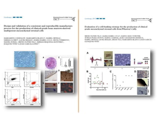
![Regenerative Medicine
Sharpe et al. in: Cell Therapy GxP (2015) [Vives&Carmona eds]](https://image.slidesharecdn.com/sessioformaciobstterapiesavancades-190603155230/85/Sessio-formacio-bst-terapies-avancades-5-320.jpg)
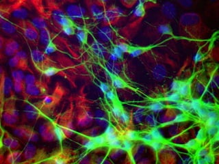
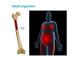

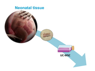


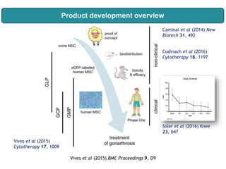
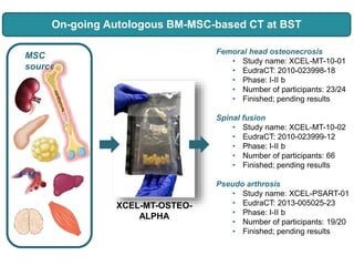
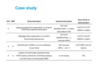
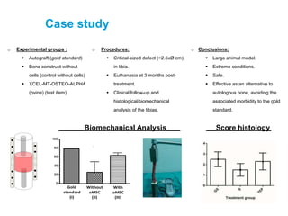
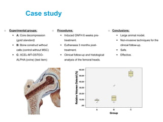
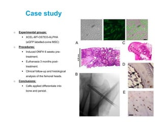
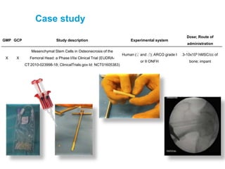
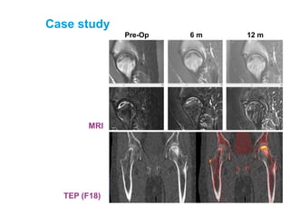

This document summarizes several case studies using mesenchymal stem cells (MSCs) for regenerative medicine applications: 1. A pre-clinical study using ovine MSCs implanted in a critical-sized bone defect in sheep tibias showed effective bone regeneration compared to autograft or no cell controls. 2. A large animal study treating induced osteonecrosis of the femoral head in sheep with ovine MSCs implanted at the lesion site showed persistence of cells differentiating into bone and effective treatment compared to controls. 3. A Phase I/IIa clinical trial in humans with osteonecrosis of the femoral head treated with implanted human MSCs showed safety and potential efficacy based on imaging follow-up


![Presentation8 16 10[1]](https://cdn.slidesharecdn.com/ss_thumbnails/presentation8-16-101-100921223521-phpapp01-thumbnail.jpg?width=640&height=640&fit=bounds)


![PERI-PROSTHETIC FRACTURE NAIL-PLATE CONSTRUCT [NPC].pptx](https://cdn.slidesharecdn.com/ss_thumbnails/drarunkumardrmohamedashrafperiprostheticfrasturenail-plateconstructnpc-260209164459-7e9d15a1-thumbnail.jpg?width=640&height=640&fit=bounds)


