1) The study characterized host vascularization of transplanted human neural stem cell grafts in porcine spinal cords. Twelve grafts of human neural progenitor cells were transplanted into three pigs' spinal cords.
2) Six weeks after transplantation, the pigs were sacrificed and the grafts were analyzed. Graft survival ranged from 0-65% and inversely correlated with host microvascular density in the grafts. More vascularized grafts had less cell survival.
3) Vessels in the grafts expressed vascular endothelial growth factor and contained proliferating endothelial cells and immune cells, which may impact graft survival. The findings could influence future immunosuppression regimens for cell transplantation.
Endothelial Cell Mediated Delay of Blood Brain Barrier Recovery Following Tra...Arthur Stem
TBI is the leading cause of death among young adults and children in the developed world, accounting for over 50,000 deaths per year. [12] TBI results in a sleuth of poor health outcomes, including hemorrhaging, seizures, neural edema, neural inflammation, and cognitive and emotional disabilities. All of these outcomes are a direct result of fundamental degradation of the BBB over a time course post TBI. [1] [12] The BBB is an integral structure that forms around the microvascular of the cerebral cavity. Endothelial cells form the basal membrane through which strictly controlled movement of molecules is observed between the extravascular and intravascular space across this basal membrane. This basal membrane is maintained by endothelial cells, having tight junctions between them to make up the pores through which transport of molecules can occur between the brain and microvasculature. These tight junctions are maintained through cross-talk between the endothelial cells and supporting neurons such as astrocytes and pericytes. [2] A multitude of proteins make up the tight junctions between the endothelial cells, including six main scaffolding structures Claudins 1, 3, and 5, ZO-1, Occludins, and Cadherins. [3] VEGF release following trauma induces endothelial cells to release matrix metalloproteinases (MMPs), in particular MMP9, which can catalyze the N-terminal amino acids that compose the tight junction protein ZO-1. [10] [11] MMP9 when in circulation is also known to activate tumor necrosis factor alpha (TNFɑ) which in turn upregulates transcription of MMP9, creating a positive feedback loop. [11] The management of MMP production is three fold, transcription, proenzyme activation, and substrate inhibition. [11] In our study, it is proenzyme activation via TP that is the focus and how that affects the overall transcription levels of the tight junction proteins within the endothelial cells and astrocytes.
Neuromics base presentation 2020 with Virus Transport MediaPete Shuster
Neuromics' is a leader in providing Biopharmas, Academic and Government with CFR compliant 2 and 3-D human primary cell assays, media and supplements for discovery. We also provide antibodies, proteins/growth factors, apoptosis kits and genetic engineering/manipulation tools. We now have FDA registered Virus Transport Media (VTM).
Los días 11 y 12 de diciembre de 2014, la Fundación Ramón Areces celebró el Simposio Internacional 'Neuropatías periféricas hereditarias. Desde la biología a la terapéutica' en colaboración con CIBERER-ISCIII y el Centro de Investigación Príncipe Felipe. El tipo más común de estas patologías es la enfermedad de Charcot-Marie-Tooth, un trastorno neuromuscular hereditario con una prevalencia estimada de 17-40 afectados por 100.000 habitantes. Durante estos dos días, investigadores mostraron sus avances en la mejora del diagnóstico y el tratamiento y, por ende, de la aproximación clínica y la calidad de vida de las personas afectadas por estas patologías.
Journal of Stem Cells Research, Reviews & Reports is a peer-reviewed, open access journal published by Austin Publishers. It provides easy access to high quality Manuscripts in all related aspects covering Stem cell research that focuses on stem cells, which have a capacity to regenerate and develop into other types of cells namely, like kidney cells, liver cells, heart cells, etc. These circulate and function to replace dysfunctional cells, naturally maintaining optimal health. The Journal encourages all the current medical research that is focused on two particular types of stem cells -- adult and embryonic stem cells that are used in various stem cell therapies against many dreadful diseases.
Austin Publishing Group is a successful host of more than hundred peer reviewed, open access journals in various fields of science and medicine with intent to bridge the gap between academia and research access.
Journal of Stem Cells Research, Reviews & Reports accepts original research articles, review articles, case reports, mini reviews, rapid communication, opinions and editorials on all the related aspects of Stem Cells and Cell-Based Therapies.
Induced Pluripotent Stem Cells and Somatic Cardiac Regeneration— An Explorato...Robert Chen
In the exploratory analysis, the functional pathway networks of nucleostemin and iPS interact via TP53 (tumor protein P53) and Fgf (fibroblast growth factor), which could be further investigated to provide clues for the future research of postnatal cardiac regeneration.
Overview of Neuromics A to Z. From high speed filling of transport media for Covid-19 testing to 2 and 3-D Cell Based Assays, defined media and supplements like FBS.
Cartilage Repair using Stem cell & OrthobiologicsVaibhav Bagaria
Regenerating Cartilage is a challenge. What's new in this field of cartilage regeneration and the current status of the stem cell use in this field is described.
(February 16, 2023) Webinar: Intracerebral Transplantation of Autologous Bone...Scintica Instrumentation
Overview:
In this webinar, Max Myers presented his work on the use of autologous bone marrow-derived stem cells injected into the cortex of rats, following a stable stroke. Max also demonstrated its lab’s findings and talked about the Aspect Imaging M7 compact MRI system as it relates to its use in this project.
Key Points:
The critical use of stem cells in stroke research
Overcoming the blood-brain barrier via intracerebral injection of stem cells
The introduction of stem cells led to improved functional recovery following an ischemic stroke
How MRI can contribute to the understanding of treatments following stroke
Paludisme grave : pourquoi doit-on développer des modèles in vitro sur le terrain ? - Conférence de la 8e édition du Cours international « Atelier Paludisme » - RAZAKANDRAINIBE Romy - Madagascar - romy@pasteur.mg
LncRNA WARS2-IT1 Functions as an Oncogene and is Associated with Poor Outcome...semualkaira
Glioblastoma multiforme (GBM) is one of the most common malignant brain tumors in adults and has high mortality and relapse rates. Over the past few years, great advances have been made in the diagnosis and treatment of GBM, but unfortunately, the five-year overall survival rate of GBM patients is approximately 5.1%. Our study aimed to investigate the new mechanism of Long noncoding RNAs (lncRNAs) WARS2-IT1 regulate the malignant progression of Glioblastoma.
Autologous Bone Marrow Cell Therapy for Autism: An Open Label Uncontrolled C...remedypublications2
The aim of this study is to assess the safety and effectiveness of autologous bone marrow
mononuclear stem cell (BMMNC) transplantation in patients with autism.
Induced Pluripotent Stem Cell & Cell Dedifferentiation: The Breakthrough of S...Vincentsia Vienna
The phenomenon of cell dedifferentiation is yet one promising trend to explore. In future, the science fiction of regenerative medicine could be turned into reality.
Autologous Bone Marrow Mononuclear Cell Therapy for Autism: An Open Label Pro...DrAlokSharma
Autism spectrum disorders (ASD) are a group of heterogeneous neurodevelopmental disorders characterized by
deficits in verbal and nonverbal communication, social
interaction, and presence of stereotypical repetitive behavior.
Autologous Bone Marrow Mononuclear Cell Therapy for Autism: An Open Label Pro...
Vascular_Poster
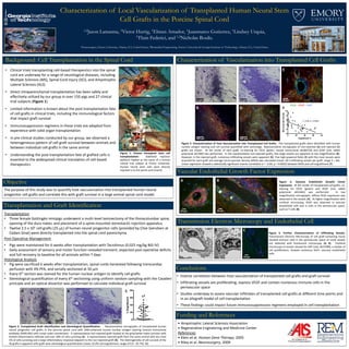
1. Characterization of Local Vascularization of Transplanted Human Neural Stem
Cell Grafts in the Porcine Spinal Cord
1,2Jason Lamanna, 1Victor Hurtig, 1Elman Amador, 1Juanmarco Gutierrez, 1Lindsey Urquia,
1Thais Federici, and 1,2Nicholas Boulis.
Background: Cell Transplantation in the Spinal Cord
• Clinical trials transplanting cell-based therapeutics into the spinal
cord are underway for a range of neurological diseases, including
Multiple Sclerosis (MS), Spinal Cord Injury (SCI), and Amyotrophic
Lateral Sclerosis (ALS)
• Direct intraparenchymal transplantation has been safely and
effectively utilized by our group in over 150 pigs and 27 clinical
trial subjects (Figure 1)
• Limited information is known about the post-transplantation fate
of cell grafts in clinical trials, including the immunological factors
that impact graft survival
• Immunosuppression regimens in these trials are adapted from
experience with solid organ transplantation
• In pre-clinical studies conducted by our group, we observed a
heterogeneous pattern of cell graft survival between animals and
between individual cell grafts in the same animal
• Understanding the post-transplantation fate of grafted cells is
essential to the widespread clinical translation of cell-based
therapeutics
Objective
The purpose of this study was to quantify host vascularization into transplanted human neural
progenitor cell grafts and correlate this with graft survival in a large animal spinal cord model.
Transplantation and Graft Identification
Funding and References
• Amyotrophic Lateral Sclerosis Association
• Regenerative Engineering and Medicine Center
References
• Klein et al. Human Gene Therapy, 2005
• Riley et al. Neurosurgery, 2009
Vascular Endothelial Growth Factor Expression
Conclusions
Characterization of Vascularization into Transplanted Cell Grafts
1Neurosurgery, Emory University, Atlanta, GA, United States; 2Biomedical Engineering, Emory University & Georgia Institute of Technology, Atlanta, GA, United States
Figure 1: Human intraspinal stem cell
transplantation. Stabilized injection
platform fixated to the spine of a human
clinical trial subject at Emory University.
Human neural stem cells were directly
injected in to the spinal cord (insert).
Transplantation
• Three female Gottingën minipigs underwent a multi-level laminectomy of the thoracolumbar spine,
opening of the dura mater, and placement of a spine-mounted stereotactic injection apparatus
• Twelve 2.5 x 10⁵ cell grafts (25 μL) of human neural progenitor cells (provided by Clive Svendsen at
Cedars Sinai) were directly transplanted into the spinal cord parenchyma
Post-Operative Management
• Pigs were maintained for 6 weeks after transplantation with Tacrolimus (0.025 mg/kg BID IV)
• Daily assessment of sensory and motor function revealed transient, expected post-operative deficits
and full recovery to baseline for all animals within 7 days
Histological Analysis
• Pig were sacrificed 6 weeks after transplantation, spinal cords harvested following transcardiac
perfusion with 4% PFA, and serially sectioned at 50 μm
• Every 6th section was stained for the human nuclear antigen to identify cell grafts
• Stereological quantification of every 6th sectioning using uniform random sampling with the Cavalieri
principle and an optical dissector was performed to calculate individual graft survival
0
20
40
60
80
%Engraftment
200 μm 200 μm
Figure 2. Transplanted Graft Identification and Stereological Quantification. Representative micrographs of transplanted human
neural progenitor cell grafts in the porcine spinal cord with DAB-enhanced human nuclear antigen staining (mouse monoclonal
antibody, MAB1281) with cresyl violet counterstain. A representative non-rejected graft located at the grey/white mater junction with
limited inflammatory infiltrate and over 30% of cells surviving (A). A representative rejected graft from the same animal with less than
5% of cells surviving and a large inflammatory response adjacent to the non-rejected graft (B). The heterogeneity of cell survival of the
36 grafts is apparent with graft-wise stereological quantification (mean 22.0% cell engraftment, range of 0.0 - 65.7%) (C).
A B C
200 μm 200 μm
A
B
0 20 40 60 80
0
2
4
6
8
10
Engraftment %
MicrovascularDensity
r = -0.60, p = 0.0002
D
EC
F
Figure 3. Characterization of Host Vascularization into Transplanted Cell Grafts. The transplanted grafts were identified with human
nuclear antigen staining and cell survival quantified with stereology. Representative micrographs of non-rejected (A) and rejected (C)
grafts are shown. At the center of each graft, co-staining for CD31 (green, mouse monoclonal ab186720) and GFAP (red, rabbit
polyclonal ab7260) was performed. In the representative non-rejected grafts, few large vessels are apparent at low magnification (B).
However, in the rejected graft, numerous infiltrating vessels were apparent (D). Five high-powered fields (E) with the most vessels were
acquired for each graft and average micro-vascular density (MVD) was calculated (mean 18.3 infiltrating vessels per graft, range 3 – 46).
Linear regression showed a statistically significant inverse correlation (r = -0.60, p = 0.0002) between MVD and cell engraftment (F).
CD31 - GFAP - DAPI
• Inverse correlation between host vascularization of transplanted cell grafts and graft survival
• Infiltrating vessels are proliferating, express VEGF and contain numerous immune cells in the
perivascular space
• Studies underway to assess vascular infiltrates of transplanted cell grafts at different time points and
in an allograft model of cell transplantation
• These findings could impact future immunosuppression regimens employed in cell transplantation
Transmission Electron Microscopy and Endothelial Cell
Proliferation
BA Figure 4. Vascular Endothelial Growth Factor
Expression. At the center of transplanted cell grafts, co-
staining for CD31 (green) and VEGF (red, rabbit
polyclonal ab53465) was performed. In low
magnification micrographs, diffuse VEGF expression was
observed in the vessels (A). At higher magnification with
confocal microscopy, VEGF was observed in vascular
endothelial cells and in cells in the perivascular space,
such as T cells (B).
Figure 5. Further Characterization of Infiltrating Vessels.
Transmission Electron Microscopy of cell graft-containing tissue
showed immune cells in the perivascular space of small vessels
not detected with fluorescent microscopy (A, B). Confocal
microscopy of vessels stained for Ki67 (red, ab15580), a marker of
cell proliferation, showed numerous Ki67+ vascular endothelial
cells.
CBA