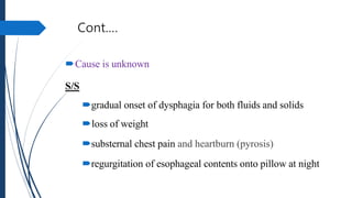
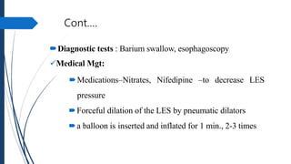
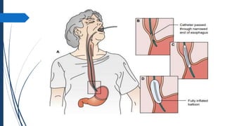
The document discusses disorders of the gastrointestinal system, beginning with a review of the anatomy and physiology of the GI tract. It describes the components and functions of the upper and lower GI tract. Key diagnostic tests are outlined including barium swallow, upper endoscopy, and colonoscopy. The disorder Achalasia is summarized, noting its causes, symptoms of dysphagia and chest pain, and treatments including medications to decrease LES pressure and forceful dilation.
The document provides information on the gastrointestinal system. It describes the structures and functions of the upper and lower gastrointestinal tract, including the mouth, esophagus, stomach, and intestines. It discusses digestion, absorption of nutrients, and the roles of the gastrointestinal tract in fluid and electrolyte balance. The document also outlines how to assess the gastrointestinal system through obtaining a patient history and performing a physical examination of the abdomen.
The document provides information on the gastrointestinal system and gastrointestinal disorders. It describes the structures and functions of the upper and lower gastrointestinal tract, including the mouth, esophagus, stomach, small intestine, and large intestine. It discusses digestion, absorption of nutrients, and the role of the gastrointestinal tract in fluid and electrolyte balance. Common gastrointestinal disorders mentioned include dysphagia, esophageal disorders, and various conditions affecting the stomach and intestines.
The document summarizes the gastrointestinal system, including the structures and functions of the upper and lower gastrointestinal tract. It describes the mouth, esophagus, stomach, small intestine, large intestine, and associated structures. It discusses the roles of these structures in digestion, absorption of nutrients, and elimination of waste. It also outlines assessments of the gastrointestinal system, including physical exams, diagnostic tests like endoscopy and radiology, and laboratory tests.
BILIARY AND GASTRO INTESTINAL CONDITIONS-1.pptxflamestart
The document discusses disorders of the digestive system and associated organs. It provides an overview of the anatomy and physiology of the digestive system and its accessory organs. It then outlines various disorders that can affect the mouth, esophagus, stomach, small intestine, large intestine, liver, gallbladder and pancreas. These include dental caries, oral thrush, gastritis, peptic ulcers, hernias, cancers, hepatitis, cirrhosis and others. Nursing management of clients with these digestive disorders is also addressed.
The digestive system consists of a tube running from the mouth to anus and accessory organs including the liver, pancreas, and salivary glands. The functions of the digestive system include ingestion, digestion, absorption of nutrients, and elimination of waste. The document describes the anatomy and functions of the oral cavity, esophagus, stomach, small intestine, large intestine, and their roles in digestion. It also discusses several diseases that can affect the digestive system.
The document summarizes key aspects of the digestive system, including:
1) It describes the general organization of the digestive tract into four layers - mucosa, submucosa, muscularis externa, and serosa.
2) It outlines the regions and functions of the small intestine - duodenum, jejunum, and ileum. Most absorption occurs in the small intestine.
3) It discusses the large intestine/colon and its regions - ascending, transverse, descending, and sigmoid colon. The colon has fewer villi and glands than the small intestine.
4) It briefly covers the liver, gallbladder, and pancreas - their locations, functions, histology
The document discusses the anatomy and functions of the digestive system, describing the tubular nature of the digestive tract and its individual organs like the liver, gallbladder, pancreas, and sections of the small and large intestines. It provides details on the histology and microscopic structure of the digestive organs and discusses their roles in digestion and absorption of nutrients. The major blood vessels supplying the digestive system and regions like the duodenum, jejunum and ileum of the small intestine are also outlined.
HAP 4 SEMESTER 2 BPHARMACY AS PER PCI SYLLABUSchristinajohn24
The document summarizes key aspects of the digestive system, including its main organs and functions. It describes the two main groups that make up the digestive system - the gastrointestinal tract and accessory digestive organs. It then provides details on the structure and functions of key organs like the stomach, small intestine, large intestine, salivary glands, and their role in digestion.
This document discusses metabolism and the gastrointestinal system. It defines metabolism as all biochemical reactions in the body and notes the gastrointestinal tract aids in ingestion and digestion of food. It then provides details on the structure and functions of the mouth, esophagus, stomach, small intestine, large intestine, and their roles in digestion and absorption of nutrients. Key points covered include the types of nutrients (carbohydrates, proteins, lipids, vitamins, minerals), digestion processes, and storage and use of nutrients throughout the body.
The digestive system breaks down ingested food into smaller molecules that can be absorbed and used by the body. It consists of the gastrointestinal tract - a long tube running from the mouth to the anus, including the mouth, esophagus, stomach, small intestine, large intestine, rectum and anus. Accessory organs like the teeth, tongue, liver, gallbladder and pancreas aid in digestion by secreting enzymes and digestive juices. The system mechanically and chemically breaks down food, absorbs nutrients, and eliminates waste through a multi-step process involving both organs and hormones.
The document discusses the anatomy and physiology of the gastrointestinal tract and how it relates to drug absorption. It describes the structures and functions of the mouth, esophagus, stomach, small intestine, large intestine, and accessory organs like the liver, gallbladder and pancreas. It explains the three phases of digestion - cephalic, gastric, and intestinal phases. It also discusses factors like gastrointestinal pH, enzymes, and presence of food that can affect the oral bioavailability of drugs.
The gastrointestinal tract has several major functions:
1. Motility through peristalsis and other contractions to move food through the tract.
2. Secretion of enzymes and hormones to aid in digestion. The stomach secretes 2-3 liters of gastric juice per day.
3. Digestion through the breakdown of food molecules by enzymes to allow absorption in the small intestine.
4. Absorption of digested nutrients through the intestinal walls into the bloodstream.
The gastrointestinal tract (GI tract) digests food and expels waste. It has 4 layers and is divided into upper and lower tracts. The upper tract includes the mouth, esophagus, and stomach. The stomach acidifies food and the lower tract, including the small and large intestines, further digests and absorbs nutrients before waste is excreted. Accessory organs like the liver, pancreas, and gallbladder produce substances like bile and enzymes to aid digestion. The kidneys filter waste from the blood to produce urine for excretion via the ureters, bladder, and urethra.
The digestive system breaks down food into nutrients that the body can use. It begins with the mouth and includes the esophagus, stomach, small and large intestines, and ends at the anus. Digestion involves both mechanical and chemical breakdown of food. The system secretes enzymes and absorbs nutrients for energy, growth, and cell repair. Waste products are eliminated through the rectum and anus.
The small intestine extends from the stomach to the large intestine and is divided into three sections - the duodenum, jejunum, and ileum. The duodenum receives partially digested food from the stomach along with pancreatic juices and bile to further break down nutrients. The jejunum and ileum then absorb these nutrients into the bloodstream. The small intestine is highly folded and contains villi to increase its surface area for maximum absorption of nutrients to nourish the body.
The stomach is a J-shaped organ located in the upper abdomen between the esophagus and small intestine. It has four regions - cardia, fundus, corpus, and pyloric part. The stomach stores and breaks down food through secretion of acids and enzymes. Motility mixes foods and empties contents into the small intestine. Diseases include peptic ulcers, gastritis, and stomach cancer.
The document summarizes the key parts and functions of the human digestive tract. It describes the mechanical and chemical breakdown of food that occurs in the mouth, stomach, and small and large intestines. Enzymes produced in the saliva, stomach, pancreas, liver, and intestines aid in digestion. The digestive tract includes the mouth, esophagus, stomach, small intestine, large intestine and rectum, each playing an important role in digesting food into smaller molecules that can be absorbed and used for energy and growth.
The document provides an overview of the human digestive system and the process of digestion. It describes each part of the digestive tract from the mouth through the esophagus, stomach, small intestine including the duodenum, jejunum and ileum. It then discusses the large intestine including the cecum, appendix, colon, rectum and anus. It explains the functions of each part and how digestion and absorption of nutrients occurs before waste is eliminated from the body.
The document provides an overview of the human digestive system and the process of digestion. It describes each part of the digestive tract from the mouth to the anus. Food enters the mouth and is broken down through chewing and saliva. It then moves to the stomach through the esophagus and is further broken down by gastric juices. Nutrients are absorbed in the small intestine before waste is passed to the large intestine, where water is reabsorbed and waste is formed into feces to be excreted from the anus.
The document provides an overview of the digestive system, including its main organs and their functions. It describes the mouth, esophagus, stomach, small intestine, large intestine, liver, gallbladder and pancreas. It explains that food is broken down mechanically and chemically by these organs, through processes like digestion and absorption, into nutrients that can be used by the body's cells. Key enzymes produced by organs like the stomach, pancreas and intestines help break down carbohydrates, proteins and fats during digestion.
The document summarizes the structure and function of the gastrointestinal tract. It describes how the gut forms from the embryonic gut tube and associated organs like the liver and pancreas. It then details the role of each section of the gut from mouth to anus in digesting and absorbing food, including mechanical and chemical breakdown by teeth, stomach acid, and enzymes from the pancreas and liver. Absorption occurs along the small intestine, with waste exiting through the large intestine and anus.
Gastroinestinal Digestion and Harmonal Interplaymeducationdotnet
This document provides an overview of the gastrointestinal hormones and their roles in regulating digestive functions. It describes the key hormones like gastrin, histamine, somatostatin, secretin, and cholecystokinin that are involved in stimulating or inhibiting secretion of acids and enzymes from various organs. The document also explains how neural and endocrine signals are integrated to precisely control digestion through feedback mechanisms involving these hormones.
The document summarizes the anatomy and physiology of the digestive tract. It describes each part of the digestive tract from the mouth to the anus. Key points include that the mouth begins digestion through chewing and saliva. Food then moves to the stomach through the esophagus. The stomach contains acid and enzymes that break down food into a liquid. This liquid moves to the small intestine where nutrients are absorbed into the bloodstream and lymph vessels. The large intestine absorbs water before waste is eliminated through the anus.
The document provides information on human anatomy and physiology, with a focus on the digestive system. It describes the structure and functions of the cells, tissues, organs and body systems. It then discusses the layers, organs and functions of the digestive system in detail. This includes the mouth, salivary glands, esophagus, stomach, small intestine, large intestine, liver and pancreas. It also covers the mechanisms of acid secretion in the stomach and factors that regulate gastric juice production. Finally, it provides an overview of acid peptic disorders like gastritis, GERD, peptic ulcers, and their causes and treatments.
With this presentation we will learn to develop an understanding of the relationships between the structures and functions of the human digestive system and digestive processes which include the processes of digestion include six activities: ingestion, propulsion, mechanical or physical digestion, chemical digestion, absorption, and defecation
The document summarizes the key parts and functions of the human digestive system. It describes the main organs of the alimentary canal including the mouth, esophagus, stomach, small intestine, large intestine and anus. It explains the processes of ingestion, digestion, absorption, assimilation and egestion. It provides details on digestion in each part of the alimentary canal and the roles of the liver, gallbladder and pancreas in aiding digestion.
The document summarizes the key processes and structures of the gastrointestinal (GI) tract. It describes the 6 basic processes of ingestion, secretion, mixing and propulsion, digestion, absorption, and defecation. It then details the layers of the GI tract and defines important vocabulary words. Specific sections are dedicated to describing the mouth, teeth, tongue, salivary glands, pharynx, esophagus, stomach, pancreas, liver, gallbladder, small intestine, large intestine, and the roles of enzymes and hormones. Key points include the 4 layers of the GI tract, the 5 types of teeth, and the 3 parts of the small intestine.
This document summarizes benign prostatic hyperplasia (BPH). It discusses the pathology and pathogenesis of BPH, including that it affects glandular epithelium, stromal cells, and causes increased growth. It also covers the symptomatology, evaluation, and various treatment options for BPH including watchful waiting, medical therapy, and prostatectomies. Surgical treatments discussed are transurethral resection of the prostate (TURP), retropubic prostatectomy (RPP), and transvesical prostatectomy (TVP).
This document provides an introduction to pathology. It defines pathology as the study of disease through scientific methods and examines the mechanisms of disease from etiology to clinical manifestation. The key points are:
1. Pathology studies the etiology, pathogenesis, morphologic changes, and functional derangements that result from disease processes.
2. Diseases are examined through diagnostic techniques including histopathology, cytopathology, and biochemical/immunological testing to identify structural and molecular alterations.
3. The natural course of a disease involves stages from initial exposure through biological onset, clinical onset, potential resolution or death.
This document provides an overview of preeclampsia and eclampsia. It begins with an introduction and outlines risk factors and classifications. It then describes clinical features such as hypertension and proteinuria. The pathophysiology section explains how abnormal placentation leads to reduced blood flow and imbalance of prostaglandins. Complications are also discussed, including renal failure, pulmonary edema, and intrauterine growth restriction. The document provides information on diagnosis and management of preeclampsia and eclampsia.
This seminar presentation discusses hypersensitivity reactions, which are exaggerated or inappropriate immune responses to benign antigens. It covers the objectives, mechanisms, classification, complications, and references related to hypersensitivity reactions. There are four main types of hypersensitivity reactions: Type I involves IgE antibodies and mast cell degranulation, Type II involves antibody-mediated cell cytotoxicity, Type III involves immune complex formation and deposition, and Type IV involves T-cell mediated reactions. The presentation provides examples and details of each type of hypersensitivity reaction and their clinical implications.
This document discusses inflammation. It defines inflammation as the body's local response to injury or infection aimed at eliminating the cause of injury and initiating repair. The cardinal signs of inflammation are redness, swelling, heat, pain, and loss of function. The early response involves vasodilation and increased permeability, causing swelling. The late response involves neutrophils in acute inflammation and macrophages in chronic cases, which work to destroy pathogens and initiate healing. Understanding inflammation is important for diagnosing conditions like appendicitis and treating diseases.
This document provides an overview of hyaline membrane disease (HMD), also known as respiratory distress syndrome (RDS), for nursing students. It defines RDS as a lack of pulmonary surfactant, outlines its pathophysiology and risk factors. The document discusses the clinical presentation of RDS, including respiratory distress, radiographic findings and laboratory abnormalities. It also covers diagnosis, differential diagnoses, treatment including surfactant replacement and supportive care, complications and prevention of RDS through antenatal corticosteroids.
1. Acute inflammation is rapid in onset and short in duration, characterized by fluid and protein exudation and neutrophil accumulation. Chronic inflammation is slower in onset and longer lasting, characterized by mononuclear cell infiltration, ongoing tissue destruction, and attempts at repair through fibrosis.
2. The key features of acute inflammation are vasodilation, increased vascular permeability, and recruitment of leukocytes from the blood vessels to the site of injury. Chronic inflammation features mononuclear cell infiltration, persistent tissue damage, and attempts to repair through fibrosis and angiogenesis.
3. Granulomatous inflammation is a pattern of chronic inflammation seen with certain infections, featuring focal collections of activated macrophages that develop an epithelial-like appearance known
Cellular injury can result in adaptation, reversible injury, irreversible injury leading to necrosis or apoptosis, or intracellular accumulation. The outcome depends on the injurious agent and cell type. Adaptations include hypertrophy, hyperplasia, atrophy, and metaplasia. Reversible injury includes fatty changes and pigment accumulation. Necrosis is cell death resulting from hypoxia, free radicals, membrane damage, or calcium influx. There are several types of necrosis including coagulative, liquefactive, fat, caseous, and gangrenous. Apoptosis is programmed cell death that does not cause inflammation.
This document discusses pelvic inflammatory disease (PID) and ectopic pregnancy. It defines PID as an infection of the upper female genital tract that spreads to involve the uterus, fallopian tubes, and ovaries. Common causes are Neisseria gonorrhoeae, Chlamydia trachomatis, and bacterial vaginosis. Risk factors include multiple sexual partners and past gynecological procedures. Symptoms can range from mild to severe abdominal pain. Diagnosis involves clinical exams and tests. Complications include infertility and ectopic pregnancy. Ectopic pregnancy is defined as implantation outside the uterus, most commonly in the fallopian tube. Causes may include anatomical obstructions or abnormalities in the fallop
The document discusses acid-base balance and disturbances. It defines the two main buffer systems - metabolic (kidneys) and respiratory (lungs) - that work to maintain blood pH between 7.35-7.45. Five primary acid-base imbalances are described: metabolic acidosis, metabolic alkalosis, respiratory acidosis, respiratory alkalosis, and mixed disturbances. Diagnosis involves blood tests including arterial blood gases and electrolytes to classify the disturbance based on pH, PCO2, and bicarbonate levels. Treatment focuses on addressing the underlying cause rather than just the pH effect.
This document provides an overview of autoimmune diseases. It defines autoimmune diseases as conditions where the immune system mistakenly attacks and destroys healthy body tissue. The causes include genetic factors, environmental triggers like infections, and defects in immunologic tolerance. Some specific autoimmune diseases discussed are rheumatoid arthritis, type 1 diabetes, Hashimoto's thyroiditis, Graves' disease, myasthenia gravis, and systemic sclerosis. The mechanisms, clinical features, pathology, and treatment options are described for each condition.
Patient safety is a fundamental principle of healthcare. Adverse events may result from problems in practice, products, procedures or systems. Improving patient safety demands a complex, system-wide effort involving performance improvement, risk management, infection control, safe clinical practices, and a safe environment of care. Unsafe injections expose millions of people to infections worldwide each year. Ensuring single-use injection devices and safety boxes are available in every healthcare facility can prevent reuse and unsafe waste disposal.
The document discusses integumentary disorders and provides information on the anatomy and functions of the skin. It describes common skin conditions like eczema, acne, and psoriasis. Eczema is characterized by redness, dryness, and itching. Acne presents as inflamed papules and pustules on the face and back. Psoriasis causes thickened red patches covered with silvery scales. The document outlines signs, causes, and management approaches for various dermatological disorders and skin lesions.
A nebulizer converts liquid medication into a mist that can be inhaled directly into the lungs, allowing for rapid onset of medication effects. There are different types of nebulizers that administer medication via mouthpiece or mask. Nebulizers are commonly used to treat conditions involving airflow obstruction like asthma. Proper use involves preparing equipment and medication, positioning the patient, administering the treatment, and monitoring for side effects.
This document provides an overview of the endocrine system, including the major glands and hormones. It describes the hypothalamus and pituitary glands which regulate many other endocrine glands. Other glands covered include the thyroid, parathyroid, adrenal, pancreas, ovaries, testes, thymus, and pineal. The document outlines how to assess endocrine disorders and lists some common laboratory studies. It also provides details on diabetes mellitus, describing the main types of diabetes including type 1, type 2, and gestational diabetes.
This document provides guidance on performing a cardiac and abdominal examination. It outlines the objectives, symptoms, and physical examination techniques for assessing the cardiovascular and abdominal systems. The cardiovascular section covers inspection of the jugular veins, palpation of pulses, auscultation of heart sounds, and measurement of blood pressure. The abdominal section reviews inspection, auscultation, percussion and palpation techniques. Proper examination order and identification of normal versus abnormal findings are emphasized.
This document summarizes several endocrine system disorders including hyperthyroidism, hypothyroidism, hyperparathyroidism, hypoparathyroidism, Cushing's syndrome, Conn's syndrome, Addison's disease, and pituitary adenomas. It provides epidemiological data on certain disorders and describes associated symptoms, diagnostic evaluations, and medical management approaches. Multiple endocrine neoplasia syndromes are also briefly discussed.
This document provides guidance on effectively breaking bad news to patients. It discusses the importance of this communication skill for healthcare professionals. The document outlines best practices for setting, perception checking, invitation, knowledge sharing, exploring the patient's response, and summarizing. Key aspects include ensuring privacy, empathy, clarity, and allowing time for the patient's questions and reactions. The SPIKES protocol is presented as a framework for structuring the discussion. Examples of both best practices and things to avoid are also highlighted.
2 Assessment of patient with respiratory disorder.pptxMohammedAbdela7
This document provides guidelines for performing a physical examination of the thorax and lungs. It begins by outlining the session objectives and general examination guidelines. It then discusses pertinent history data to obtain, such as cough characteristics and sputum type/color. The physical exam involves inspection, palpation, percussion, and auscultation of the chest. Inspection evaluates breathing patterns, respiratory distress signs, and overall appearance. Palpation assesses tracheal position, chest expansion, tactile fremitus, and tenderness. Percussion and auscultation are also performed to evaluate the lungs. Proper equipment, patient positioning, and exam techniques are emphasized throughout.
This document provides an overview of critical thinking, evidence-based medicine, and how to practice evidence-based medicine. It defines critical thinking as the process of conceptualizing and evaluating information to guide beliefs and actions. Evidence-based medicine is defined as integrating the best research evidence with clinical expertise and patient values/circumstances. The history of evidence-based medicine is discussed, from Cochrane's work in the 1970s highlighting gaps between research and practice, to Guyatt coining the term "evidence-based medicine" in 1991 and Sackett explaining the combination of research, expertise, and patient factors in 1996. The five steps to practice evidence-based medicine are described as developing questions, finding evidence, appraising evidence, integrating
This presentation gives information on the pharmacology of Prostaglandins, Thromboxanes and Leukotrienes i.e. Eicosanoids. Eicosanoids are signaling molecules derived from polyunsaturated fatty acids like arachidonic acid. They are involved in complex control over inflammation, immunity, and the central nervous system. Eicosanoids are synthesized through the enzymatic oxidation of fatty acids by cyclooxygenase and lipoxygenase enzymes. They have short half-lives and act locally through autocrine and paracrine signaling.
STUDIES IN SUPPORT OF SPECIAL POPULATIONS: GERIATRICS E7shruti jagirdar
Unit 4: MRA 103T Regulatory affairs
This guideline is directed principally toward new Molecular Entities that are
likely to have significant use in the elderly, either because the disease intended
to be treated is characteristically a disease of aging ( e.g., Alzheimer's disease) or
because the population to be treated is known to include substantial numbers of
geriatric patients (e.g., hypertension).
Nutritional deficiency Disorder are problems in india.
It is very important to learn about Indian child's nutritional parameters as well the Disease related to alteration in their Nutrition.
The biomechanics of running involves the study of the mechanical principles underlying running movements. It includes the analysis of the running gait cycle, which consists of the stance phase (foot contact to push-off) and the swing phase (foot lift-off to next contact). Key aspects include kinematics (joint angles and movements, stride length and frequency) and kinetics (forces involved in running, including ground reaction and muscle forces). Understanding these factors helps in improving running performance, optimizing technique, and preventing injuries.
Pictorial and detailed description of patellar instability with sign and symptoms and how to diagnose , what investigations you should go with and how to approach with treatment options . I have presented this slide in my 2nd year junior residency in orthopedics at LLRM medical college Meerut and got good reviews for it
After getting it read you will definitely understand the topic.
Spontaneous Bacterial Peritonitis - Pathogenesis , Clinical Features & Manage...Jim Jacob Roy
In this presentation , SBP ( spontaneous bacterial peritonitis ) , which is a common complication in patients with cirrhosis and ascites is described in detail.
The reference for this presentation is Sleisenger and Fordtran's Gastrointestinal and Liver Disease Textbook ( 11th edition ).
Osvaldo Bernardo Muchanga-GASTROINTESTINAL INFECTIONS AND GASTRITIS-2024.pdfOsvaldo Bernardo Muchanga
GASTROINTESTINAL INFECTIONS AND GASTRITIS
Osvaldo Bernardo Muchanga
Gastrointestinal Infections
GASTROINTESTINAL INFECTIONS result from the ingestion of pathogens that cause infections at the level of this tract, generally being transmitted by food, water and hands contaminated by microorganisms such as E. coli, Salmonella, Shigella, Vibrio cholerae, Campylobacter, Staphylococcus, Rotavirus among others that are generally contained in feces, thus configuring a FECAL-ORAL type of transmission.
Among the factors that lead to the occurrence of gastrointestinal infections are the hygienic and sanitary deficiencies that characterize our markets and other places where raw or cooked food is sold, poor environmental sanitation in communities, deficiencies in water treatment (or in the process of its plumbing), risky hygienic-sanitary habits (not washing hands after major and/or minor needs), among others.
These are generally consequences (signs and symptoms) resulting from gastrointestinal infections: diarrhea, vomiting, fever and malaise, among others.
The treatment consists of replacing lost liquids and electrolytes (drinking drinking water and other recommended liquids, including consumption of juicy fruits such as papayas, apples, pears, among others that contain water in their composition).
To prevent this, it is necessary to promote health education, improve the hygienic-sanitary conditions of markets and communities in general as a way of promoting, preserving and prolonging PUBLIC HEALTH.
Gastritis and Gastric Health
Gastric Health is one of the most relevant concerns in human health, with gastrointestinal infections being among the main illnesses that affect humans.
Among gastric problems, we have GASTRITIS AND GASTRIC ULCERS as the main public health problems. Gastritis and gastric ulcers normally result from inflammation and corrosion of the walls of the stomach (gastric mucosa) and are generally associated (caused) by the bacterium Helicobacter pylor, which, according to the literature, this bacterium settles on these walls (of the stomach) and starts to release urease that ends up altering the normal pH of the stomach (acid), which leads to inflammation and corrosion of the mucous membranes and consequent gastritis or ulcers, respectively.
In addition to bacterial infections, gastritis and gastric ulcers are associated with several factors, with emphasis on prolonged fasting, chemical substances including drugs, alcohol, foods with strong seasonings including chilli, which ends up causing inflammation of the stomach walls and/or corrosion. of the same, resulting in the appearance of wounds and consequent gastritis or ulcers, respectively.
Among patients with gastritis and/or ulcers, one of the dilemmas is associated with the foods to consume in order to minimize the sensation of pain and discomfort.
Giloy in Ayurveda - Classical Categorization and SynonymsPlanet Ayurveda
Giloy, also known as Guduchi or Amrita in classical Ayurvedic texts, is a revered herb renowned for its myriad health benefits. It is categorized as a Rasayana, meaning it has rejuvenating properties that enhance vitality and longevity. Giloy is celebrated for its ability to boost the immune system, detoxify the body, and promote overall wellness. Its anti-inflammatory, antipyretic, and antioxidant properties make it a staple in managing conditions like fever, diabetes, and stress. The versatility and efficacy of Giloy in supporting health naturally highlight its importance in Ayurveda. At Planet Ayurveda, we provide a comprehensive range of health services and 100% herbal supplements that harness the power of natural ingredients like Giloy. Our products are globally available and affordable, ensuring that everyone can benefit from the ancient wisdom of Ayurveda. If you or your loved ones are dealing with health issues, contact Planet Ayurveda at 01725214040 to book an online video consultation with our professional doctors. Let us help you achieve optimal health and wellness naturally.
Discover the benefits of homeopathic medicine for irregular periods with our guide on 5 common remedies. Learn how these natural treatments can help regulate menstrual cycles and improve overall menstrual health.
Visit Us: https://drdeepikashomeopathy.com/service/irregular-periods-treatment/
Congestive Heart failure is caused by low cardiac output and high sympathetic discharge. Diuretics reduce preload, ACE inhibitors lower afterload, beta blockers reduce sympathetic activity, and digitalis has inotropic effects. Newer medications target vasodilation and myosin activation to improve heart efficiency while lowering energy requirements. Combination therapy, following an assessment of cardiac function and volume status, is the most effective strategy to heart failure care.
2. Review of anatomy and physiology of GIT
The GI tract is a 23- to 26-foot-long pathway that extends from
the mouth through the esophagus, stomach, and intestines to
the anus.
The esophagus is located in the mediastinum in the thoracic
cavity, anterior to the spine and posterior to the trachea and
heart.
It passes through the diaphragm at an opening called the
diaphragmatic hiatus.
The remaining portion of the GI tract is located within the
peritoneal cavity.
3. UPPER GIT
Consists of structures that aid in the ingestion and digestion of food.
includes the mouth, esophagus, stomach, duodenum
Hypothalamus
is responsible for notifying the body that it is satisfied or
has received sufficient food
4. Cont.…
Lower GIT
Consists of the small and large intestines
Digestion is completed in the small intestine , and most
nutrients are absorbed in this part of the GIT
The large intestine serves primarily to absorb water and
electrolytes and to eliminate the waste products of digestion
through the feces
5. Mouth
Salivation
the “thought” of food initiates saliva production
a.) Serous secretions;-contain ptyalin for starch digestion –
produced by parotid and submaxillary glands
b.) Mucous secretions- for lubrication of food – produced by the
buccal, sublingual and submaxillary glands
6. Cont.…
Mastication
chewing of food
teeth - for initial breakdown of food to small particles
it helps prevent excoriation of the lining of the tract and
increase rate of digestion
7. Cont.…
Major Structures in the Mouth
teeth – to grind the food
salivary glands – moisten food and mucous
membranes and begin carbohydrate digestion
8. Esophagus
is a hollow tube, the upper 1/3 is composed of skeletal muscles, the
rest is smooth muscle
lined with mucous membrane – secretes mucoid substance for
protection
the bolus of food arrives at the cardiac sphincter of the
stomach w/in 5-10 secs. after ingestion
the lower esophageal sphincter (LES) prevents reflux of food in the
stomach back into the lower esophagus
9. Cont.…
Swallowing(deglutition)
3phases:
1.) tongue forces the bolus of food into the pharynx
2.) the food moves into the upper esophagus
3.) the food moves down into the stomach
* Food is prevented from passing into the trachea by closing of the
epiglottis.
10. Stomach
Made up of 5 layers of smooth muscle 2
types of contractions:
1.) tonus contractions – continuous contractions
2.) rhythmic contractions – may be slow ( q2-3 mins.) or fast
– responsible for the mixing of food and peristaltic movement
Vagus nerve – supplies the nervous stimulation for the stomach
11. Cont.…
movement of food through the stomach and intestines is by
peristalsis the alternate contraction and relaxation of the muscle
fibers that pushes the food in a wave-like motion
chyme – food in the stomach
- is pumped through the pyloric sphincter into the duodenum
chyme – food in the stomach
- is pumped through the pyloric sphincter into the duodenum
12. Cont.…
Digestive Function of the Stomach:
Pepsin – needed for protein digestion
HCL acid – aids in pre-digestion of food
13. Intestines
Small Intestine
2.5 cm.(1 inch) wide and 6 meters (20 feet) long–fills most of
the abdomen
3 parts :
a) duodenum – which connects to the stomach (10 inches)
b) jejunum – middle portion (8 feet long)
c) ileum – with connects to the large intestine (12 feet long)
14. Cont.….
Large Intestine
6 cm. (2 ½ in.) wide and 1.5 meters (5 feet long)
3 parts :
a.) cecum – which connects to the small intestines
b.) colon – 4 parts (ascending, transverse, descending,
sigmoid colon)
c.) rectum – 17-20 cm. (7-8 inches) long, anal canal
15. Cont.…
ileocecal valve – prevents backward flow of fecal contents
from the large intestine to the small intestine
vermiform appendix – has no function , near the ileocecal
valve anus – anal opening, is controlled by a smooth muscle
internal sphincter and a striated muscle external sphincter
chyme is propelled toward the anus by peristalsis, also mixes
the intestinal contents
in the colon, the feces is pushed forward by mass movements
stimulated by gastrocolic reflexes initiated when food enters
the duodenum from the stomach.
16. Cont.…
Defecation reflex
when feces enter the rectum and cause distention of wall of
the rectum - send impulses to the sacral segment of the spinal
cord – then back to the colon, sigmoid and rectum - initiate
relaxation of the internal anal sphincter -relaxation or
17. cont…
Secretion and Digestion
major portion of digestion occurs in the small intestines by the action of
pancreatic and intestinal secretions (enzymes) and bile
a.) Carbohydrate digestion
start in the mouth Ptyalin – breakdown polysaccharides to
disaccharides
intestinal enzymes (maltase, lactase, sucrase)
🡲 breakdown disaccharides to monosaccharides (glucose, galactose
fructose)
b.) Protein digestion
- start in the stomach pepsin – breakdown of proteins to polypeptides
- small intestines trypsin – breakdown of polypeptides into peptides and
amino acids
c.) Fat digestion
- fats require emulsification into small droplets before it can be broken down
into glycerol and fatty acids
18. cont.…
Absorption
• the intestinal wall has many folds which are covered by fingerlike
projections called (villi) -increase the absorptive area of the small intestines
• in the center of the villi are capillaries, veins, small arteries for absorption of nutrients into
the blood vessel system
• 90% of absorption occurs within the small intestines by active transport or diffusion
• amino acids, monosaccharides, Na+, Ca++ are transported by active transport w/ the
expenditure or use of energy
• other nutrients, fatty acids and H2O – diffuse passively across the cell membrane
• reabsorption of H2O, electrolytes and bile occurs mainly in the ascending colon
19. cont.…
• GIT role in Fluid and Electrolytes Balance
GIT secretions contain electrolytes
severe fluid and electrolyte imbalance may occur with excessive losses
of gastrointestinal fluids
Ex. 1.) Na+ and K+ deficits : vomiting, diarrhea, gastric suctioning, intestinal
fistula
2.) Ca++ & Mg++ deficits: malnutrition, malabsorption, intestinal fistula 3.)
Metabolic alkalosis : loss of gastric acid by suctioning or persistent
vomiting
4.) Metabolic acidosis : loss of bicarbonate-rich intestinal secretions by severe
diarrhea or fistula
• Other functions of the GIT
• the GIT supports bacterial growth and has a role in antibody formation
• intestinal bacteria synthesize Vit. K required for production of clotting
factors II (Prothrombin), VII, IX,X
21. Assessment of the GIT
Nursing History : Subjective Data
1.General Data
a. presence of dental prosthesis, comfort of usage
b. difficulty eating or digesting food
c. nausea or vomiting
d. weight loss
e. pain – may be caused by distention or sudden contraction of
any part of the GIT
specify the area, describe the pain
22. Cont.….
2.Specific data if symptoms are present
situations or events that effect symptoms
onset, possible cause, location, duration, character
of symptoms
relationship of specific foods, smoking or alcohol to
severity
23. Cont.….
3. Normal pattern of bowel elimination
a. frequency and character of stool
b. use of laxatives, enemas
24. Cont.…
Recent changes in normal patterns
changes in character of stool (constipation, diarrhea, or alternating
constipation and diarrhea)
changes in color of stool
melena - black tarry stool (upper GI bleeding)
hematochezia – fresh blood in the stool (lower GI bleeding)
c. drugs /medications being taken
d. measures taken to relieve symptoms
25. Physical Examination : Objective Data
a.) Mouth and Pharynx
1. lips – color, moisture, swelling, cracks or lesions
2. teeth – completeness (20 in children, 32 in adults), caries,
loose teeth, absence of teeth impair adequate chewing
3. gums – color, redness, swelling, bleeding, pain (gingivitis)
4. mucosa – color (light pink)
26. Cont.….
examine for moisture, white spots or patches, areas of bleeding,
or ulcers
white patches – due to candidiasis (oral thrush)
white plaques w/in red patches may be malignant lesions
tongue – color, mobility, symmetry, ulcerations / lesions or nodules
pharynx – observe the uvula, soft palate, tonsils, posterior pharynx
signs of inflammation (redness, edema, ulceration, thick
yellowish secretions), assess also for symmetry of uvula and
tonsil.
27. Cont.…
b.)Abdomen
assess for the presence or absence of tenderness, organ enlargement,
masses, spasm or rigidity of the abdominal muscles, fluid or air in the
abdominal cavity.
Anatomic Location of Organs
RUQ – liver, gallbladder, duodenum, right kidney, hepatic flexure of colon
RLQ- cecum, appendix, right ovary and fallopian tube
LUQ – stomach, spleen, left kidney, pancreas, splenic flexure of colon
LLQ – sigmoid colon, left ovary and tube
28. Cont.…
1. Inspection
assess the skin for color, texture, scars, striae, engorged
veins, visible peristalsis (intestinal obstruction), visible
pulsations (abdominal aorta), visible masses (hernia)
assess contour (flat, protuberant, globular)
abdominal distension, measure abdominal girth or
circumference at the level of umbilicus or 2-5 cm. below
29. Cont.…
2. Auscultation
presence or absence of peristalsis or bowel sounds
Normoactive – every 5-20 secs.
Hypoactive – 1 or 2 sounds in 2 mins.
Absent – no sounds in 3-5 mins.
peritonitis, paralytic ileus,
Hyperactive – 5-6 sounds in less than 30 sec.
diarrhea, gastroenteritis, early intestinal obstruction
30. Cont.…
3.Percussion
done to confirm the size of various organs
to determine presence of excessive amounts of air or fluid
Normal tympany
dullness or flatness – area of liver and spleen, solid structure.
Tumor
31. Cont.…
4.Palpation
to determine size of liver, spleen, uterus, kidneys – if
enlarged determine presence and chac. of abdominal masses
determine degree of tenderness and muscle rigidity
(rebound or direct).
32. Cont.…
c.) Rectum
perineal skin and perianal skin
assess for presence of pruritus, fissures, externa
hemorrhoids, rectal prolapse
33. Diagnostic Tests
1. Stool examination (fecalysis)
Stool for occult blood
o GI bleeding
o No red meat, turnips, horseradish, steroids, NSAIDS, iron
Stool for Ova and parasites
proper collection of specimen should not be mixed with water or
urine, should be sent immediately to the laboratory
34. Cont.…
2. CEA (Carcinoembryonic antigen)
(+) colon cancer and other forms of cancer
it is useful as in indicator of the effects of therapy
CEA - recurrence or spread of tumor
effectiveness of therapy
A blood sample is withdrawn or sent to laboratory
36. Radiologic Tests
visualization of the GIT by barium swallow, upper GI
series or barium enema
Barium – is a radiopaque substance that when
ingested or given by enema in solution, outlines the
passage ways of the GIT for viewing by x-ray or
fluoroscopy
37. Cont.….
1. Barium swallow/UGIS
for identification of disorders of esophagus, stomach, duodenum –
esophageal lesions, hiatal hernia, esophageal reflux, tumors, ulcers,
inflammation
Pt. swallows a flavored barium solution and the radiologist observes
the progress of the barium through the esophagus and take x-ray films
NPO for 6-8 hrs.
Post procedure:
o Increase fluid intake
Laxative
o Stool – white for 24-72 hrs.
o Observe for: impaction, distended abdomen.
38. Cont.…
2. Barium Enema/LGIS
Purpose: to visualize the colon to detect tumors, polyps,
inflammation, obstruction
Prep.
o low residue diet (1-2 days), clear liquid diet (evening meal)
o Laxative, cleansing enema in AM
Post
o Laxative or enema
o Same as UGIS
39. Other analysis
a.) Gastric analysis
to quantify gastric acidity Normal 1-5 mEq / L
gastric acid : gastric cancer, pernicious anemia
gastric acid : duodenal ulcer Normal gastric
acid : gastric ulcer
40. Cont.…
NPO for 12 hours
an NGT is inserted and gastric contents are aspirated, connected to suction
41. Cont.…
b.)Biopsy
Upper GI biopsy – biopsy of the oral cavity or tongue, or any lesion or
ulcerated area
- local anesthesia assess site for bleeding , give oral
hygiene
Biopsy of stomach - done during endoscopy
Rectal biopsy–biopsy of lesions, polyps, tumors of the lower sigmoid
colon, rectum and anal canal during sigmoidoscopy
monitor for signs of bleeding
42. Cont.…
Endoscope
directly visualize the GIT by the use of a fiberscape
fiberscope – has a thin, flexible shaft that can pass through and
around bends in the GIT, transmit light and the image can be seen in
the monitor
Colonoscopy
to visualize the colon
useful to identify tumors, colonic cancer, colonic polyps
not done when there is active bleeding or inflammatory disease
44. Achalasia
Absent or ineffective peristalsis of the distal esophagus accompanied by
failure of the esophageal sphincter to relax in response to swallowing.
Narrowing of the esophagus just above the stomach results in a
gradually increasing dilation of the esophagus in the upper chest.
Achalasia may progress slowly and occurs most often in people 40
years of age or older
45. Cont.…
Cause is unknown
S/S
gradual onset of dysphagia for both fluids and solids
loss of weight
substernal chest pain and heartburn (pyrosis)
regurgitation of esophageal contents onto pillow at night
46. Cont.…
Diagnostic tests : Barium swallow, esophagoscopy
Medical Mgt:
Medications–Nitrates, Nifedipine –to decrease LES
pressure
Forceful dilation of the LES by pneumatic dilators
a balloon is inserted and inflated for 1 min., 2-3 times
47.
48. Cont.…
Nursing mgt
Encourage pt. To drink fluids with meals and use the valsalva
Maneuver (bearing down with a closed glottis) while swallowing
To help push the food
Advise soft diet
Elevate head during sleepingto prevent regurgitation
After esophageal surgery, monitor for signs of esophageal
perforation as evidenced by chest pain, shock, dyspnea and fever
49. Gastritis
Gastritis(inflammation of the gastric or stomach mucosa) is a common GI
problem.
Gastritis may be acute, lasting several hours to a few days, or chronic,
resulting from repeated exposure to irritating agents or recurring episodes of
acute gastritis.
50. Cont.…
Gastritis is an inflammation of the stomach lining due to either erosion or
atrophy.
Erosive causes include stresses such as physical illness or medications such
as nonsteroidal anti-inflammatory drugs (NSAIDs).
Atrophic causes include a history of prior surgery (such as gastrectomy),
alcohol use, or Helicobacter pylori infection.
51. Pathophysiology
In gastritis, the gastric mucous membrane becomes edematous
and hyperemic (congested with fluid and blood) and undergoes
superficial erosion .
It secretes a scanty amount of gastric juice, containing very little
acid but much mucus.
Superficial ulceration may occur and can lead to hemorrhage.
52. Cont.…
Risk factors
Bacterial infection: Helicobacter pylori (H. pylori), Salmonella,
Streptococci, Staphylococci.
Family member with H. pylori infection
Family history of gastritis
Prolonged use of NSAIDs, corticosteroids (stops prostaglandin
synthesis)
Excessive alcohol use
Bile reflux disease
Advanced age
54. SIGNS AND SYMPTOMS
The patient with acute gastritis
may have
abdominal discomfort and
distension
headache
tiredness
nausea
anorexia
vomiting
hiccupping.
heartburn after eating
belching
a sour taste in the mouth
55. Cont.…
Epigastric tenderness on palpation due to gastric irritation
Bleeding from irritation of the gastric mucosa
Hematemesis—possible coffee ground emesis due to partial digestion
of blood
Melena—black, tarry stool
56. Laboratory Tests
Noninvasive tests
CBC to check for anemia (in women, Hgb less than 12 g/dL and RBC
less than 4.2 cells/mcL; in men, Hgb less than 14 g/dL and RBC less
than 4.7 cells/mcL)
Serum and stool antibody/antigen test for presence of H. pylori .
Diagnostic Procedures
■ Upper endoscopy
A small flexible scope is inserted through the mouth into the esophagus,
stomach, and duodenum to visualize the upper digestive tract.
This procedure allows for a biopsy, cauterization, removal of polyps,
dilation, or diagnosis.
57. INTERPRETING TEST RESULTS
Hemoglobin and hematocrit decrease.
Anemia (iron deficiency) due to chronic, slow blood loss.
Fecal occult blood positive.
Helicobacter pylori may be positive.
Upper endoscopy shows inflammation, allows biopsy
58. Medical TREATMENT
Administer antacids
anti acid syrup or MTS
Administer histamine 2 blockers:
ranitidine, famotidine, cimetidine
Administer proton pump inhibitors:
omeprazole and pantoprazole
Eradicate Helicobacter pylori infection if present.
triple treatment
59. Nursing Care
Monitor fluid intake and urine output.
Administer IV fluids as prescribed.
Monitor electrolytes (diarrhea and vomiting may deplete
electrolytes and cause dehydration).
Assist the client in identifying foods that are triggers.
Provide small, frequent meals and encourage the client to
eat slowly.
60. Cont.…
Advise the client to avoid alcohol, caffeine, and foods that may cause
gastric irritation.
Assist the client in identifying ways to reduce stress.
Monitor for indications of gastric bleeding (coffee-ground emesis; black,
tarry stools).
Monitor for findings of anemia (tachycardia, hypotension, fatigue,
shortness of breath, pallor, feeling light-headed or dizzy, chest pain).
61. Gastric and Duodenal Ulcers(PUD)
Normally, the gastric and duodenal mucosa is protected from acid and pepsin
by mucusand bicarbonate(base) that are secreted by surface epithetical cells.
Peptic ulcer
is a sharply defined break or ulceration in the protective mucosal lining of the
lower esophagus, stomach or duodenum which may involve the submucosa
and muscular layers
such breaks may expose the submucosal layers to gastric acid secretions and
pepsin and cause Autodigestion
True ulcers extend through the muscularis mucosa and damage blood
vessels, causing bleeding or may lead to perforation of the GIT wall
62. Cont.…
Peptic ulcers include:
Gastric ulcers that occur on the inside of the stomach
Duodenal ulcers that occur on the inside of the upper
portion of your small intestine (duodenum)
63. Pathophysiology
Peptic ulcers occur mainly in the gastroduodenal mucosa because
this tissue cannot withstand the digestive action of gastric acid
(HCl) and pepsin. The erosion is caused by the increased
concentration or activity of acid-pepsin, or by decreased resistance
of the mucosa. A damaged mucosa cannot secrete enough mucus to
act as a barrier against HCl.
64.
65. Cause of PUD
The most common causes of peptic ulcers are infection
with the bacterium Helicobacter pylori (H. pylori).
long-term use of nonsteroidal anti-inflammatory drugs
(NSAIDs) .
It’s a common misconception that coffee and
spicy foods can cause ulcers.
66. SIGNS AND SYMPTOMS
Epigastric area pain:
Worse just after eating as acid increases with gastric ulcer
Worse when stomach is empty (with duodenal ulcer); may
awaken during the night due to pain
67. Cont.…
Bleeding from ulcer causes:
Hematemesis (vomiting bloody fluid—red,
maroon); more likely with gastric ulcer
Coffee-ground emesis (partially digested blood)
Melena (tarry stool) more likely with duodenal
ulcer
68. Cont.…
Perforation of ulcer causes:
Sudden, sharp pain
Tender, rigid, board-like abdomen
Knee-chest position reduces pain
Hypovolemic shock
70. INTERPRETING TEST RESULTS
Anemia due to bleeding.
Stool for occult blood positive due to bleeding.
H. pylori testing positive.
Upper GI or barium swallow shows areas of ulceration—not
done if perforation suspected.
Upper endoscopy shows ulcer.
Abdominal x-rays show free air in perforation
71. MEDICAL TREATMENT
Administer antacids
Administer histamine-2 blockers:
• famotidine, ranitidine, nizatidine
Administer proton pump inhibitors:
• omeprazole and pantoprazole
Treat H. pylori infection if present with combination therapy:
• Proton pump inhibitor plus clarithromycin plus amoxicillin or
• Proton pump inhibitor plus metronidazole plus clarithromycin or
72. NURSING INTERVENTION
Monitor vital signs.
Monitor intake and output.
Assess abdomen for bowel sounds, tenderness, rigidity, rebound pain,
guarding.
Monitor stool for change in color, consistency, blood.
Teach patient about home care:
Diet modification to avoid acidic foods, caffeine, alcohol.
Eat more frequent, small meals.
Avoid nonsteroidal anti-inflammatory medication.
Stop smoking
73. Gastroenteritis
An acute inflammation of the gastric and intestinal mucosa which is most
commonly due to bacterial, viral, protozoal, or parasitic infection.
It may also be caused by irritation due to chemical or toxin exposure or
allergic response.
Symptoms may be self-limiting or may need prescription medication to
resolve the illness.
Older or debilitated patients may have more severe symptoms or require
hospitalization due to dehydration.
74. CAUSES
Viruses
such as caliciviruses, rotaviruses, astroviruses and adenoviruses.
Bacteria – such as the Campylobacter bacterium
Parasites – such as Entamoeba histolytica, Giardia lamblia and
Cryptosporidium
Bacterial toxins – poisonous by products caused by bacteria can
contaminate food
Chemicals – lead poisoning, for example, can trigger
gastroenteritis.
75. SIGNS AND SYMPTOMS
Nausea and vomiting due to gastric irritation
Diarrhea—watery, soft, may be mixed with mucous or
blood
Abdominal pain due to intestinal irritation
Abdominal distention
Fever due to infection
76. Cont.…
Anorexia due to gastric irritation
Malaise due to infection
Headache due to viral illness
Signs of dehydration—dry, flushed skin and mucous
membranes, decreased urine output, tachycardia, poor skin
turgor, orthostatic blood pressure changes
78. INTERPRETING TEST RESULTS
CBC may show leukocytosis or eosinophilia (parasites).
Electrolytes show imbalance due to GI loss.
BUN and creatinine elevated due to dehydration.
Stool for ova and parasites show positive with parasitic
infection.
80. Cont.…
Administer antidiarrheal medications for symptom relief:
• loperamide
• diphenoxylate
• kaolin-pectin
• bismuth subsalicylate
Need to allow organism one way out of gastrointestinal
system (either antiemetic or antidiarrheal.
81. PREVENTION
Wash hands thoroughly with soap and water after activities
Wash your hands thoroughly with soap and water before preparing food
or eating
Use disposable paper towels to dry your hands rather than cloth towels.
Make sure foods are thoroughly cooked
Clean the toilet and bathroom regularly, especially the toilet seat, door
handles and taps
82. Stomatitis
DEFINITION: Stomatitis is an inflammation of the mucous lining
of the mouth , which may involve the cheeks, gums ,tongue ,lips ,
and roof or floor of the mouth. The word“ stomatitis “ literally
means inflammation of the mouth
85. Sign & symptoms
1. Pain or discomfort in the mouth.
2. The presence of open sores or ulcers in the mouth.
3. Fever
4. Irritability and restlessness
5. Blisters in the mouth
6. Swollen gums , which may be irritated and bleed.
7. Drooling.
8. Dysphagia.
9. Foul-smelling breath.
86. Management of Stomatitis
1. Medical management :- bismuth salicylate , sucralfate, antacids
• Water –Soluble lubricants from mouth and lips
• Topical analgesics, such as benzamine hydrochloride
• Topical anesthetics, such as lidocaine viscous
• Oral or parenteral analgesics, including opioids if needed, for
pain not controlled with above
• Topical corticosteroids.
2. Other management :-
• Antiseptic mouth wash
• Avoid excessive brushing
• Denture hygiene measures
87. Nursing Management
Maintain integrity of the oral mucosa.
• Instruct the client to brush and floss his teeth and massage his
gums several times daily.
• Advise the client to use gauze or a sponge tooth to clean the oral
mucosa when pain prevents the use of a toothbrush.
• Recommend the use of water, saline, or a dilute solution of
hydrogen peroxide instead of toothpaste or mouthwash.
88. Cont.…
Promote adequate food and fluid intake.
• Advise the client to eat a bland diet.
• Suggest that the client consume lukewarm, or cold food and
fluids, which may minimize discomfort and result in
increased intake.
89.
90. Prevention /Management
1. Good oral hygiene
2. Regular dental visit
3. Good health practices
4. Brush twice a day after meal and snack
5. Use soft brush or electric toothbrush
6. Use electric brush and floss daily
7. Stop smoking
92. DENTAL PLAQUE AND CARIES
Tooth decay is an erosive process that begins with the action of bacteria on
fermentable carbohydrates in the mouth, which produces acids that dissolve tooth
enamel.
The extent of damage to the teeth depends on the following:
The presence of dental plaque
The strength of the acids and the ability of the saliva to neutralize them
The length of time the acids are in contact with the teeth
The susceptibility of the teeth to decay
93. Dental plaque is a thick, gelatin-like substance that adheres to the teeth.
The initial action that causes damage to a tooth occurs under dental
plaque.
Dental decay begins with a small hole, usually in a fissure (a break in the
tooth’s enamel) or in an area that is hard to clean.
Left unchecked, the affected area penetrates the enamel into the dentin.
94. Because dentin is not as hard as enamel, decay progresses more
rapidly and in time reaches the pulp.
When the blood, lymph vessels, and nerves are exposed, they become
infected and an abscess may form, either within the tooth or at the tip
of the root.
Soreness and pain usually occur with an abscess.
95. Cont.…
The dentist can determine by x-ray studies the extent of damage and
the type of treatment needed.
Treatment for dental caries includes fillings, dental implants, and
extractions. If treatment is not successful, the tooth may need to be
extracted.
In general, dental decay is associated with young people, but older
adults are subject to decay as well, particularly from drug-induced or
age-related oral dryness (see the accompanying Gerontologic
Considerations box).
97. Sign and symptom
The patient’s face may swell
Pulsating pain
may feel tenderness
pain when eating or drinking something hot
cold or sweet.
grey, brown or black spots appearing on your teeth
bad breath
unpleasant taste in your mouth
98. Medical treatment
Treatment for dental caries includes
fillings,
dental implants
extractions. If treatment is not successful, the
tooth may need to be extracted.
Antibiotics
Analgesics
99. Prevention
Measures used to prevent and control dental caries include
practicing effective mouth care,
Reducing the intake of starches and sugars (refined
carbohydrates),
applying fluoride to the teeth or drinking fluoridated water,
refraining from smoking,
controlling diabetes, and
using pit and fissure sealants
100. Cont…
Brush teeth using a soft toothbrush at least two times
daily.
Hold toothbrush at a 45-degree angle between the brush
and the gums and teeth.
A small brush is better than a large brush. Gums and
tongue surface should be brushed.
Floss at least once daily.
101. Cont…
Use an antiplaque mouth rinse.
Visit a dentist at least every 6 months, or when you have a
chipped tooth, a lost filling, an oral sore that persists longer
than 2 weeks, or a toothache.
Avoid alcohol and tobacco products, including smokeless
tobacco.
Maintain adequate nutrition and avoid sweets.
Replace toothbrush at first signs of wear, usually every 2
months.
102. Disorders of the Salivary Glands
The salivary glands consist of the parotid glands, one on each side of the
face below the ear; the submandibular and sublingual glands, both in
the floor of the mouth; and the buccal gland, beneath the lips.
About 1200 mL of saliva are produced daily.
The glands’ primary functions are lubrication, protection against
harmful bacteria, and digestion.
103. PAROTITIS
Parotitis(inflammation of the parotid gland) is the most common
inflammatory condition of the salivary glands, although inflammation can
occur in the other salivary glands as well.
Mumps (epidemic parotitis), a communicable disease caused by viral
infection and most commonly affecting children, is an inflammation of a
salivary gland, usually the parotid.
104. Elderly, acutely ill, or debilitated people with decreased salivary flow
from general dehydration or medications are at high risk for parotitis.
The infecting organisms travel from the mouth through the salivary duct.
The organism is usually Staphylococcus aureus (except in mumps).
The onset of this complication is sudden, with an exacerbation of both
the fever and the symptoms of the primary condition.
The gland swells and becomes tense and tender.
The patient feels pain in the ear, and swollen glands interfere with
swallowing.
The swelling increases rapidly, and the overlying skin soon becomes red
and shiny
105. Cont…
Preventive measures are essential and include advising the patient to have
necessary dental work performed before surgery.
In addition, maintaining adequate nutritional and fluid intake, good oral
hygiene, and discontinuing medications (e.g., tranquilizers, diuretics) that can
diminish salivation may help prevent the condition.
If parotitis occurs, antibiotic therapy is necessary.
Analgesics may also be prescribed to control pain.
If antibiotic therapy is not effective, the gland may need to be drained by a
surgical procedure known as parotidectomy.
This procedure may be necessary to treat chronic parotitis.
106. SIALADENITIS
Sialadenitis (inflammation of the salivary glands) may be caused by
dehydration, radiation therapy, stress, malnutrition, salivary gland calculi
(stones), or improper oral hygiene.
The inflammation is associated with infection by S. aureus, Streptococcus
viridans, or pneumococcus.
In hospitalized or institutionalized patients the infecting organism may be
methicillin-resistant S. aureus (MRSA) (McQuone, 1999).
Symptoms include pain, swelling, and purulent discharge.
Antibiotics are used to treat infections.
Massage, hydration, and corticosteroids frequently cure the problem.
Chronic sialadenitis with uncontrolled pain is treated by surgical drainage of
the gland or excision of the gland and its duct.
Editor's Notes
sympathetic nervous system is a network of nerves that helps your body activate its “fight-or-flight” response..
It could be called your “automatic” nervous system, as it is responsible for many functions that you don’t have to think about to control.
This can include control of your heart rate, blood pressure, digestion, urination and sweating, among other functions.
Your sympathetic nervous system is best known for its role in responding to dangerous or stressful situations.
In response to danger or stress, your sympathetic nervous system may affect your:
Eyes: Enlarge your pupils to let more light in and improve your vision.
Heart: Increase your heart rate to improve the delivery of oxygen to other parts of your body.
Lungs: Relax your airway muscles to improve oxygen delivery to your lungs.
Digestive tract: Slow down your digestion so its energy is diverted to other areas of your body.
Liver: Activate energy stores in your liver to an energy that can be used quickly.
The word “vagus” means wandering in Latin. This is a very appropriate name, as the vagus nerve is the longest cranial nerve. It runs from the brain stem to part of the colon.
Motor functions of the vagus nerve include:
stimulating muscles in the pharynx, larynx, and the soft palate, which is the fleshy area near the back of the roof of the mouth
stimulating muscles in the heart, where it helps to lower resting heart rate
stimulating involuntary contractions in the digestive tract, including the esophagus, stomach, and most of the intestines, which allow food to move through the tract
chyme: mixture of food with saliva, salivary enzymes, and gastric secretions that is produced as the food passes through the mouth, esophagus, and stomach