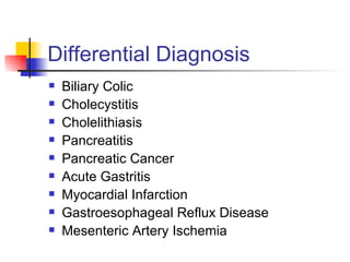
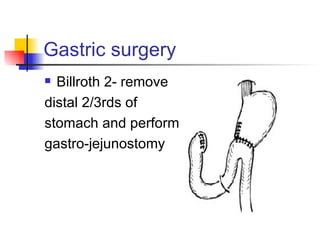
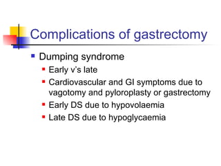
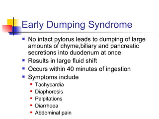
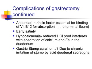
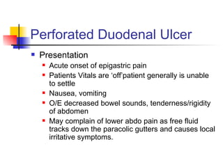
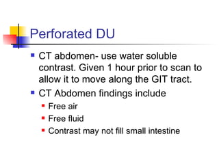
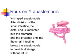
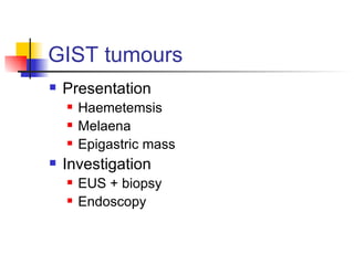
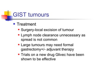
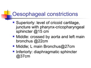
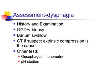
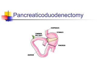
Upper GI surgery Ms. N. Bambury 12/02/2009
Overview Stomach Duodenum Oesophagus Pancreas
Anatomy of the stomach
Physiology of the stomach Reservoir for digestion Endocrine functions Secretion of gastrin(from antral cells) Gastrin acts on parietal cells to increase acid production Exocrine functions Acid secretion
Peptic Ulcer Disease Peptic ulcers are defects in the gastric or duodenal mucosa that extend through the muscularis mucosa.  Occur when defensive mechanisms ie. tight intercellular junctions, mucus, and epithelial renewal,   are overcome by aggressive factors
PUD Aggressive factors/Causitive factors NSAIDS H.Pylori Severe physiologic stress Burns   CNS trauma   Surgery Hypersecretory states  Gastrinoma (Zollinger-Ellison syndrome) multiple endocrine neoplasia (MEN-I)
Patient Factors 40-70 years Men Duodenal ulcers>gastric ulcers
Gastric ulcers Same age group Males Located in prepyloric pyloric or coexist 70% located in lesser curvature Risk factors Smoking Alcolhol Burns  Trauma  NSAIDS/steroids
Gastric ulcers Symptoms Epigastric pain Transiently relieved by food May be associated with nausea , vomiting and decreased appetite
Duodenal ulcers Same age,sex Located mostly 2cm from pylorus-duodenal bulb Caused by increase in gastric acid
Duodenal ulcers Symptoms Burning or aching several hours after a meal Haematemesis,FOB+ve Back pain Nausea, vomiting,decreased appetite
Differential Diagnosis Biliary Colic   Cholecystitis   Cholelithiasis Pancreatitis Pancreatic Cancer Acute   Gastritis  Myocardial Infarction Gastroesophageal Reflux Disease Mesenteric Artery Ischemia
Diagnosing ulcers Outpatient setting Bloods OGD In the case of gastric ulcers biopsy should always be taken to outrule carcinoma. Patient should be treated and rescoped in approx 6 weeks to ensure healing. Biopsies are taken on all walls at 2 cm intervals
Medical treatment Treat H pylori infection with triple therapy  PPI Clarithromycin penecillin PPI Metronidazole Ampicillin PPI Metronidazole Clarithromycin Those not found to be H. Pylori free Treat with PPI or H2 receptor blockers
Complications of PUD Bleeding Perforation Pain Obstruction Secondary to repeated inflammation and subsequent scarring namely gastric outlet obstruction
Indications for surgery Similar in both types of ulceration Refractory to treatment Haemorrhage Perforation Obstructive symptoms Surgery is less common now since the introduction of PPIs
Surgical management of Gastric ulcers Principles Remove ulcer remove gastrin secreting antrum Two operations described
Gastric Surgery Billroth 1-remove distal third  of stomach and anastomose remainder to duodenum
Gastric surgery Billroth 2- remove  distal 2/3rds of  stomach and perform gastro-jejunostomy
Surgical management of Duodenal ulcers Principles Reduce acid secretion by dividing the vagus nerve  called a  vagotomy. Vagotomy denervates the stomach and therefore the pylorus which will lead to gastric outlet obstruction. Therefore a drainage procedure is performed called a pyloroplasty. 2 surgical operations Truncal vagotomy and pyloroplasty Selective vagotomy and pyloroplasty
Vagotomy complications Decreased acid secretion (aim of the game) Faster gastric emptying (loss of vagally mediated) Diarrhoea Dumping syndrome Gastric outlet obstruction(unless pyloroplasty performed)
Vagotomy complications Remember proximal vagotomy denervates from the stomach to the distal transverse colon including the pancreas and gallbladder Gallbladder denervation leads to stasis and which increases the chance of gallstones. Decrease in pancreatic and gallbladder secreations leading to undigested fats-steatorrhoea
Complications of gastrectomy Dumping syndrome Early v’s late Cardiovascular and GI symptoms due to vagotomy and pyloroplasty or gastrectomy Early DS due to hypovolaemia Late DS due to hypoglycaemia
Early Dumping Syndrome No intact pylorus leads to dumping of large amounts of chyme,biliary and pancreatic secretions into duodenum at once Results in large fluid shift Occurs within 40 minutes of ingestion Symptoms include Tachycardia Diaphoresis Palpitations Diarrhoea Abdominal pain
Late Dumping Syndrome Due to rebound hypoglycaemia  Occurs 2-4 hours post op  CHO  enterglucagon  sensitizes islet cells  overproduction of insulin Symptoms include Tachycardia Palpitations Diaphoresis Dizziness
Complications of gastrectomy continued Anaemia( Intrinsic factor essential for binding of Vit B12 for absorption in the terminal ileum) Early satiety Hypocalcaemia- reduced HCl prod interferes with absorption of calcium and Fe in the duodenum Gastric Stump carcinoma? Due to chronic irritation of stump by acid duodenal secretions
Specific management Patient presenting with  Haematemesis History and Examination Vital signs 2 large bore cannulae Bloods including FBC  U&E  Coag screen  Group and X match for 6 units Inflammatory markers Amylase
Specific management If greater than 6 units required urgent scope warranted. Oversewing of bleeding vessel. In the case of gastric ulceration distal gastrectomy may be warranted to excise the ulcer. No vagotomy required
Perforated Duodenal Ulcer Presentation Acute onset of epigastric pain Patients Vitals are ‘off’patient generally is unable to settle Nausea, vomiting O/E decreased bowel sounds, tenderness/rigidity of abdomen May complain of lower abdo pain as free fluid tracks down the paracolic gutters and causes local irritative symptoms.
Management of Perforated DU 2 large bore cannulae Fluid resuscitation NG insertion Urinary catheter insertion  Analgesia and antibiotics Bloods as before Erect CXR-Free air PFA- may see free air also
Perforated DU CT abdomen- use water soluble contrast. Given 1 hour prior to scan to allow it to move along the GIT tract.  CT Abdomen findings include Free air Free fluid Contrast may not fill small intestine
Surgical Management Graham patch Mini laparotomy from below umbilicus to epigastrium Stomach is identified and duodenum Perforation sought Omentum mobilised so that it is easily applied to site of perforation Suturedin 2 layers to peration site Closure
Definitions you should know Cushings Ulcer- ulcer associated with trauma, tumour or neurology Curlings ulcer- associated with major burns Marginal ulcer- ulceration at the site of a GI anastomosis Dieulafoys ulcer-underlying gastric malformation
Gastric Ca Adenocarcinoma Risk factors Blood group A, male Pernicious anaemia (Autoimmune disease) Hypogammaglobulinaemia  Previous partial gastrectomy  Helicobacter pylori  infection  Atrophic gastritis  Intestinal metaplasia
Gastric Ca Presentation Age group>60 Weight loss Loss of appetite Palpable epigastric mass Melaena Most present late and are not amenable to radical surgery
Gastric Ca Investigations/Staging  CEA OGD confirms diagnosis with a tissue biopsy Endoscopic ultrasound may allow assessment of intramural tumour penetration  CT will assess nodal spread and extent of metastatic disease  Laparoscopy will identify peritoneal seeding  Survival Overall 5 year survival is approximately 5%
Gastric Ca Surgical options No real role for adjuvant therapy Billroth 2 or Roux en Y anastomosis Antrum-Distal subtotal(75%) gastrectomy Body- total gastrectomy Proximal- total gastrectomy
Roux en Y anastomosis Y-shaped anastomosis After division of the small intestine,the distal end is implanted into the stomach  and the proximal end into the small intestine below the anastomosis to provide drainage without reflux.
Gastric Ca Virchow’s node Metastatic gastric carcinoma in the left supraclavicular fossa Krukenburg tumour Gastric ca which has metastasized to the ovaries
Gastric lymphoma The Stomach is the commonest extranodal primary site for non-Hodgkin's lymphoma  Secondary Lymphoma is commonly seen in stomach from another site Accounts for 5% of gastric malignancies  Presentation is similar to gastric carcinoma Anaemia and epigastric mass are common Age of presentation is approx 60 years Investigations EUS is the best modality
Gastric Lymphoma Treatment  70% of tumours are resectable  5-year survival is approximately 25%  Treatment modalities are discussed on a case by case basis at an MDT Treatments range from chemotherapy alone, H pylori eradication or surgery and chemotherapychemotherapy may be useful
MALT lymphoma A type of primary gastric lymphoma The stomach does not usually contain lymphoid tissue MALT follicles found in the stomach are associated with H pylori infection Patients can be completely cured by H pylori eradication or else use it in conjunction with chemotherapy
GIST tumours Gastrointestinal sromal tumours Previously classified as Leiomyosarcomas, Leiomyomas or sarcomatous lesions ie they originate from smooth muscle. There are classified according to the degree of differentiation towards different cells
GIST tumours Classification Benign Differentiation toward muscle cells Differentiation toward neural elements Malignant Dual differentiation Lacking differentiation 1% of stomach cancers
GIST tumours Presentation Haemetemsis Melaena Epigastric mass Investigation EUS + biopsy Endoscopy
GIST tumours Treatment Surgery-local excision of tumour Lymph node clearance unnecessary as spread is not common Large tumours may need formal gastrectomy+/- adjuvant therapy Trials on a new drug Glivec have been shown to be effective
Sister Mary Joseph Nodule A 'nodule' in the umbilicus often associated with advanced malignancy  Presents as firm, red, non-tender nodule  Results from spread of tumour within the falciform ligament  90% of tumours are adenocarcinomas  Commonest primaries are stomach and ovary  Primary tumour is almost invariably inoperable
Oesophageal Anatomy Superior 1/3rd-smooth muscle Middle 1/3rd-mixed Inferior 1/3rd-smooth muscle Length -25cm
Oesophageal constrictions Superiorly: level of cricoid cartilage, juncture with pharynx-cricopharyngeal sphincter @15 cm Middle: crossed by aorta and left main bronchus @22cm Middle; L main Bronchus@27cm Inferiorly: diaphragmatic sphincter @37cm
Why are constrictions important Areas where foreign bodies lodge Common sites of carcinoma Difficulty passing scope on OGD may occur
Dysphagia Definition-difficulty swallowing Typical presentation in oesophageal Ca is one of progressive dysphagia starting with solids followed by fluids Odynophagia- pain on swallowing
Differential diagnosis -dysphagia Asnatomical causes Intrinsic to wall Carcinoma Cricoid web  Inflammatory lesions Extrinsic lesions Bronchial Ca Mitral stenosis leading to L atrial enlargement
Differential diagnosis -dysphagia Functional lesions Neurological causes Post CVA MND Globus hystericus-constriction of the lower part of the oeophagus associated with anxiety Dysmotility Achalasia Diffuse oesophageal spasm Scleroderma
Assessment-dysphagia History and Examination OGD+/-biopsy Barium swallow CT if suspect extrinsic compression is the cause. Other tests Oesophageal manometry pH studies
Oesophageal manometry NG tube passed into oesophagus Pressure transducer on tip of it measures resting and squeezing pressures at different levels of the oesophagus Normal peristaltic waves travel at a rate of 5cm/sec through oesophagus Normal resting pressure of LOS is 10-15mmHg Squeeze pressures should generate up to 100mmHg
pH studies Naos oesophageal wire containing a pH probe is left in oesophagus for a 24 hour period If oesophageal pH is greater than 4 for >4% of the time this indicates reflux
GORD 3 factors exist to keep gastric juices out of the oesophagus LOS competence Oesophageal motility Clearance into stomach
GORD presentation Symptoms Pain Epigastric Retrosternal Interscapular Odynophagia Reflux of food especially on bending Pulmonary aspiration Nocturnal coughing Hoarse voice
GORD Investigations pH monitoring is the gold standard investigation Rule out MI OGD + biopsy-5cm above GOJ shows increased eosinophils and hypoplasia See if patient has a hiatus hernia (1/3 of patients with h.h have GORD)
Complications of GORD Oesophageal stricture Commonest cause Treat by balloon dilatation via OGD Surgery options-Lap Nissan fundoplication Barrett’s oesophagus Increased risk of malignant transformation
Treatment of GORD Conservative mgt- Antacids, lose weight, raise head of bed etc. Metoclopramide,H2 blockers, PPIs Surgical Nissan fundoplication Fundus of stomach is mobilised,wrapped around the oesophagus
Motility disorders Primary versus secondary Primary Achalasia Diffuse oesophageal spasm Secondary Autoimmune rheumatic disorders(scleroderma) Chagas disease(chronic infection with T cruzi associated with mega disorders) DM Amyloid
Achalasia HighLOS pressure leading to failure of the sphincter to relax  poor peristalsis Presents with dysphagia and retrosternal chest pain Affects 30-60 years age group Investigation;Barium swallow   bird’s beak, lack of gastric air bubble, contrast may not enter stomach
Achalasia Treatment Balloon dilatation Heller’s cardiomyopathy-release of muscle at the GOJ, reflux common post op Injection of botulinum toxin at ultrasound
Diffuse oesophageal spasm Part of the differential of MI Symptoms Retrosternal chest pain radiating to jaw Invest; Manometry-nutcracker oesophagus Management Nifedipine
Scleroderma CREST syndrome Calcinosis Raynauds Oesphagitis Scleroderma Telangiectasia
Scleroderma Srticture formation occurs due to inflammation and GORD Investigations Manometry- incompetent LOS Treatment Partial fundoplication
Autoimmune diseases Rheumatoid Arthritis SLE Dermatomyositis Polymyositis May all be associated with oesophageal dysmotility
Oesophageal carcinoma Adenocarcinoma>SCC Adeno seen in lower 1/3rd SCCs can be any site Males>Females Age;>40 Most common at sites of physiological narrowing Lower 1/3rd and GOJ most common sites
Oesophageal carcinoma Risk factors Adeno Barretts GORD Obesity Alcohol Cigarette smoking
Oesophageal carcinoma Risk factors SCC Alcohol Smoking Coeliac Achalasia PUD
Oesophageal carcinoma Symptoms Dysphagia Retrosternal pain Coughing during eating Pseudo-achalasia
Oesophageal carcinoma Pre-operative U&Es, FBC Optimise nutrition-NG feed Stop smoking etc
Oesophageal carcinoma Surgical management Ivor Lewis procedure Laparotomy and mobilisation of stomach Right thoracotomy- resection of tumour and reanastomosis of stomach to healthy oesophagus 5 year survival 25% and only 30% patients suitable for surgery!
Oesophageal carcinoma Palliative treatment Intubation with metal stent under radiological control Chemotherapy-adeno Radiotherapy-SCC
Pancreatic Anatomy The pancreas is a retroperitoneal organ Consists of head body neck and tail Duct of Wirsung drains most of the pancreas Duct of Santorini drains the uncinate process into the 2nd part of the duodenum Note the pancreas shares a common blood supply with the duodenum!
Structure of the pancreas Exocrine  Serous secretory cells make up lobules which drain via ductules into main ducts Endocrine Islets of Langerhans secrete insulin(Beta), glucagon(Alpha) and somatostatin (Delta) directly into circulation
Pancreatitis Presentation Epigastric pain Radiating through to back or interscapular area Pain score 10/10 May be associated with nausea, vomiting diaphoresis
Acute Pancreatitis Signs Jaundice Ascites Grey-Turners sign Cullens sign Shock(Hypovolaemia) ARDS Renal failure(hypovolaemia)
Acute Pancreatitis Aetiology Gallstones Alcohol Trauma Steroids Mumps Autoimmune(SLE) Hypothermia. ↑Ca,↑lipids/TRG ERCP Drugs-Azathioprine,diuretics,thiazides, NSAIDS
Acute Pancreatitis Investigations FBC U&E, Magnesium, Calcium Amylase, glucose LFTs Coag screen Albumin
Amylase and its role in diagnosis Amylase is a digestive enzyme that normally acts extracellularly to cleave starch into monosaccharides,.  The pancreas and the salivary glands account for almost all serum amylase Used as a diagnostic tool 10% of cases amylase is normal, especially in acute on chronic pancreatitis Low specificity
Amylase and its role in diagnosis Renal failure and liver disease result in decreased metabolic clearance IBD, mesenteric infarction- ↑amylase absorption Gut perforation-leaks into peritoneum and absorbed across inflamed peritoneum Ectopic amylase production by lung, ovary, pancreas, and colon malignanciesand breast cancer (increased pancreatic amylase) are miscellaneous
Acute pancreatitis Investigations Erect CXR, PFA May see ‘sentinel node’ of proximal jejunum CT pancreatic protocal Look for fat streaking around the pancreas, fluid in the lesser sac, necrosis, pseudocyst U/S to o/r gallstones as cause ABG-hypoxia, lactic acidosis
Acute Pancreatitis Scoring systems Ranson’s criteria At admission time and 48 hours into admission A higher score is associated with a poorer prognosis
Ranson’s admission criteria Glucose >11 Age >55 LDH >350 AST>250 WCC>16
Ranson’s criteria 48 hours later Calcium <2 Haematocrit   10% pO2<60 Base deficit >4 BUN>1.8 >3 positive  severe attack
Sequelae Hypo/Hyperglycaemia Hypocalcaemia- saponification of omental fat by pancreatic lipases chelates calcium Hypomagnesaemia Chronicity DM
Sequelae ARDS/MODS Abscess formation Pancreatic necrosis Pseudocysts
Acute pancreatitis Management Oxygen 2 large bore cannulae Fluid resuscitation Analgesia NG(large bore) for drainage if vomiting Urinary catheter- measure input output Bloods
Pancreatic Ca Management Antibiotics only if evidence of pyrexia or positive blood cultures Imipenem antibiotic of choice Surgery indicated only in presence of abscess /pseudocyst or massive infected pancreatic necrosis Mortality significanty increased in the context of laparotomy
Pancreatic Carcinoma Ductal adenocarcinoma 60-80 years Risk factors Smokers DM FAP Gardner syndrome
Pancreatic Carcinoma 70% occur at the head 20% in the body 10% in the tail Presentation Weight loss Jaundice Pain Pancreatitis Trousseaus sign-superficial thrombophlebitis assoc with Panc Ca
Pancreatic Ca Head tumours Present with painless jaundice and earlier Palpable non tender gallbladder(Courvosier’s sign) Body and tail Presents later Tends to present as pain, weight loss and hepatomegaly
Pancreatic Ca Investigations Contrast enhanced CT-pancreatic protocol ERCP- useful for insertion of stent in context of jaundice and tissue diagnosis MRI to seek extent of local invasion Endoscopic ultrasound Laparoscopy
Surgical management Head of pancreas/peri-ampullary tumours Whipples operation Resect specimen including distal stomach duodenum  to jejunum head of pancreas Gastroenterostomy Choledochojejunostomy Pancreaticojejunostomy
Pancreaticoduodenectomy
Surgical management Body and Tail tumours Distal resection Resection rate <7% No place for adjuvant therapy Tissue diagnosis important Neo adjuvant/adjuvant chemotherapy and radiotherapy have a role in the management of both tumours
Palliative care Alleviate symptoms only Stenting via ERCP Bypass procedures Chemotherapy Radiotherapy
Other definitions you should know Annular pancreas Pancreas surrounding the duodenum Congenital May present with duodenal obstruction Pancreatic divisum Failure of the two ducts to fuse
Benign Panc Tumours MCQ stuff! Gastrinoma Tumour of non beta islet cells May be multiple Overproduction of gastrin Leads to multiple metastases in some instances May also occur in duodenum 60% are malignant at diagnosis 30% are associated with MEN1
Gastrinoma Presentation Peptic ulcers May be treatment resistant Diarrhoea Haematemesis Perforation
Gastrinoma Investigations Pentagastrin secretory studies Fasting serum gastrin levels U/S, CT-localizes tumour Treatment PPIS Resection only if localised Total gastrectomy if poorly defined
Insulinoma Solitary tumours of Beta cells of the pancreas Presentation Usually related to hypogylcaemia Altered consciousness Uncharacteristic behaviour
Insulinoma Diagnosis Glucose  Glucagon test Plasma insulin U/S, CT Treatment Resection as malignant potential Distal pancreatectomy if tumour cannot be found
Next week Upper GI surgery to include Liver Spleen Gallbladder and Biliary tree Small intestine


Fwd: Bambury tutorial Upper GI Surgery

  • 1.
    Upper GI surgeryMs. N. Bambury 12/02/2009
  • 2.
    Overview Stomach DuodenumOesophagus Pancreas
  • 3.
  • 4.
  • 5.
    Physiology of thestomach Reservoir for digestion Endocrine functions Secretion of gastrin(from antral cells) Gastrin acts on parietal cells to increase acid production Exocrine functions Acid secretion
  • 6.
    Peptic Ulcer DiseasePeptic ulcers are defects in the gastric or duodenal mucosa that extend through the muscularis mucosa. Occur when defensive mechanisms ie. tight intercellular junctions, mucus, and epithelial renewal, are overcome by aggressive factors
  • 7.
    PUD Aggressive factors/Causitivefactors NSAIDS H.Pylori Severe physiologic stress Burns CNS trauma Surgery Hypersecretory states Gastrinoma (Zollinger-Ellison syndrome) multiple endocrine neoplasia (MEN-I)
  • 8.
    Patient Factors 40-70years Men Duodenal ulcers>gastric ulcers
  • 9.
    Gastric ulcers Sameage group Males Located in prepyloric pyloric or coexist 70% located in lesser curvature Risk factors Smoking Alcolhol Burns Trauma NSAIDS/steroids
  • 10.
    Gastric ulcers SymptomsEpigastric pain Transiently relieved by food May be associated with nausea , vomiting and decreased appetite
  • 11.
    Duodenal ulcers Sameage,sex Located mostly 2cm from pylorus-duodenal bulb Caused by increase in gastric acid
  • 12.
    Duodenal ulcers SymptomsBurning or aching several hours after a meal Haematemesis,FOB+ve Back pain Nausea, vomiting,decreased appetite
  • 13.
    Differential Diagnosis BiliaryColic Cholecystitis Cholelithiasis Pancreatitis Pancreatic Cancer Acute Gastritis Myocardial Infarction Gastroesophageal Reflux Disease Mesenteric Artery Ischemia
  • 14.
    Diagnosing ulcers Outpatientsetting Bloods OGD In the case of gastric ulcers biopsy should always be taken to outrule carcinoma. Patient should be treated and rescoped in approx 6 weeks to ensure healing. Biopsies are taken on all walls at 2 cm intervals
  • 15.
    Medical treatment TreatH pylori infection with triple therapy PPI Clarithromycin penecillin PPI Metronidazole Ampicillin PPI Metronidazole Clarithromycin Those not found to be H. Pylori free Treat with PPI or H2 receptor blockers
  • 16.
    Complications of PUDBleeding Perforation Pain Obstruction Secondary to repeated inflammation and subsequent scarring namely gastric outlet obstruction
  • 17.
    Indications for surgerySimilar in both types of ulceration Refractory to treatment Haemorrhage Perforation Obstructive symptoms Surgery is less common now since the introduction of PPIs
  • 18.
    Surgical management ofGastric ulcers Principles Remove ulcer remove gastrin secreting antrum Two operations described
  • 19.
    Gastric Surgery Billroth1-remove distal third of stomach and anastomose remainder to duodenum
  • 20.
    Gastric surgery Billroth2- remove distal 2/3rds of stomach and perform gastro-jejunostomy
  • 21.
    Surgical management ofDuodenal ulcers Principles Reduce acid secretion by dividing the vagus nerve called a vagotomy. Vagotomy denervates the stomach and therefore the pylorus which will lead to gastric outlet obstruction. Therefore a drainage procedure is performed called a pyloroplasty. 2 surgical operations Truncal vagotomy and pyloroplasty Selective vagotomy and pyloroplasty
  • 22.
    Vagotomy complications Decreasedacid secretion (aim of the game) Faster gastric emptying (loss of vagally mediated) Diarrhoea Dumping syndrome Gastric outlet obstruction(unless pyloroplasty performed)
  • 23.
    Vagotomy complications Rememberproximal vagotomy denervates from the stomach to the distal transverse colon including the pancreas and gallbladder Gallbladder denervation leads to stasis and which increases the chance of gallstones. Decrease in pancreatic and gallbladder secreations leading to undigested fats-steatorrhoea
  • 24.
    Complications of gastrectomyDumping syndrome Early v’s late Cardiovascular and GI symptoms due to vagotomy and pyloroplasty or gastrectomy Early DS due to hypovolaemia Late DS due to hypoglycaemia
  • 25.
    Early Dumping SyndromeNo intact pylorus leads to dumping of large amounts of chyme,biliary and pancreatic secretions into duodenum at once Results in large fluid shift Occurs within 40 minutes of ingestion Symptoms include Tachycardia Diaphoresis Palpitations Diarrhoea Abdominal pain
  • 26.
    Late Dumping SyndromeDue to rebound hypoglycaemia Occurs 2-4 hours post op  CHO  enterglucagon  sensitizes islet cells  overproduction of insulin Symptoms include Tachycardia Palpitations Diaphoresis Dizziness
  • 27.
    Complications of gastrectomycontinued Anaemia( Intrinsic factor essential for binding of Vit B12 for absorption in the terminal ileum) Early satiety Hypocalcaemia- reduced HCl prod interferes with absorption of calcium and Fe in the duodenum Gastric Stump carcinoma? Due to chronic irritation of stump by acid duodenal secretions
  • 28.
    Specific management Patientpresenting with Haematemesis History and Examination Vital signs 2 large bore cannulae Bloods including FBC U&E Coag screen Group and X match for 6 units Inflammatory markers Amylase
  • 29.
    Specific management Ifgreater than 6 units required urgent scope warranted. Oversewing of bleeding vessel. In the case of gastric ulceration distal gastrectomy may be warranted to excise the ulcer. No vagotomy required
  • 30.
    Perforated Duodenal UlcerPresentation Acute onset of epigastric pain Patients Vitals are ‘off’patient generally is unable to settle Nausea, vomiting O/E decreased bowel sounds, tenderness/rigidity of abdomen May complain of lower abdo pain as free fluid tracks down the paracolic gutters and causes local irritative symptoms.
  • 31.
    Management of PerforatedDU 2 large bore cannulae Fluid resuscitation NG insertion Urinary catheter insertion Analgesia and antibiotics Bloods as before Erect CXR-Free air PFA- may see free air also
  • 32.
    Perforated DU CTabdomen- use water soluble contrast. Given 1 hour prior to scan to allow it to move along the GIT tract. CT Abdomen findings include Free air Free fluid Contrast may not fill small intestine
  • 33.
    Surgical Management Grahampatch Mini laparotomy from below umbilicus to epigastrium Stomach is identified and duodenum Perforation sought Omentum mobilised so that it is easily applied to site of perforation Suturedin 2 layers to peration site Closure
  • 34.
    Definitions you shouldknow Cushings Ulcer- ulcer associated with trauma, tumour or neurology Curlings ulcer- associated with major burns Marginal ulcer- ulceration at the site of a GI anastomosis Dieulafoys ulcer-underlying gastric malformation
  • 35.
    Gastric Ca AdenocarcinomaRisk factors Blood group A, male Pernicious anaemia (Autoimmune disease) Hypogammaglobulinaemia Previous partial gastrectomy Helicobacter pylori infection Atrophic gastritis Intestinal metaplasia
  • 36.
    Gastric Ca PresentationAge group>60 Weight loss Loss of appetite Palpable epigastric mass Melaena Most present late and are not amenable to radical surgery
  • 37.
    Gastric Ca Investigations/Staging CEA OGD confirms diagnosis with a tissue biopsy Endoscopic ultrasound may allow assessment of intramural tumour penetration CT will assess nodal spread and extent of metastatic disease Laparoscopy will identify peritoneal seeding Survival Overall 5 year survival is approximately 5%
  • 38.
    Gastric Ca Surgicaloptions No real role for adjuvant therapy Billroth 2 or Roux en Y anastomosis Antrum-Distal subtotal(75%) gastrectomy Body- total gastrectomy Proximal- total gastrectomy
  • 39.
    Roux en Yanastomosis Y-shaped anastomosis After division of the small intestine,the distal end is implanted into the stomach and the proximal end into the small intestine below the anastomosis to provide drainage without reflux.
  • 40.
    Gastric Ca Virchow’snode Metastatic gastric carcinoma in the left supraclavicular fossa Krukenburg tumour Gastric ca which has metastasized to the ovaries
  • 41.
    Gastric lymphoma TheStomach is the commonest extranodal primary site for non-Hodgkin's lymphoma Secondary Lymphoma is commonly seen in stomach from another site Accounts for 5% of gastric malignancies Presentation is similar to gastric carcinoma Anaemia and epigastric mass are common Age of presentation is approx 60 years Investigations EUS is the best modality
  • 42.
    Gastric Lymphoma Treatment 70% of tumours are resectable 5-year survival is approximately 25% Treatment modalities are discussed on a case by case basis at an MDT Treatments range from chemotherapy alone, H pylori eradication or surgery and chemotherapychemotherapy may be useful
  • 43.
    MALT lymphoma Atype of primary gastric lymphoma The stomach does not usually contain lymphoid tissue MALT follicles found in the stomach are associated with H pylori infection Patients can be completely cured by H pylori eradication or else use it in conjunction with chemotherapy
  • 44.
    GIST tumours Gastrointestinalsromal tumours Previously classified as Leiomyosarcomas, Leiomyomas or sarcomatous lesions ie they originate from smooth muscle. There are classified according to the degree of differentiation towards different cells
  • 45.
    GIST tumours ClassificationBenign Differentiation toward muscle cells Differentiation toward neural elements Malignant Dual differentiation Lacking differentiation 1% of stomach cancers
  • 46.
    GIST tumours PresentationHaemetemsis Melaena Epigastric mass Investigation EUS + biopsy Endoscopy
  • 47.
    GIST tumours TreatmentSurgery-local excision of tumour Lymph node clearance unnecessary as spread is not common Large tumours may need formal gastrectomy+/- adjuvant therapy Trials on a new drug Glivec have been shown to be effective
  • 48.
    Sister Mary JosephNodule A 'nodule' in the umbilicus often associated with advanced malignancy Presents as firm, red, non-tender nodule Results from spread of tumour within the falciform ligament 90% of tumours are adenocarcinomas Commonest primaries are stomach and ovary Primary tumour is almost invariably inoperable
  • 49.
    Oesophageal Anatomy Superior1/3rd-smooth muscle Middle 1/3rd-mixed Inferior 1/3rd-smooth muscle Length -25cm
  • 50.
    Oesophageal constrictions Superiorly:level of cricoid cartilage, juncture with pharynx-cricopharyngeal sphincter @15 cm Middle: crossed by aorta and left main bronchus @22cm Middle; L main Bronchus@27cm Inferiorly: diaphragmatic sphincter @37cm
  • 51.
    Why are constrictionsimportant Areas where foreign bodies lodge Common sites of carcinoma Difficulty passing scope on OGD may occur
  • 52.
    Dysphagia Definition-difficulty swallowingTypical presentation in oesophageal Ca is one of progressive dysphagia starting with solids followed by fluids Odynophagia- pain on swallowing
  • 53.
    Differential diagnosis -dysphagiaAsnatomical causes Intrinsic to wall Carcinoma Cricoid web Inflammatory lesions Extrinsic lesions Bronchial Ca Mitral stenosis leading to L atrial enlargement
  • 54.
    Differential diagnosis -dysphagiaFunctional lesions Neurological causes Post CVA MND Globus hystericus-constriction of the lower part of the oeophagus associated with anxiety Dysmotility Achalasia Diffuse oesophageal spasm Scleroderma
  • 55.
    Assessment-dysphagia History andExamination OGD+/-biopsy Barium swallow CT if suspect extrinsic compression is the cause. Other tests Oesophageal manometry pH studies
  • 56.
    Oesophageal manometry NGtube passed into oesophagus Pressure transducer on tip of it measures resting and squeezing pressures at different levels of the oesophagus Normal peristaltic waves travel at a rate of 5cm/sec through oesophagus Normal resting pressure of LOS is 10-15mmHg Squeeze pressures should generate up to 100mmHg
  • 57.
    pH studies Naosoesophageal wire containing a pH probe is left in oesophagus for a 24 hour period If oesophageal pH is greater than 4 for >4% of the time this indicates reflux
  • 58.
    GORD 3 factorsexist to keep gastric juices out of the oesophagus LOS competence Oesophageal motility Clearance into stomach
  • 59.
    GORD presentation SymptomsPain Epigastric Retrosternal Interscapular Odynophagia Reflux of food especially on bending Pulmonary aspiration Nocturnal coughing Hoarse voice
  • 60.
    GORD Investigations pHmonitoring is the gold standard investigation Rule out MI OGD + biopsy-5cm above GOJ shows increased eosinophils and hypoplasia See if patient has a hiatus hernia (1/3 of patients with h.h have GORD)
  • 61.
    Complications of GORDOesophageal stricture Commonest cause Treat by balloon dilatation via OGD Surgery options-Lap Nissan fundoplication Barrett’s oesophagus Increased risk of malignant transformation
  • 62.
    Treatment of GORDConservative mgt- Antacids, lose weight, raise head of bed etc. Metoclopramide,H2 blockers, PPIs Surgical Nissan fundoplication Fundus of stomach is mobilised,wrapped around the oesophagus
  • 63.
    Motility disorders Primaryversus secondary Primary Achalasia Diffuse oesophageal spasm Secondary Autoimmune rheumatic disorders(scleroderma) Chagas disease(chronic infection with T cruzi associated with mega disorders) DM Amyloid
  • 64.
    Achalasia HighLOS pressureleading to failure of the sphincter to relax  poor peristalsis Presents with dysphagia and retrosternal chest pain Affects 30-60 years age group Investigation;Barium swallow  bird’s beak, lack of gastric air bubble, contrast may not enter stomach
  • 65.
    Achalasia Treatment Balloondilatation Heller’s cardiomyopathy-release of muscle at the GOJ, reflux common post op Injection of botulinum toxin at ultrasound
  • 66.
    Diffuse oesophageal spasmPart of the differential of MI Symptoms Retrosternal chest pain radiating to jaw Invest; Manometry-nutcracker oesophagus Management Nifedipine
  • 67.
    Scleroderma CREST syndromeCalcinosis Raynauds Oesphagitis Scleroderma Telangiectasia
  • 68.
    Scleroderma Srticture formationoccurs due to inflammation and GORD Investigations Manometry- incompetent LOS Treatment Partial fundoplication
  • 69.
    Autoimmune diseases RheumatoidArthritis SLE Dermatomyositis Polymyositis May all be associated with oesophageal dysmotility
  • 70.
    Oesophageal carcinoma Adenocarcinoma>SCCAdeno seen in lower 1/3rd SCCs can be any site Males>Females Age;>40 Most common at sites of physiological narrowing Lower 1/3rd and GOJ most common sites
  • 71.
    Oesophageal carcinoma Riskfactors Adeno Barretts GORD Obesity Alcohol Cigarette smoking
  • 72.
    Oesophageal carcinoma Riskfactors SCC Alcohol Smoking Coeliac Achalasia PUD
  • 73.
    Oesophageal carcinoma SymptomsDysphagia Retrosternal pain Coughing during eating Pseudo-achalasia
  • 74.
    Oesophageal carcinoma Pre-operativeU&Es, FBC Optimise nutrition-NG feed Stop smoking etc
  • 75.
    Oesophageal carcinoma Surgicalmanagement Ivor Lewis procedure Laparotomy and mobilisation of stomach Right thoracotomy- resection of tumour and reanastomosis of stomach to healthy oesophagus 5 year survival 25% and only 30% patients suitable for surgery!
  • 76.
    Oesophageal carcinoma Palliativetreatment Intubation with metal stent under radiological control Chemotherapy-adeno Radiotherapy-SCC
  • 77.
    Pancreatic Anatomy Thepancreas is a retroperitoneal organ Consists of head body neck and tail Duct of Wirsung drains most of the pancreas Duct of Santorini drains the uncinate process into the 2nd part of the duodenum Note the pancreas shares a common blood supply with the duodenum!
  • 78.
    Structure of thepancreas Exocrine Serous secretory cells make up lobules which drain via ductules into main ducts Endocrine Islets of Langerhans secrete insulin(Beta), glucagon(Alpha) and somatostatin (Delta) directly into circulation
  • 79.
    Pancreatitis Presentation Epigastricpain Radiating through to back or interscapular area Pain score 10/10 May be associated with nausea, vomiting diaphoresis
  • 80.
    Acute Pancreatitis SignsJaundice Ascites Grey-Turners sign Cullens sign Shock(Hypovolaemia) ARDS Renal failure(hypovolaemia)
  • 81.
    Acute Pancreatitis AetiologyGallstones Alcohol Trauma Steroids Mumps Autoimmune(SLE) Hypothermia. ↑Ca,↑lipids/TRG ERCP Drugs-Azathioprine,diuretics,thiazides, NSAIDS
  • 82.
    Acute Pancreatitis InvestigationsFBC U&E, Magnesium, Calcium Amylase, glucose LFTs Coag screen Albumin
  • 83.
    Amylase and itsrole in diagnosis Amylase is a digestive enzyme that normally acts extracellularly to cleave starch into monosaccharides,. The pancreas and the salivary glands account for almost all serum amylase Used as a diagnostic tool 10% of cases amylase is normal, especially in acute on chronic pancreatitis Low specificity
  • 84.
    Amylase and itsrole in diagnosis Renal failure and liver disease result in decreased metabolic clearance IBD, mesenteric infarction- ↑amylase absorption Gut perforation-leaks into peritoneum and absorbed across inflamed peritoneum Ectopic amylase production by lung, ovary, pancreas, and colon malignanciesand breast cancer (increased pancreatic amylase) are miscellaneous
  • 85.
    Acute pancreatitis InvestigationsErect CXR, PFA May see ‘sentinel node’ of proximal jejunum CT pancreatic protocal Look for fat streaking around the pancreas, fluid in the lesser sac, necrosis, pseudocyst U/S to o/r gallstones as cause ABG-hypoxia, lactic acidosis
  • 86.
    Acute Pancreatitis Scoringsystems Ranson’s criteria At admission time and 48 hours into admission A higher score is associated with a poorer prognosis
  • 87.
    Ranson’s admission criteriaGlucose >11 Age >55 LDH >350 AST>250 WCC>16
  • 88.
    Ranson’s criteria 48hours later Calcium <2 Haematocrit  10% pO2<60 Base deficit >4 BUN>1.8 >3 positive  severe attack
  • 89.
    Sequelae Hypo/Hyperglycaemia Hypocalcaemia-saponification of omental fat by pancreatic lipases chelates calcium Hypomagnesaemia Chronicity DM
  • 90.
    Sequelae ARDS/MODS Abscessformation Pancreatic necrosis Pseudocysts
  • 91.
    Acute pancreatitis ManagementOxygen 2 large bore cannulae Fluid resuscitation Analgesia NG(large bore) for drainage if vomiting Urinary catheter- measure input output Bloods
  • 92.
    Pancreatic Ca ManagementAntibiotics only if evidence of pyrexia or positive blood cultures Imipenem antibiotic of choice Surgery indicated only in presence of abscess /pseudocyst or massive infected pancreatic necrosis Mortality significanty increased in the context of laparotomy
  • 93.
    Pancreatic Carcinoma Ductaladenocarcinoma 60-80 years Risk factors Smokers DM FAP Gardner syndrome
  • 94.
    Pancreatic Carcinoma 70%occur at the head 20% in the body 10% in the tail Presentation Weight loss Jaundice Pain Pancreatitis Trousseaus sign-superficial thrombophlebitis assoc with Panc Ca
  • 95.
    Pancreatic Ca Headtumours Present with painless jaundice and earlier Palpable non tender gallbladder(Courvosier’s sign) Body and tail Presents later Tends to present as pain, weight loss and hepatomegaly
  • 96.
    Pancreatic Ca InvestigationsContrast enhanced CT-pancreatic protocol ERCP- useful for insertion of stent in context of jaundice and tissue diagnosis MRI to seek extent of local invasion Endoscopic ultrasound Laparoscopy
  • 97.
    Surgical management Headof pancreas/peri-ampullary tumours Whipples operation Resect specimen including distal stomach duodenum to jejunum head of pancreas Gastroenterostomy Choledochojejunostomy Pancreaticojejunostomy
  • 98.
  • 99.
    Surgical management Bodyand Tail tumours Distal resection Resection rate <7% No place for adjuvant therapy Tissue diagnosis important Neo adjuvant/adjuvant chemotherapy and radiotherapy have a role in the management of both tumours
  • 100.
    Palliative care Alleviatesymptoms only Stenting via ERCP Bypass procedures Chemotherapy Radiotherapy
  • 101.
    Other definitions youshould know Annular pancreas Pancreas surrounding the duodenum Congenital May present with duodenal obstruction Pancreatic divisum Failure of the two ducts to fuse
  • 102.
    Benign Panc TumoursMCQ stuff! Gastrinoma Tumour of non beta islet cells May be multiple Overproduction of gastrin Leads to multiple metastases in some instances May also occur in duodenum 60% are malignant at diagnosis 30% are associated with MEN1
  • 103.
    Gastrinoma Presentation Pepticulcers May be treatment resistant Diarrhoea Haematemesis Perforation
  • 104.
    Gastrinoma Investigations Pentagastrinsecretory studies Fasting serum gastrin levels U/S, CT-localizes tumour Treatment PPIS Resection only if localised Total gastrectomy if poorly defined
  • 105.
    Insulinoma Solitary tumoursof Beta cells of the pancreas Presentation Usually related to hypogylcaemia Altered consciousness Uncharacteristic behaviour
  • 106.
    Insulinoma Diagnosis Glucose Glucagon test Plasma insulin U/S, CT Treatment Resection as malignant potential Distal pancreatectomy if tumour cannot be found
  • 107.
    Next week UpperGI surgery to include Liver Spleen Gallbladder and Biliary tree Small intestine
  • 108.
  • 109.