Download as PDF, PPTX

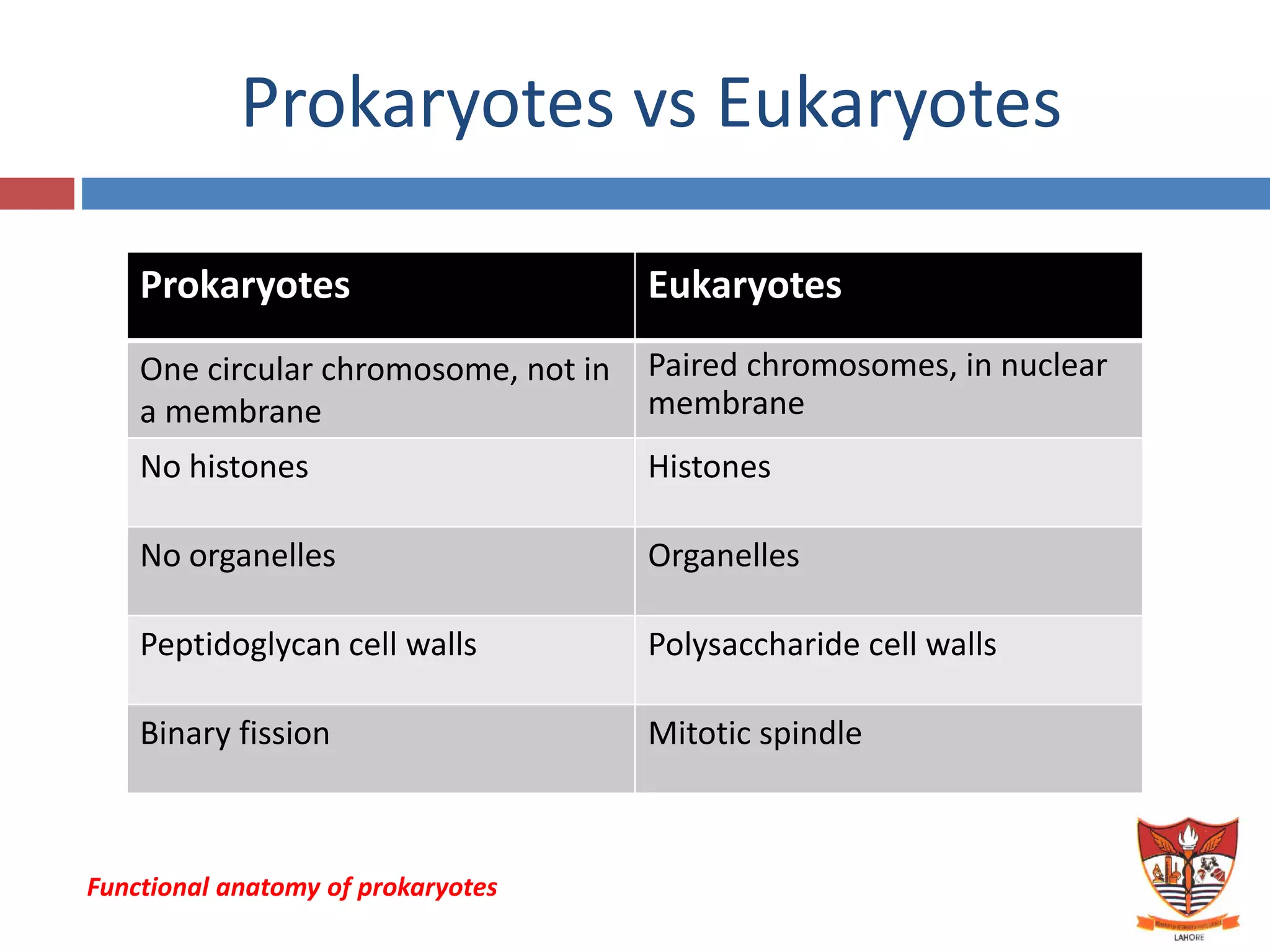
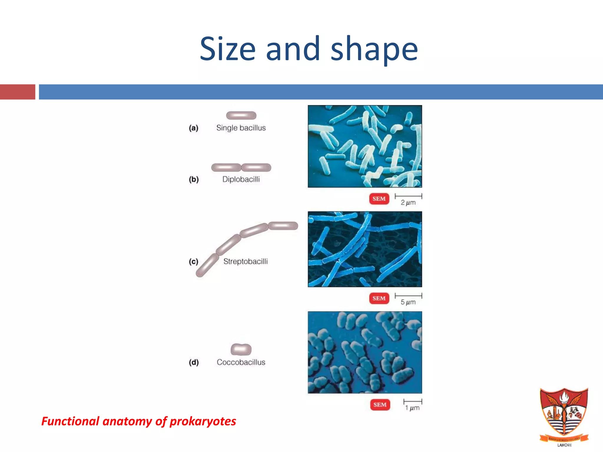
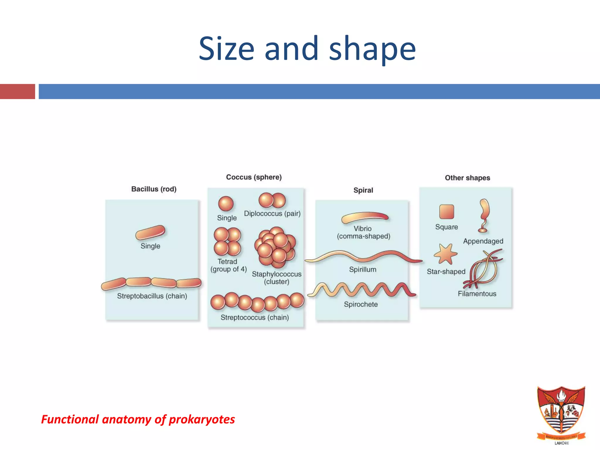
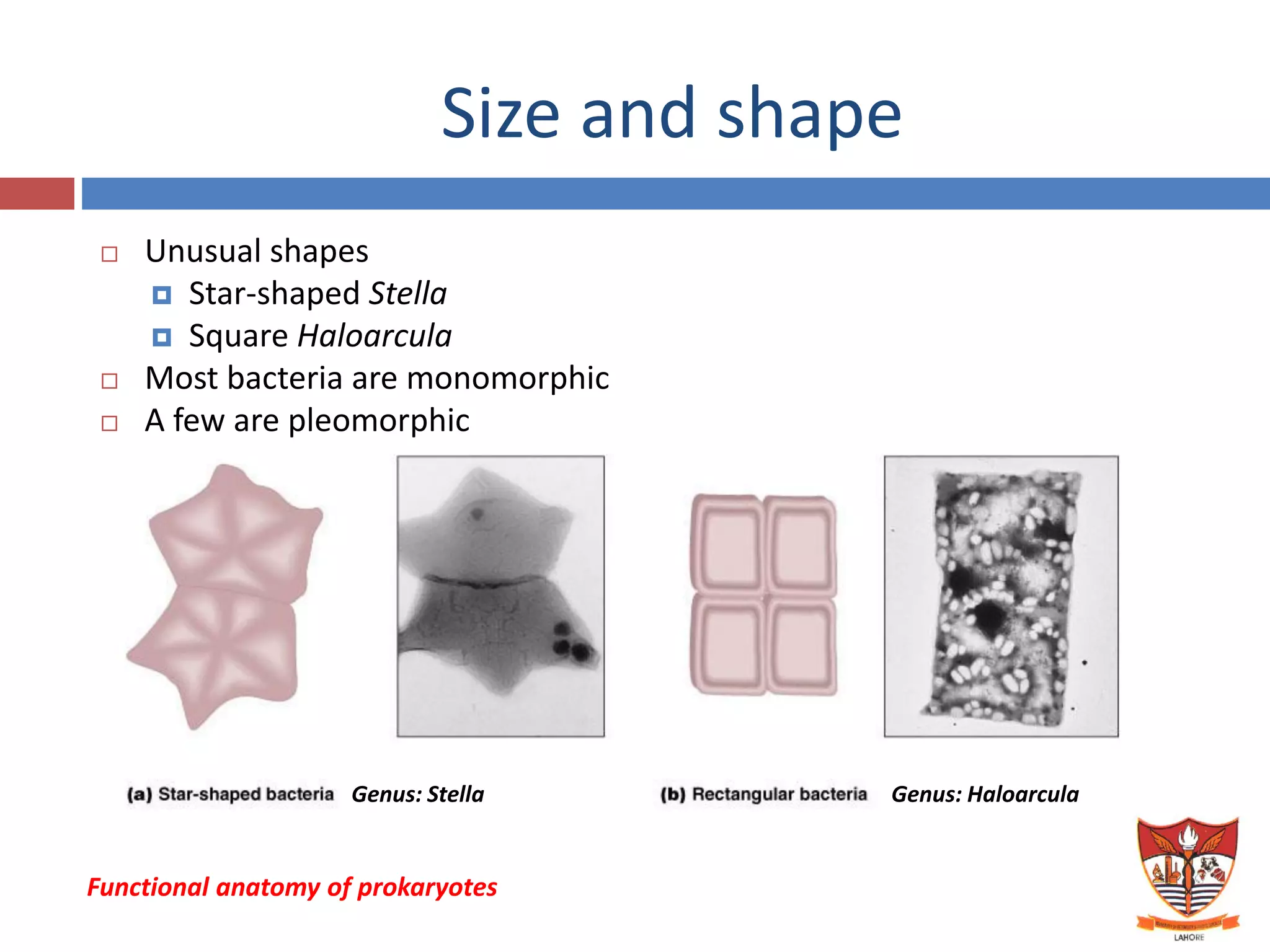
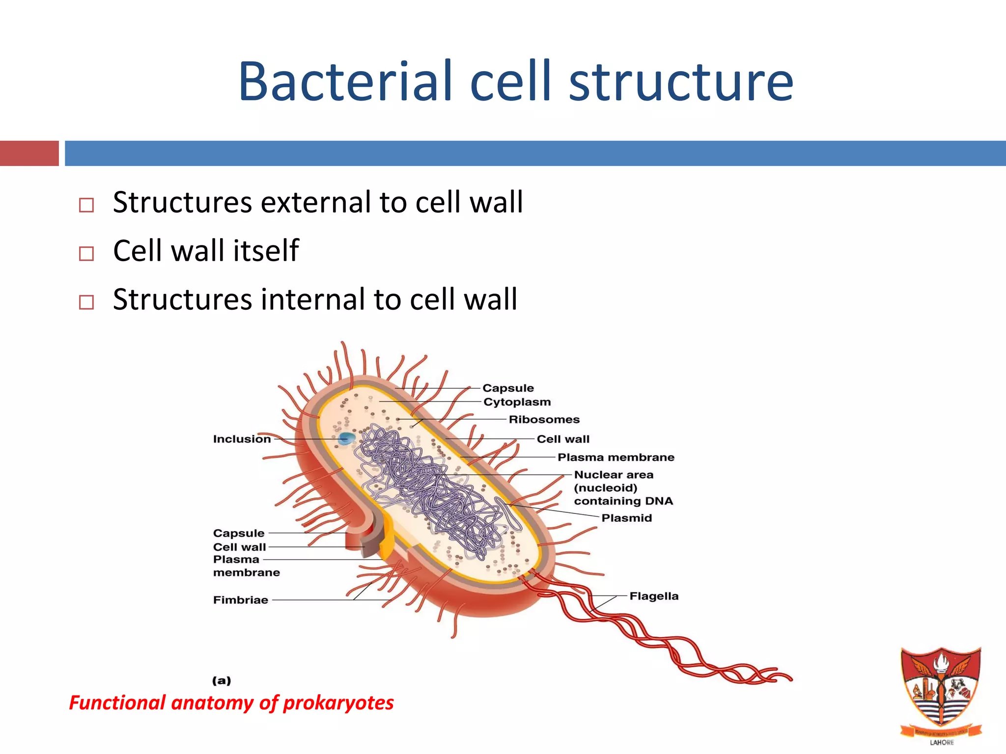

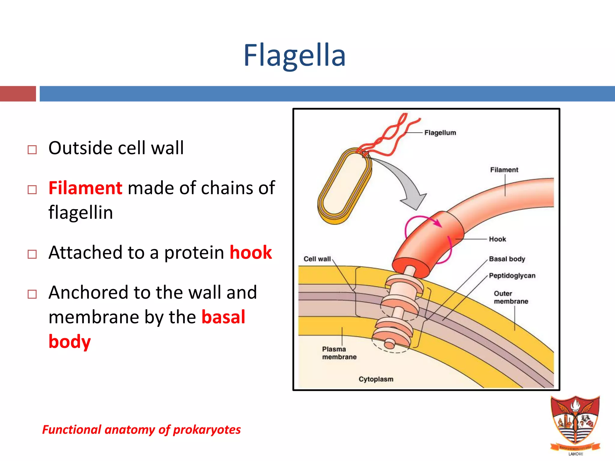


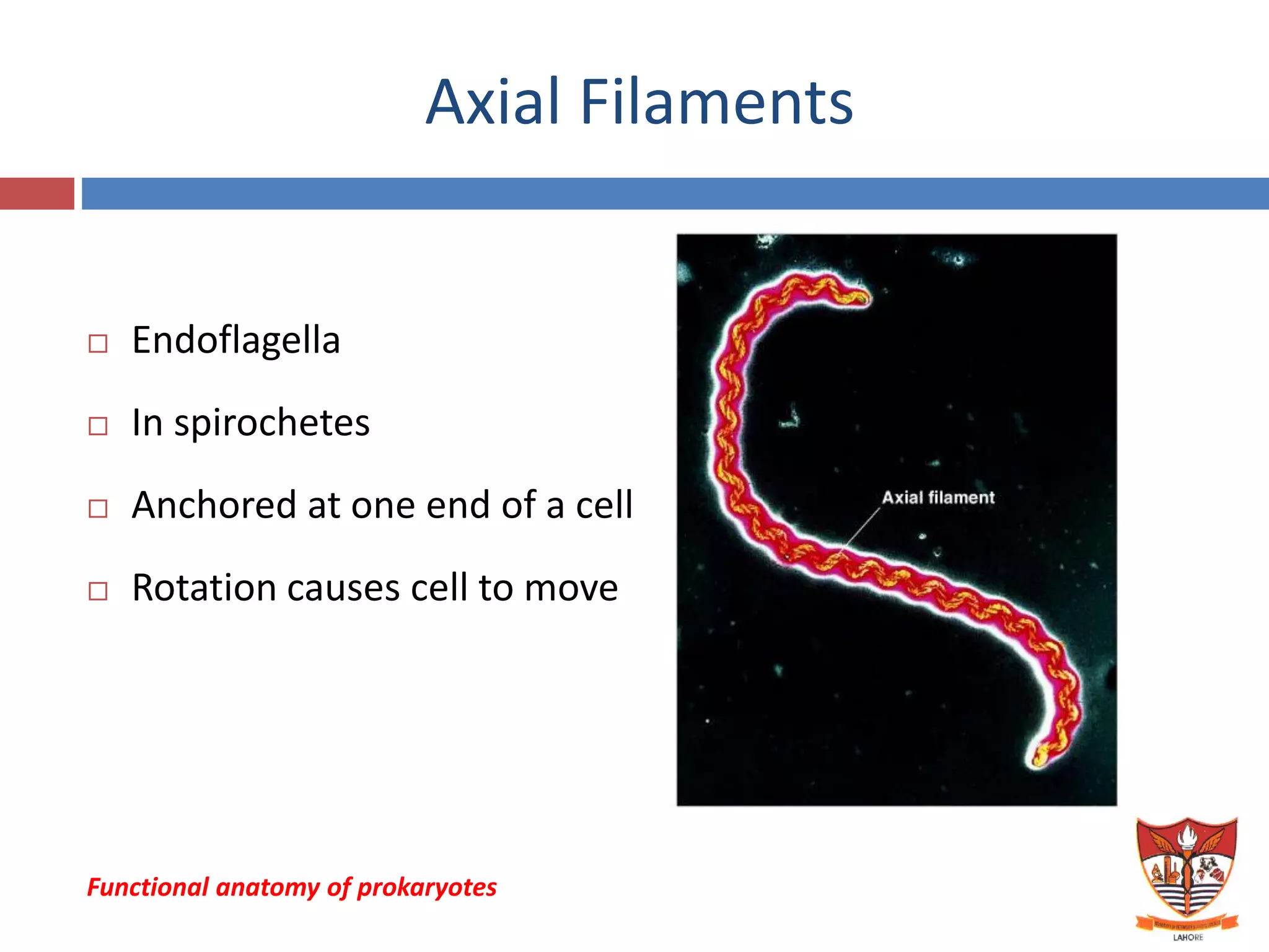

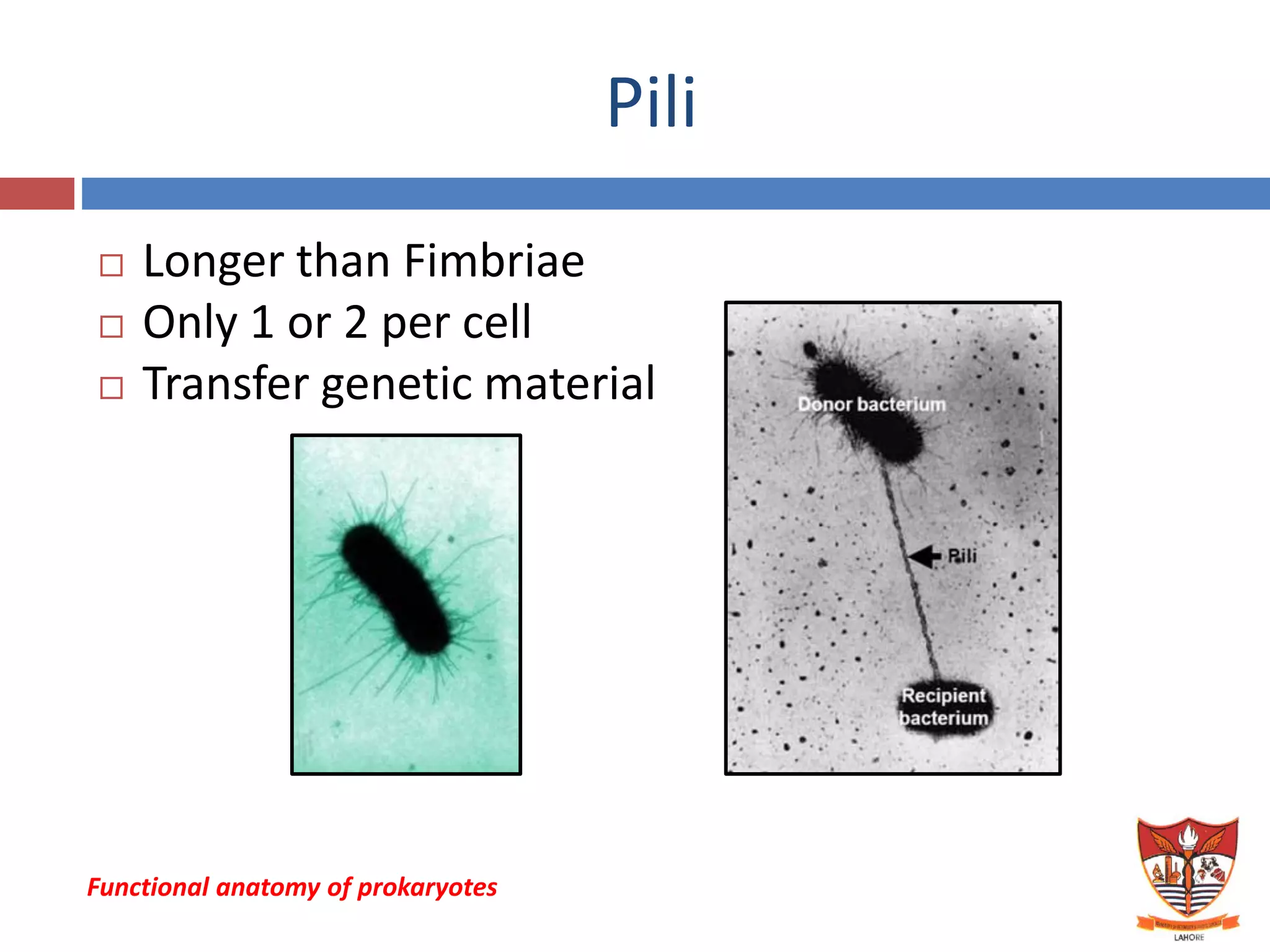

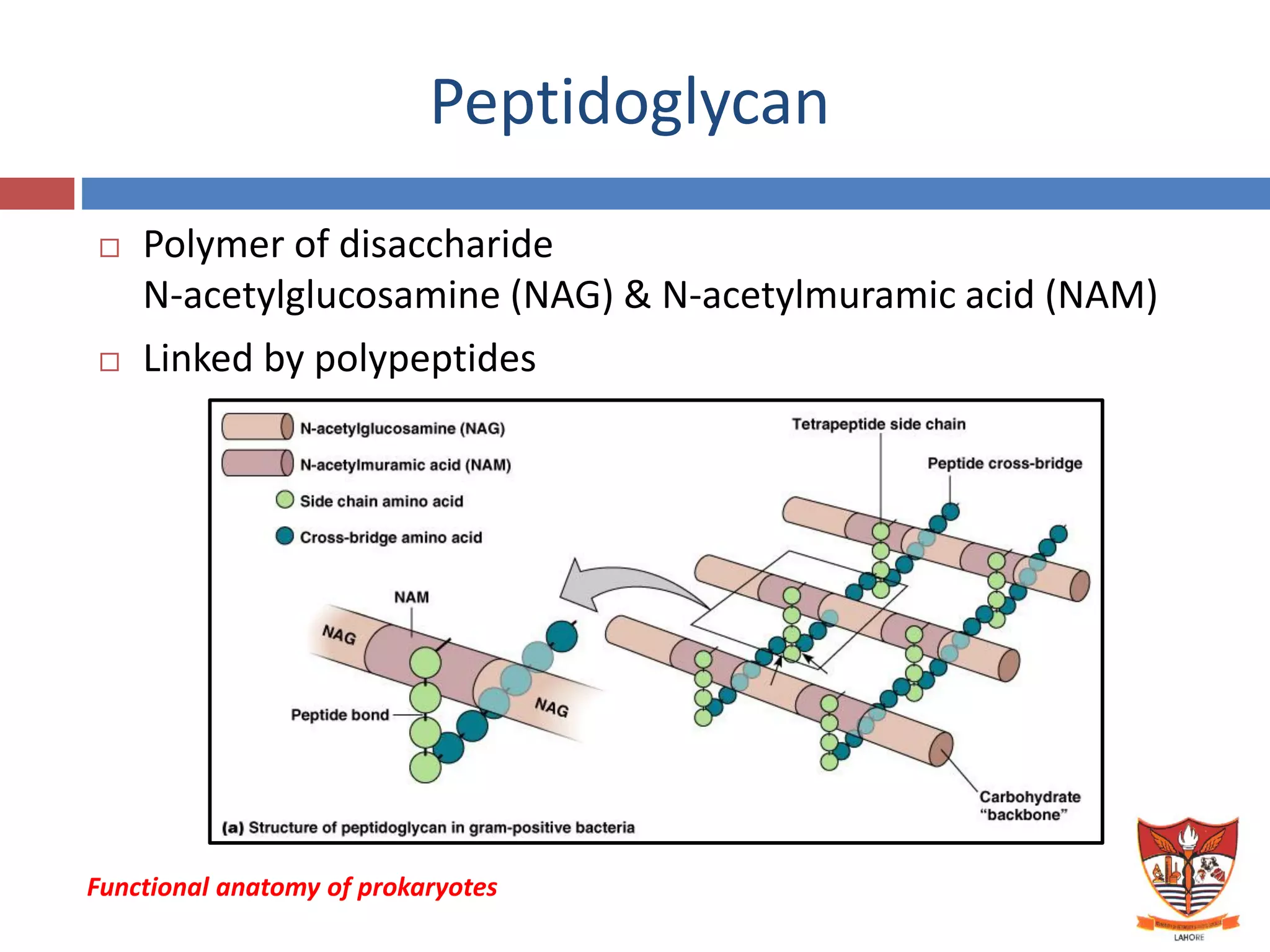


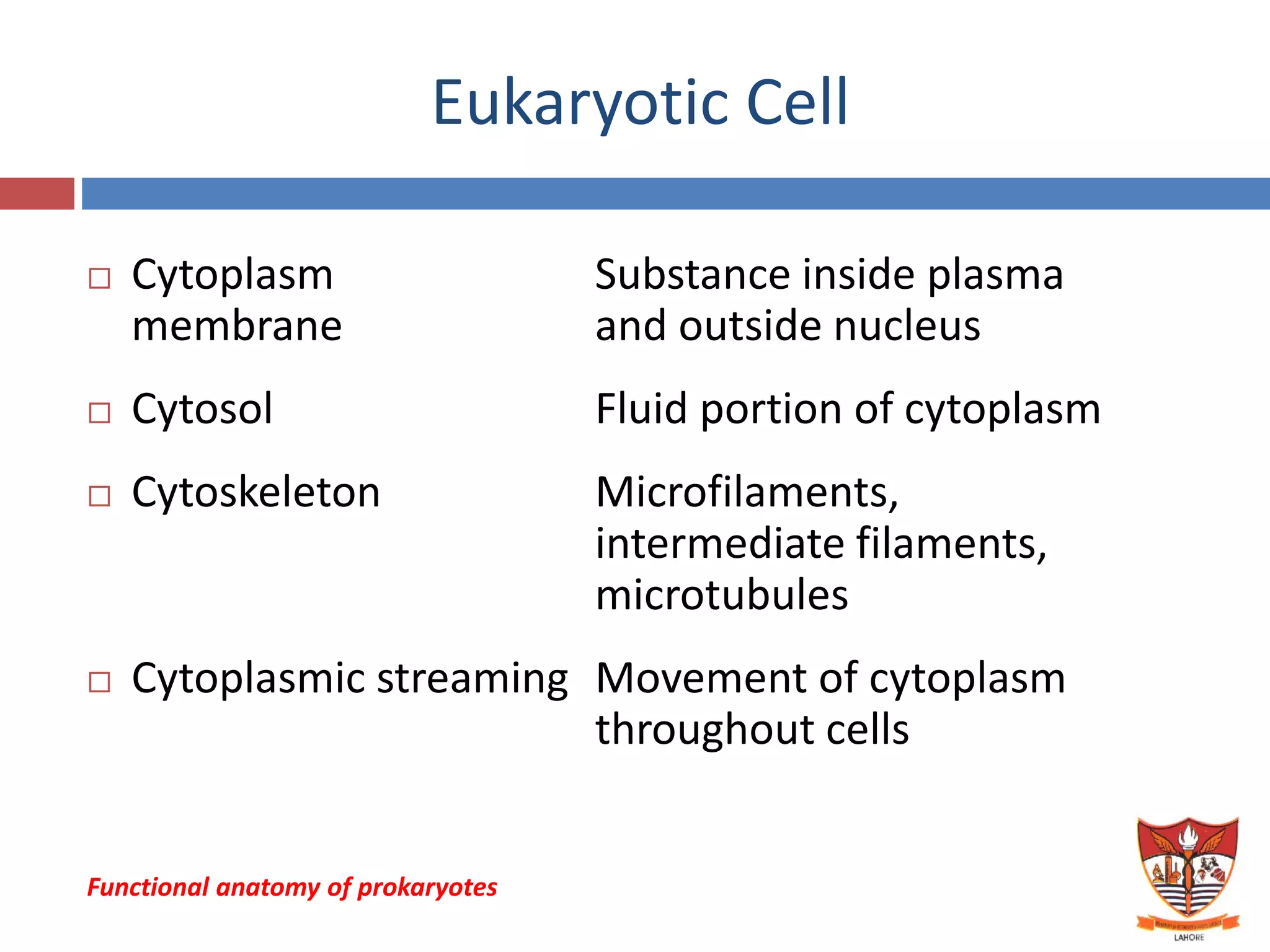


This document provides an overview of the functional anatomy of prokaryotes and eukaryotes. It describes that prokaryotes like bacteria have no nucleus or organelles, and a circular chromosome. Their cell walls are made of peptidoglycan and they divide through binary fission. Eukaryotes have membrane-bound organelles like the nucleus, mitochondria and chloroplasts. They have paired linear chromosomes within the nucleus. The document outlines the structures of bacterial and eukaryotic cells in detail.


