Downloaded 103 times


![Fibular fracture
Maisonneuve fracture - a spiral fracture of the
proximal third of the fibula associated with a tear
of the distal tibiofibular syndesmosis and the
interosseous membrane.
Le Fort fracture of ankle - a vertical fracture of
the antero-medial part of
the distal fibula with avulsion of the anterior
tibiofibular ligament.
Bosworth fracture - a fracture with an associated
fixed posterior dislocation of the proximal fibular
fragment which becomes trapped behind
the posterior tibial tubercle. The injury is caused
by severe external rotation of the ankle.[10]](https://image.slidesharecdn.com/fracturesites-140706064917-phpapp01/85/FRACTURE-Sites-25-320.jpg)


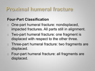
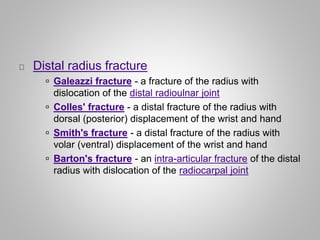
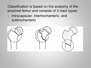
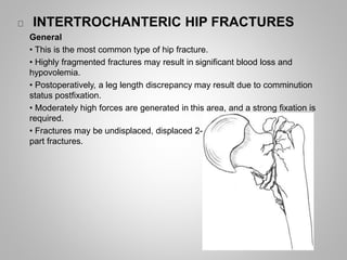
Maxillofacial fractures usually occur as the result of massive facial trauma and can include fractures of the mandible, nasal bones, maxilla, and zygomatic bones. Cervical spine fractures include fractures of C1-C2 as well as burst, compression, and teardrop fractures of the lower cervical vertebrae. Humerus fractures are classified as one, two, three, or four-part fractures. Distal radius fractures include Colles', Smith's, Barton's, and Galeazzi fractures. Hip fractures are classified as femoral neck, intertrochanteric, or subtrochanteric fractures. Common foot fractures are Lisfranc fractures and fractures of the metatarsals


