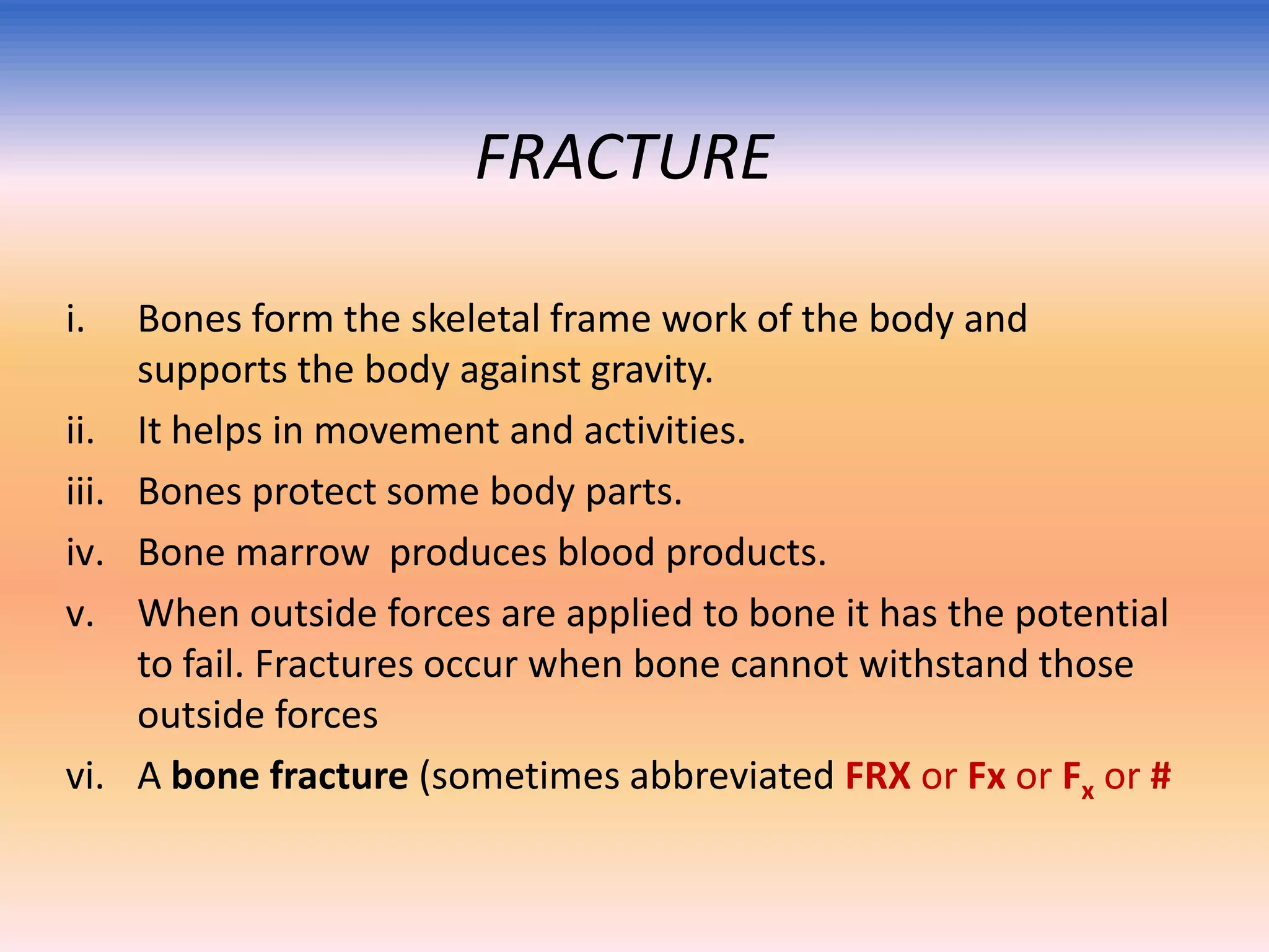
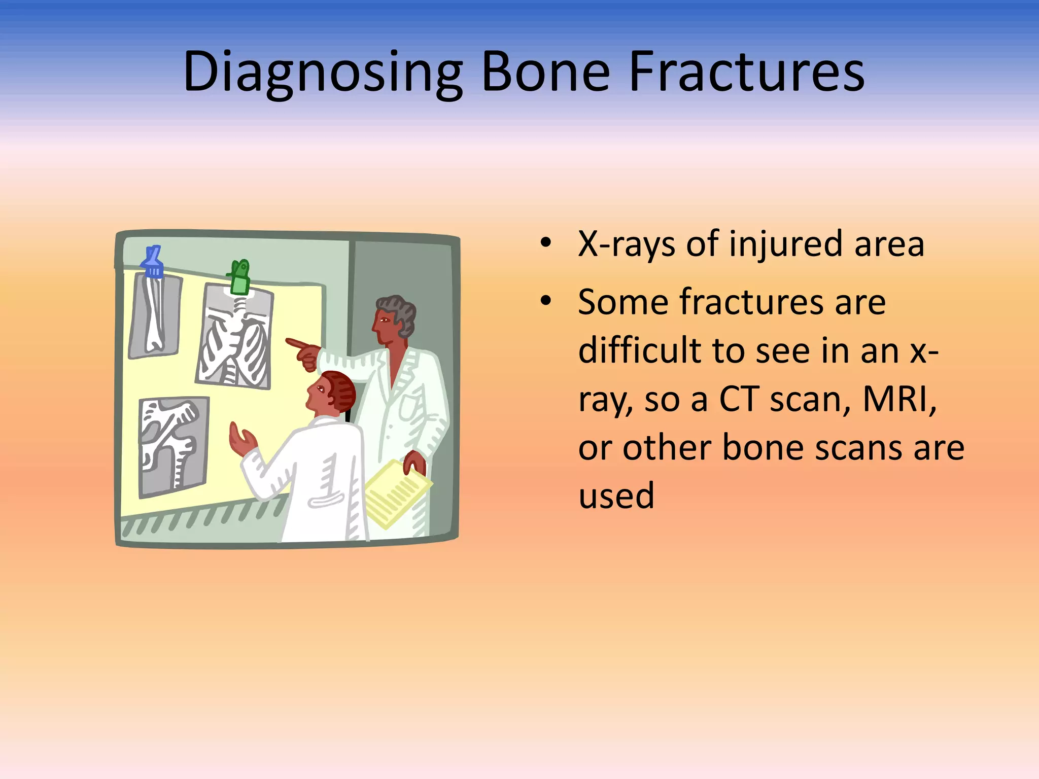
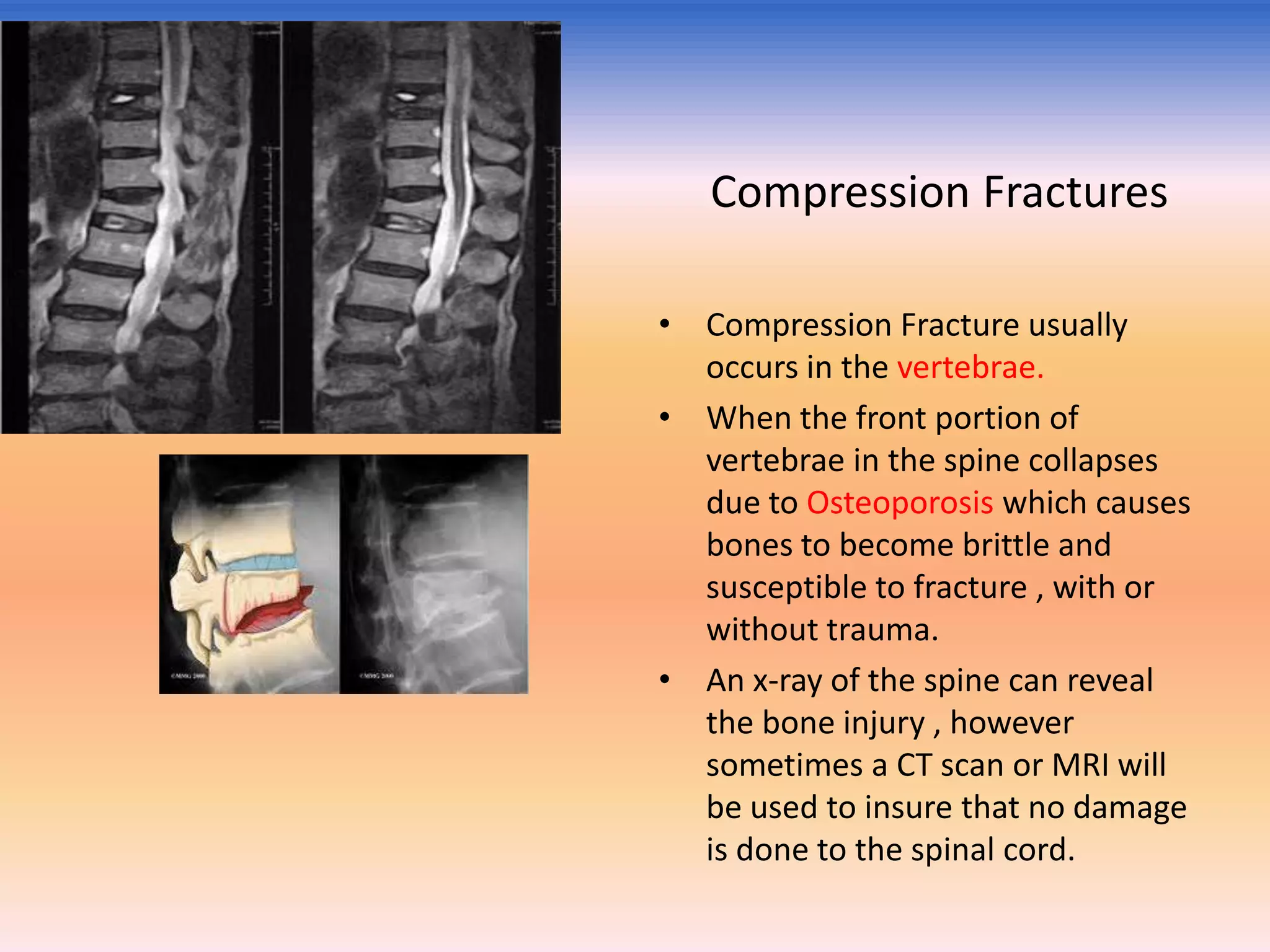
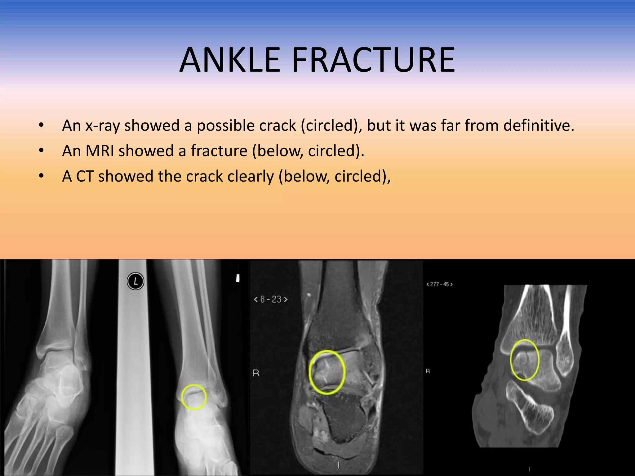
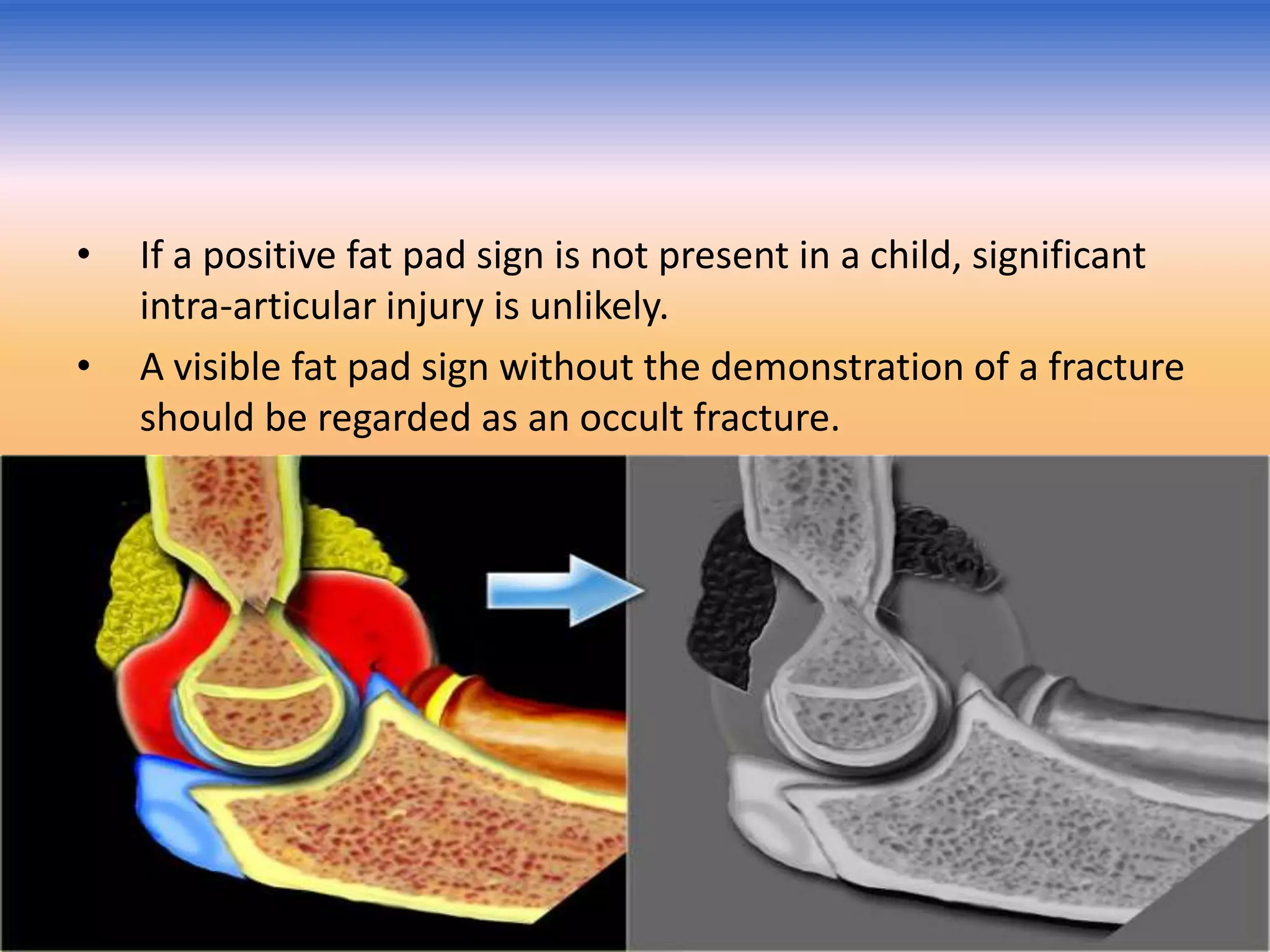
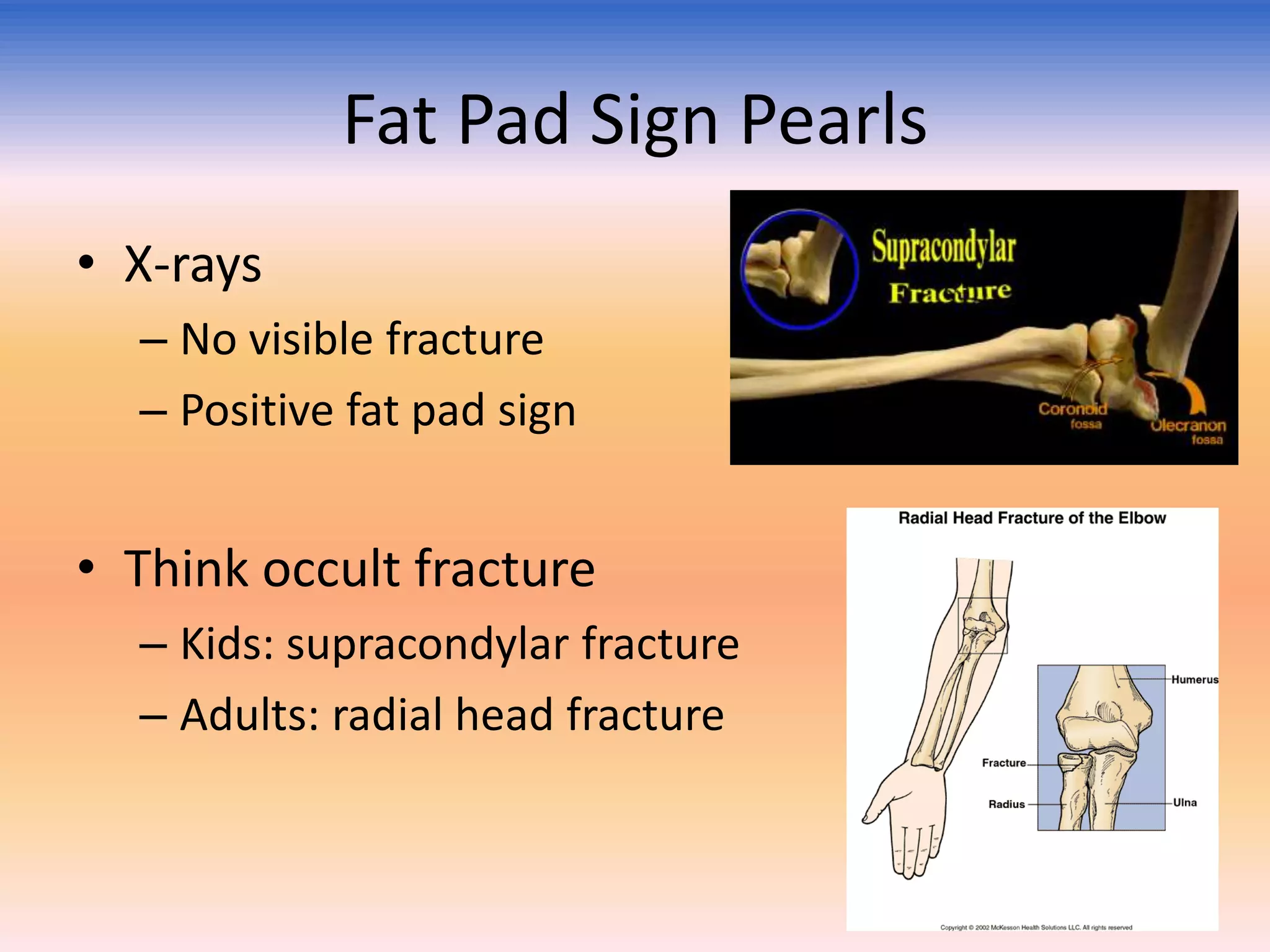
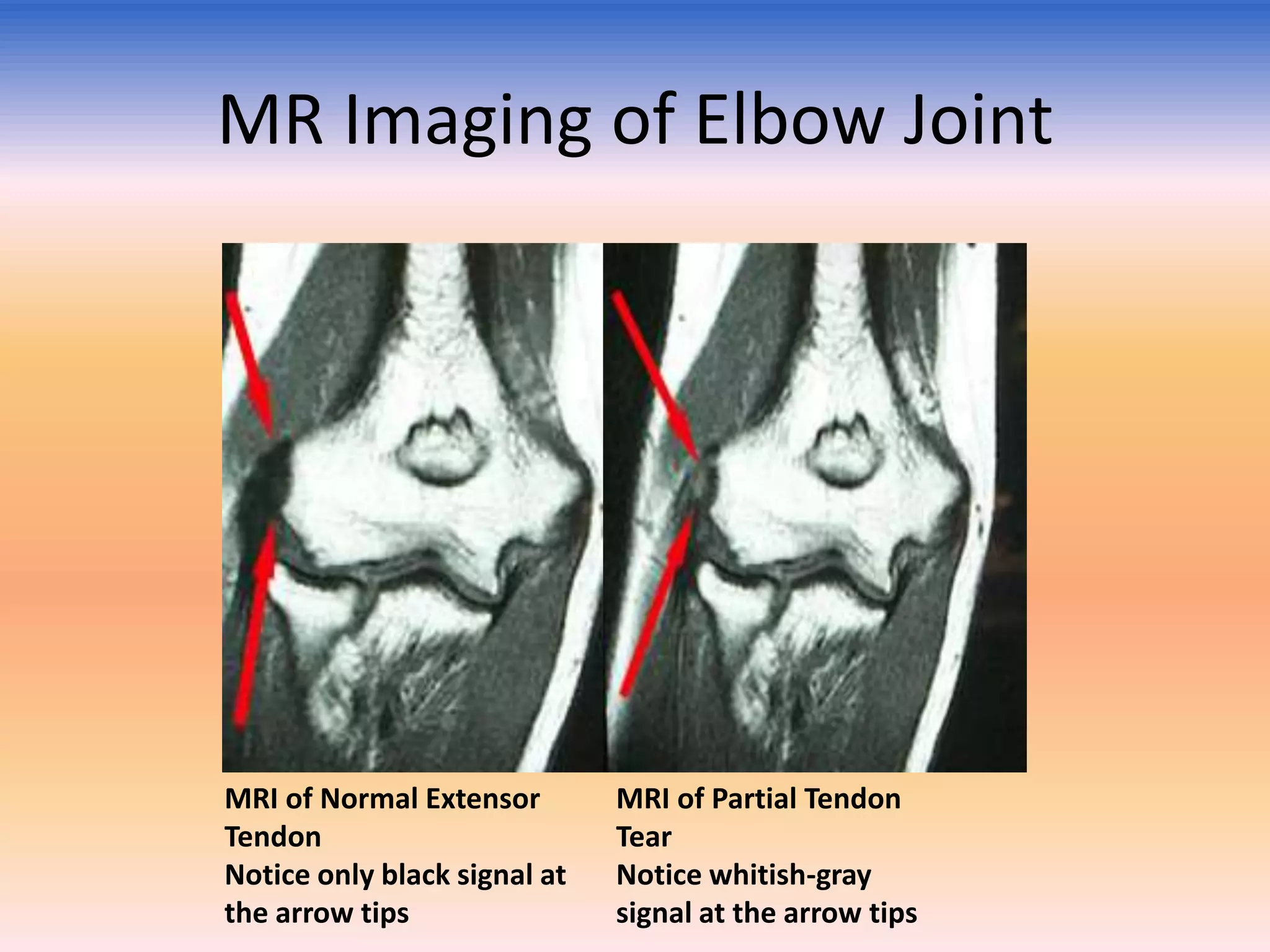
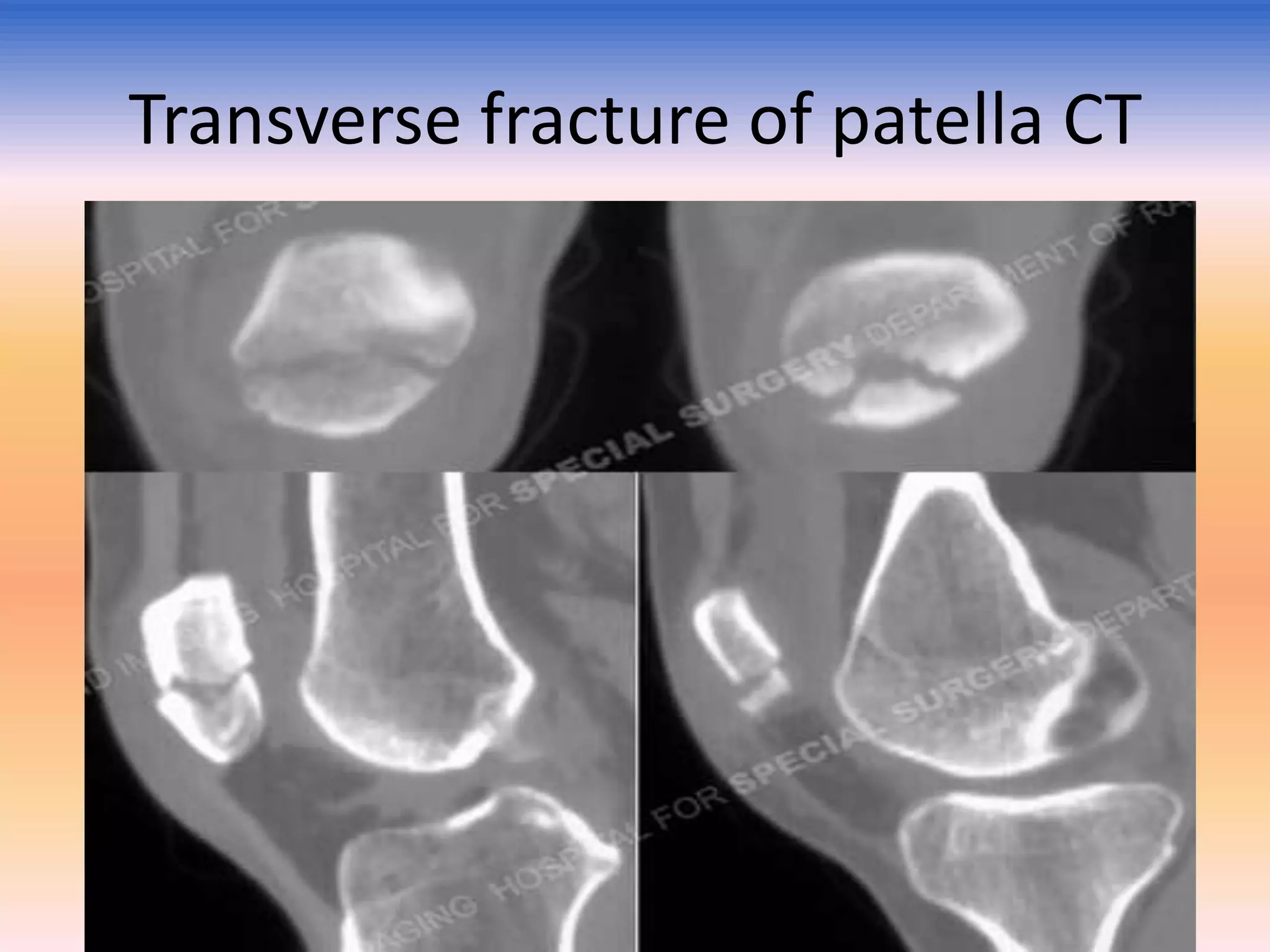
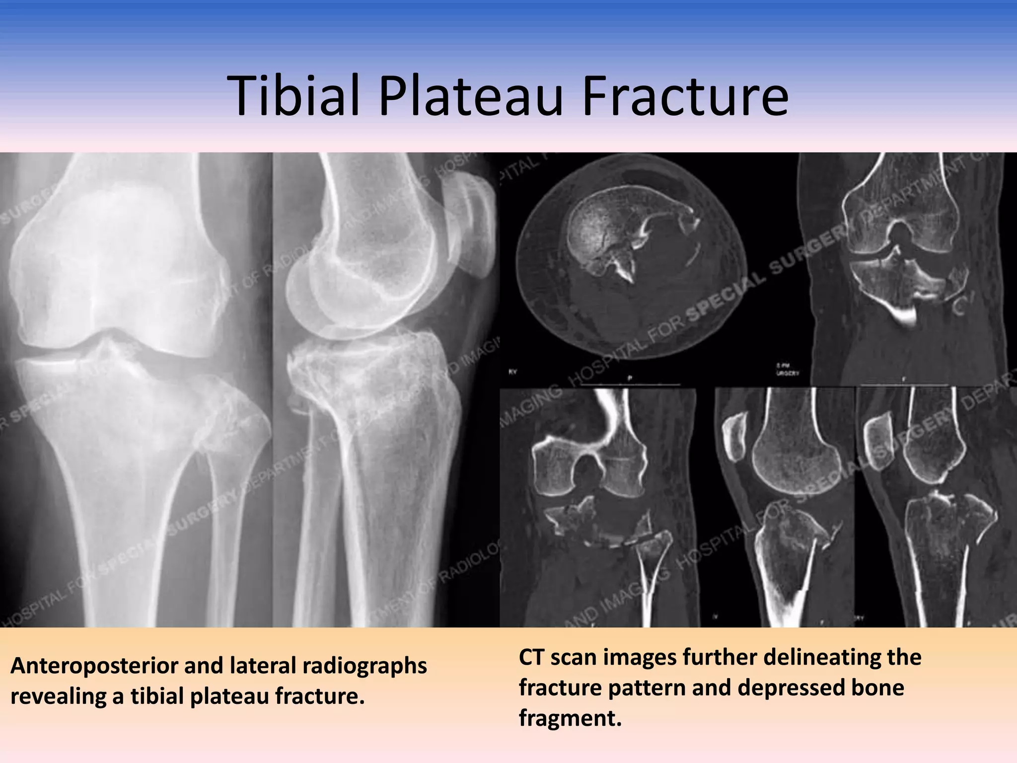
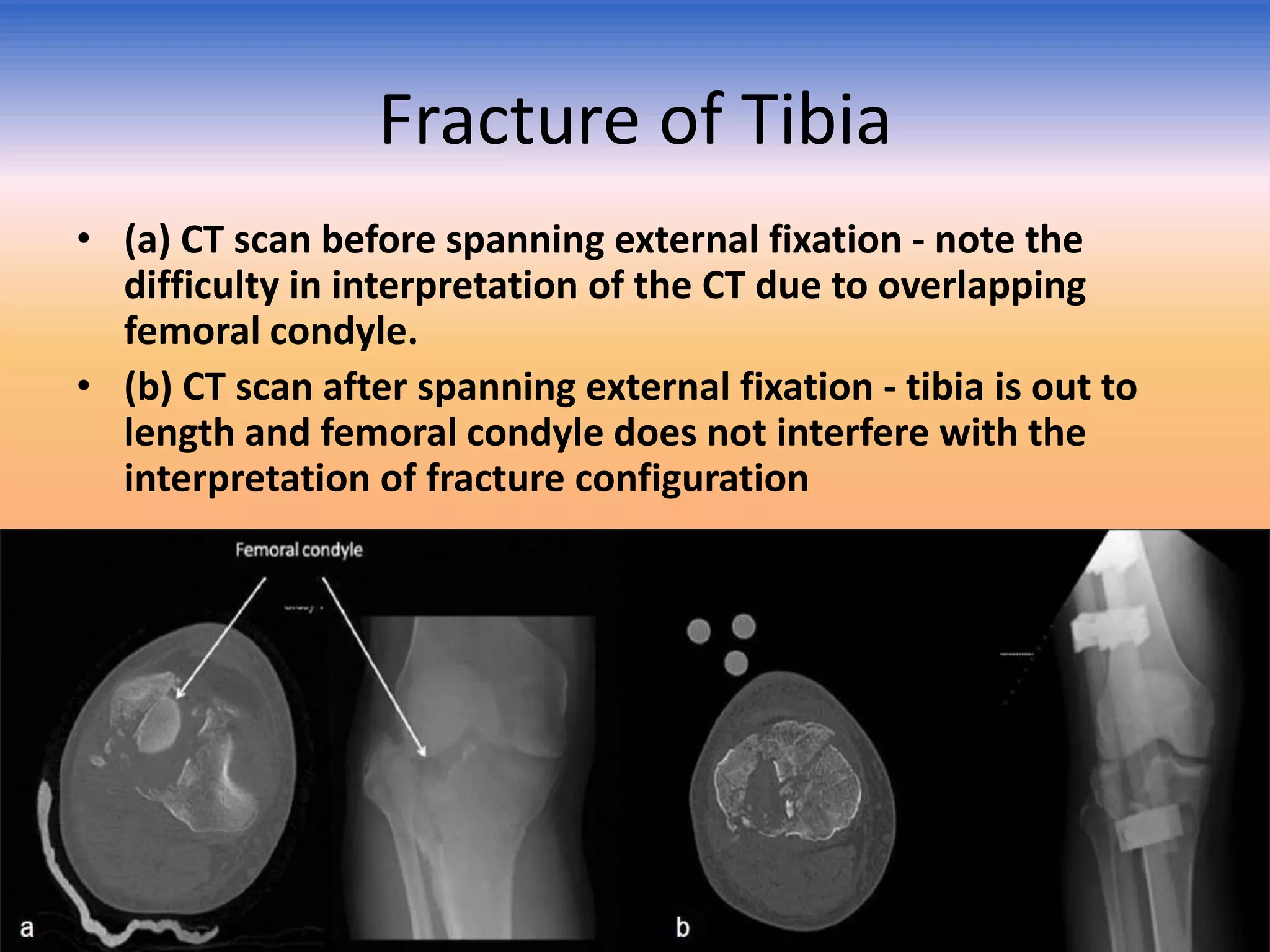
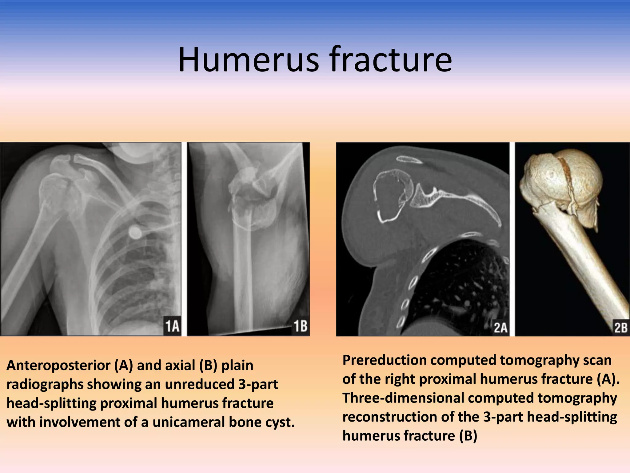
1) Imaging plays an important role in the diagnosis of fractures by identifying the location and type of fracture. X-rays are usually the initial study but CT or MRI may be needed in some cases.
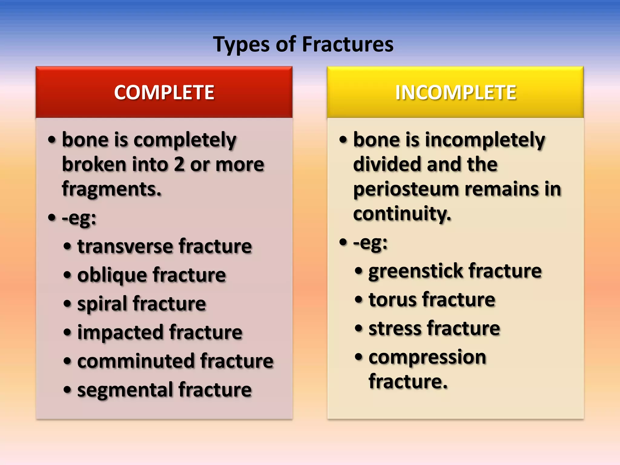
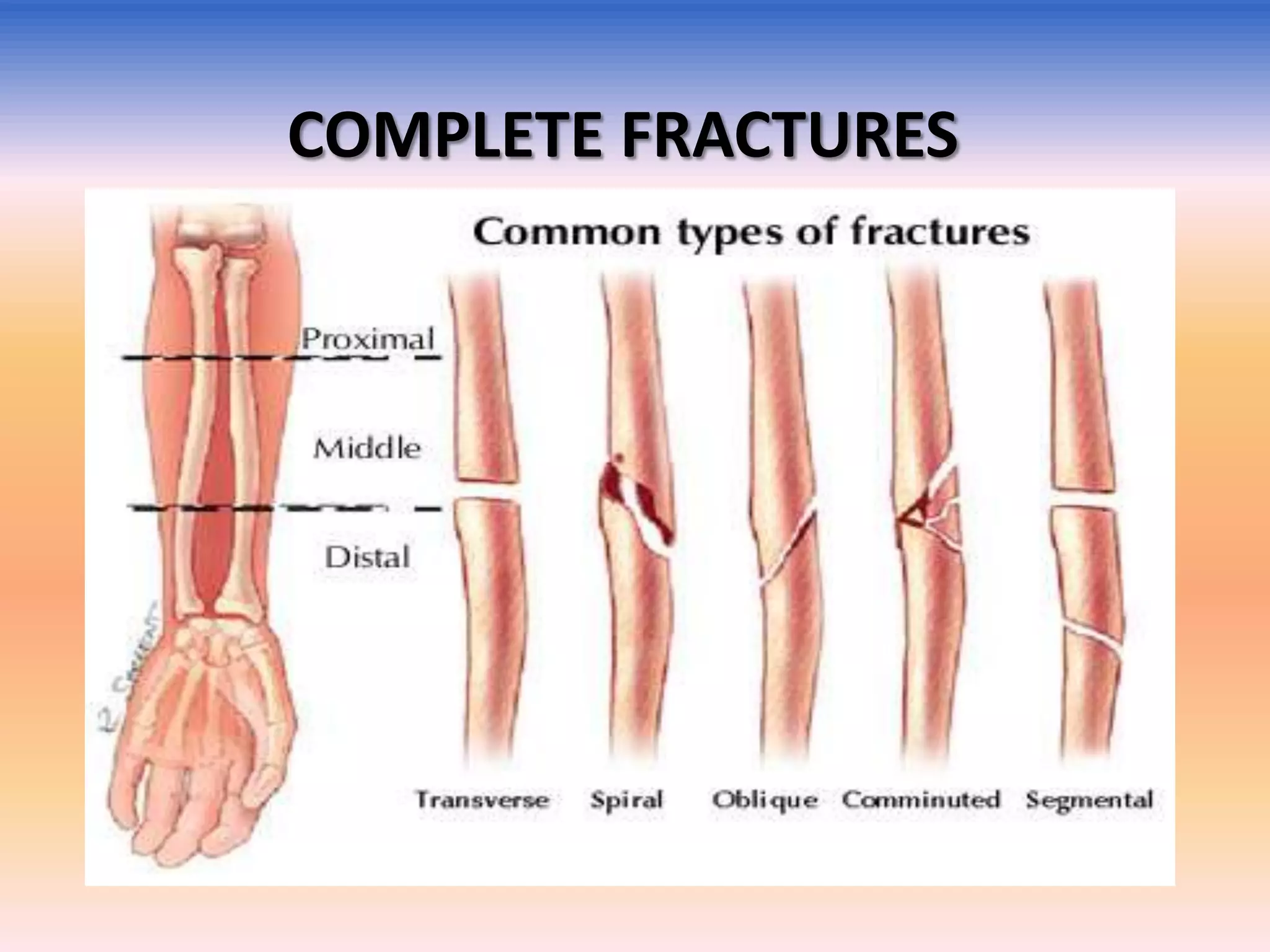
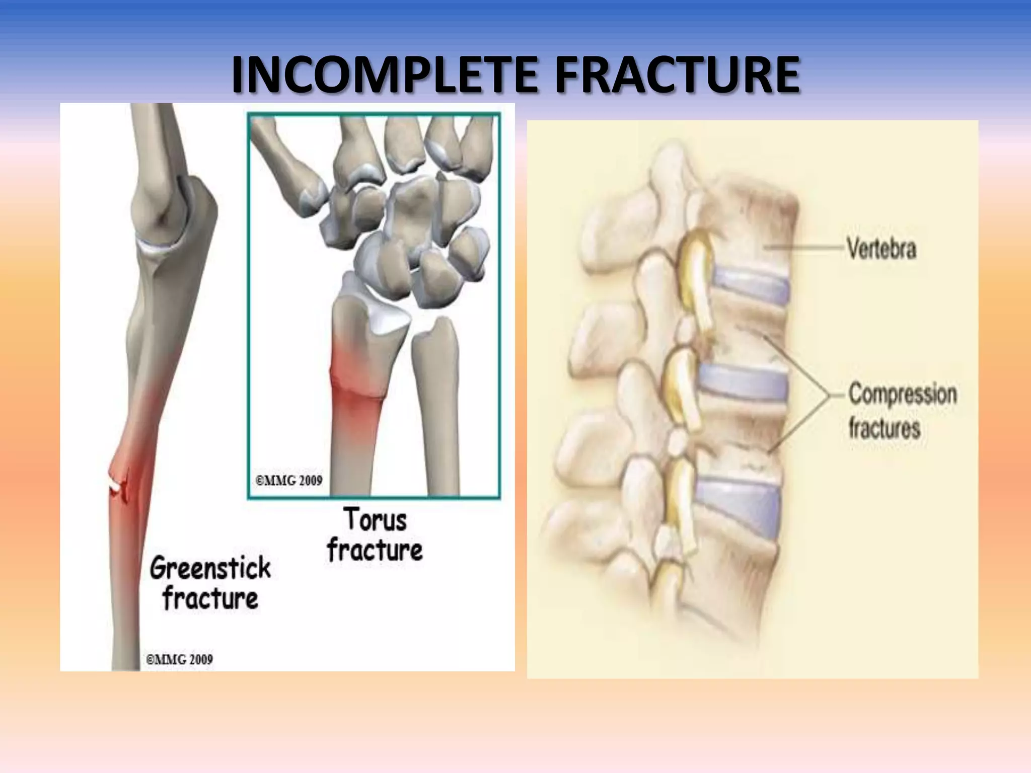
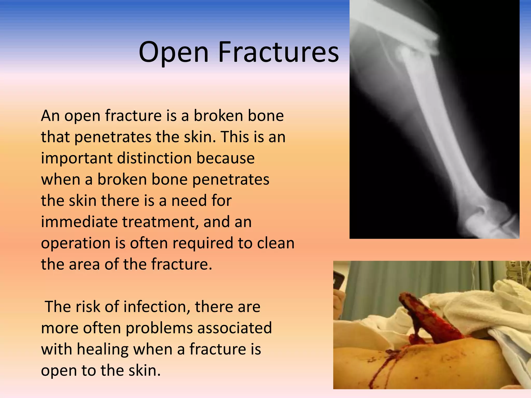
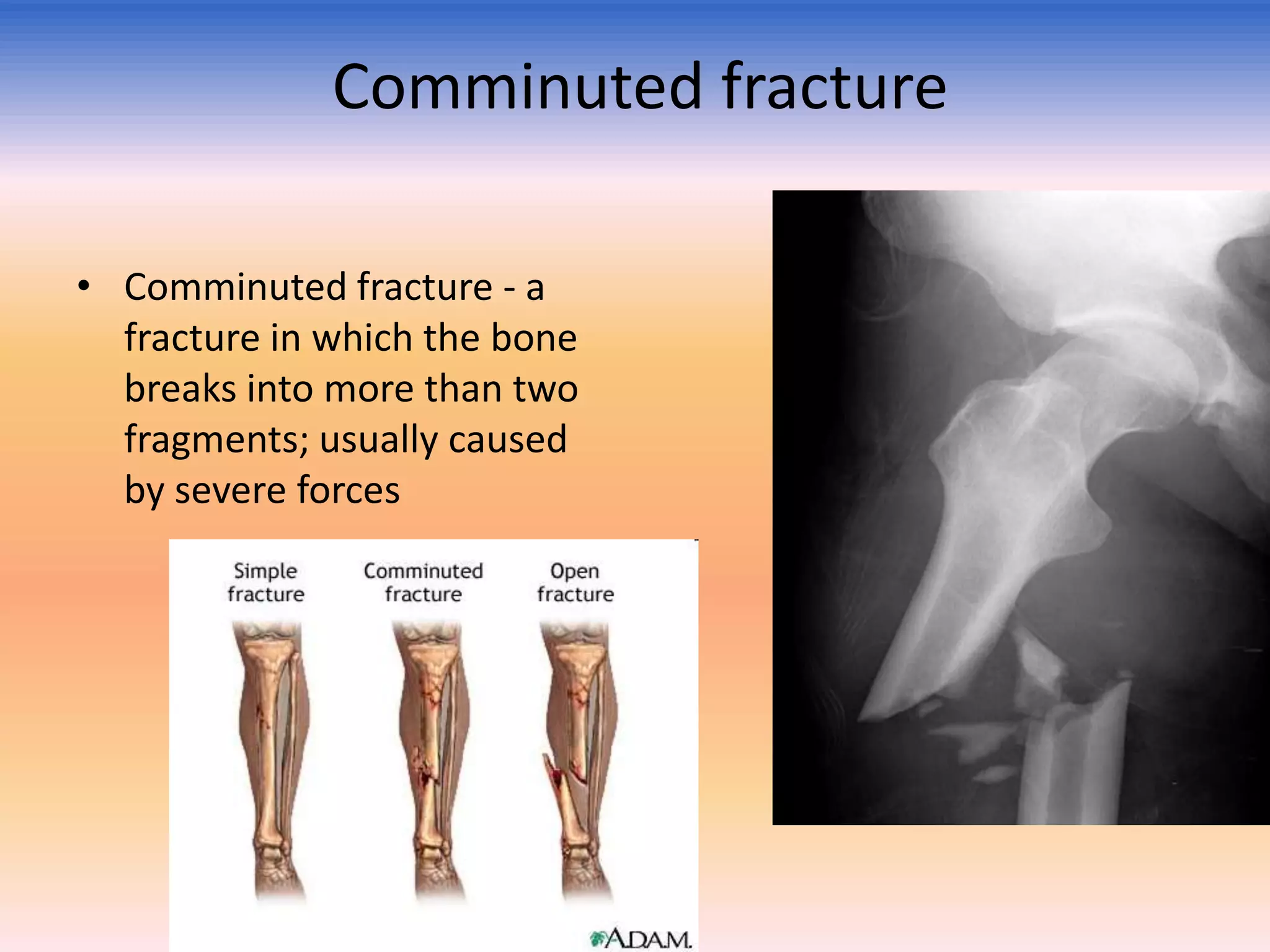
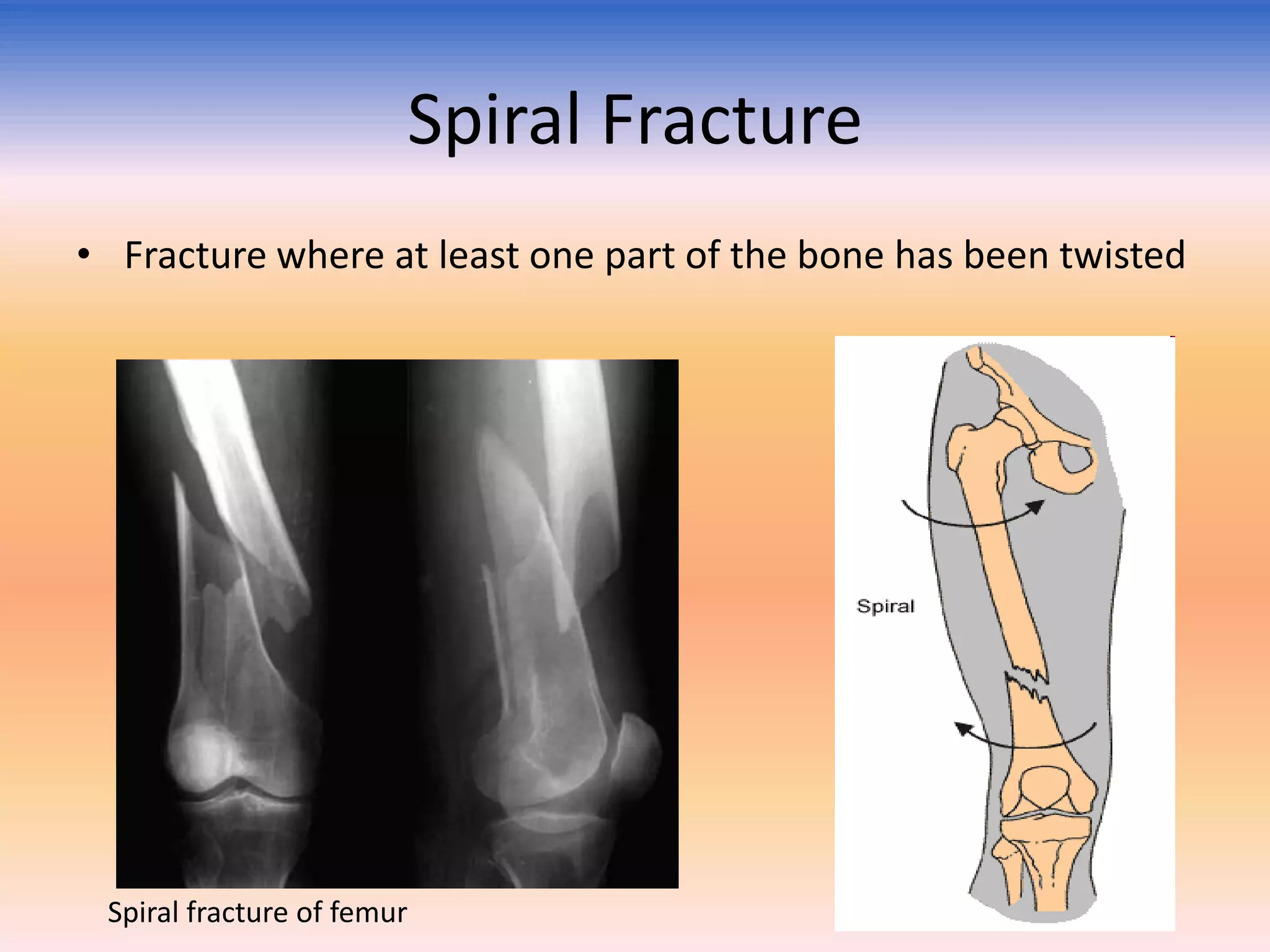
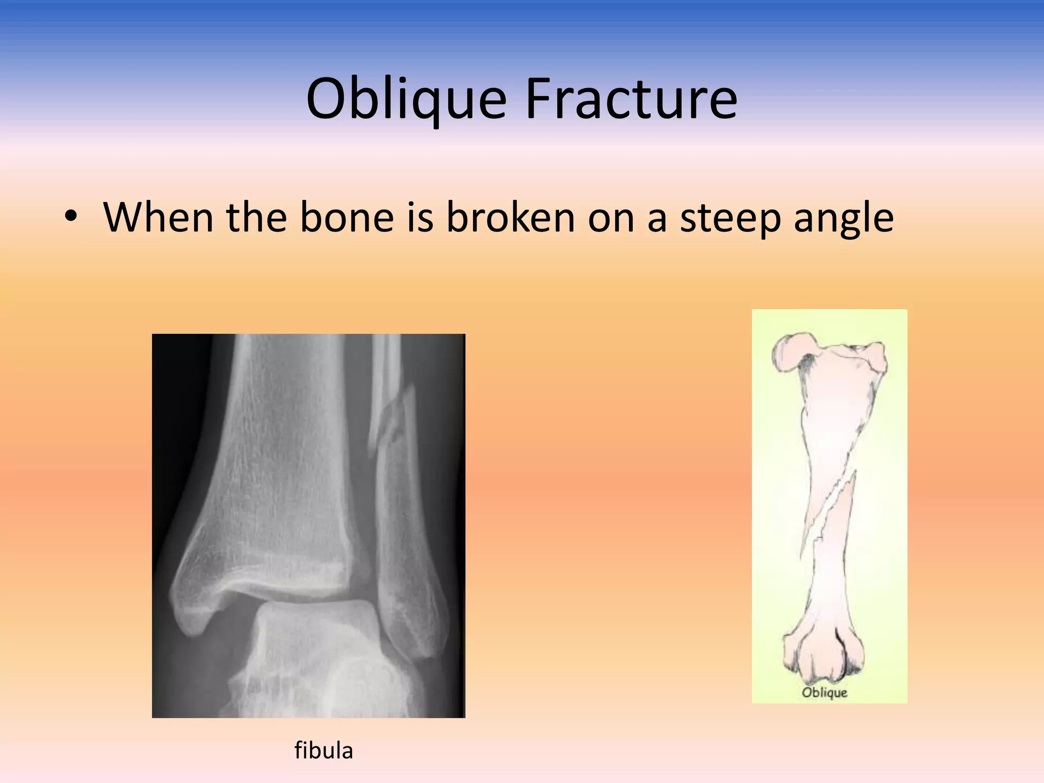
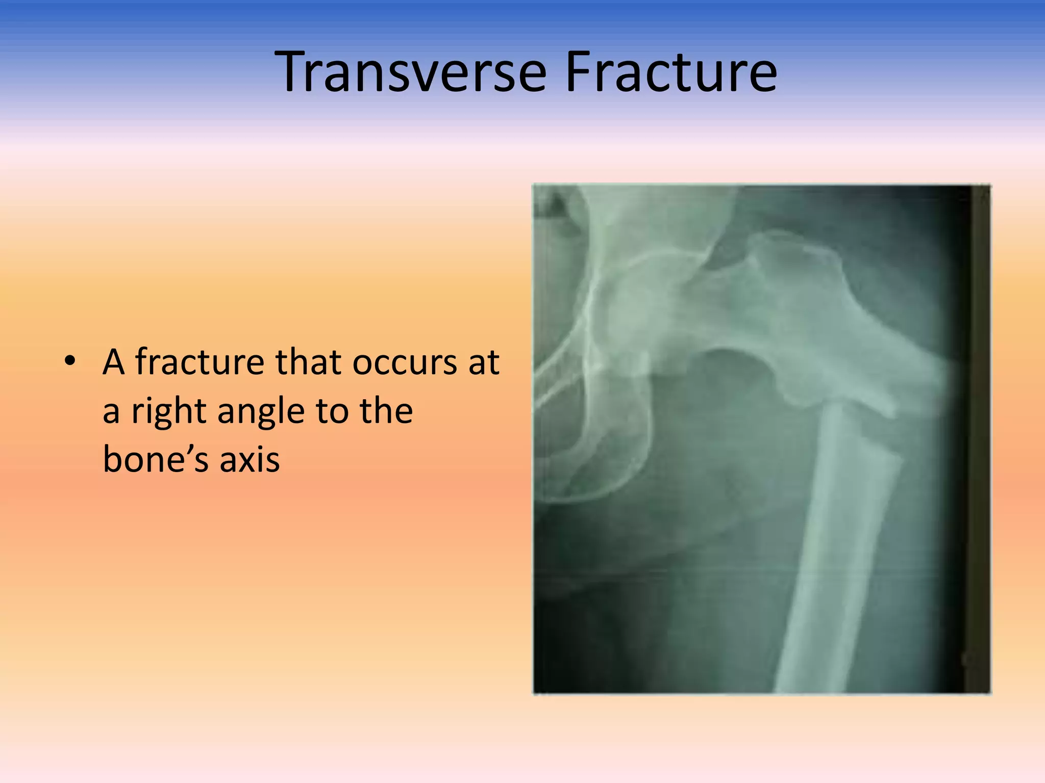
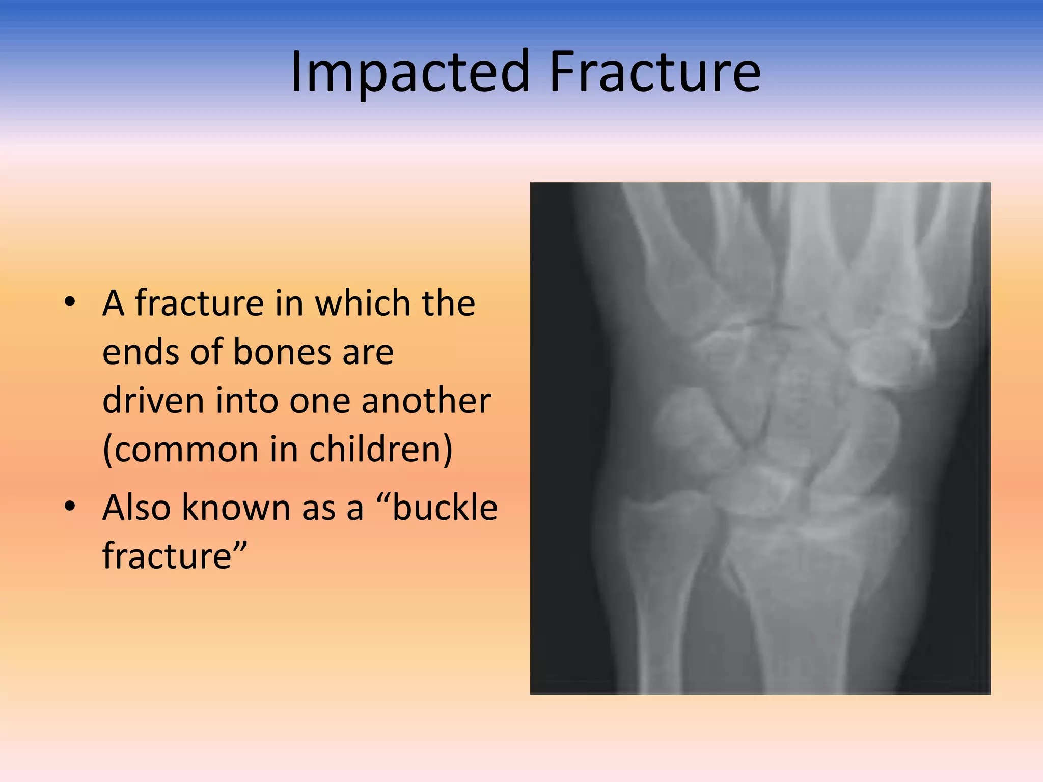
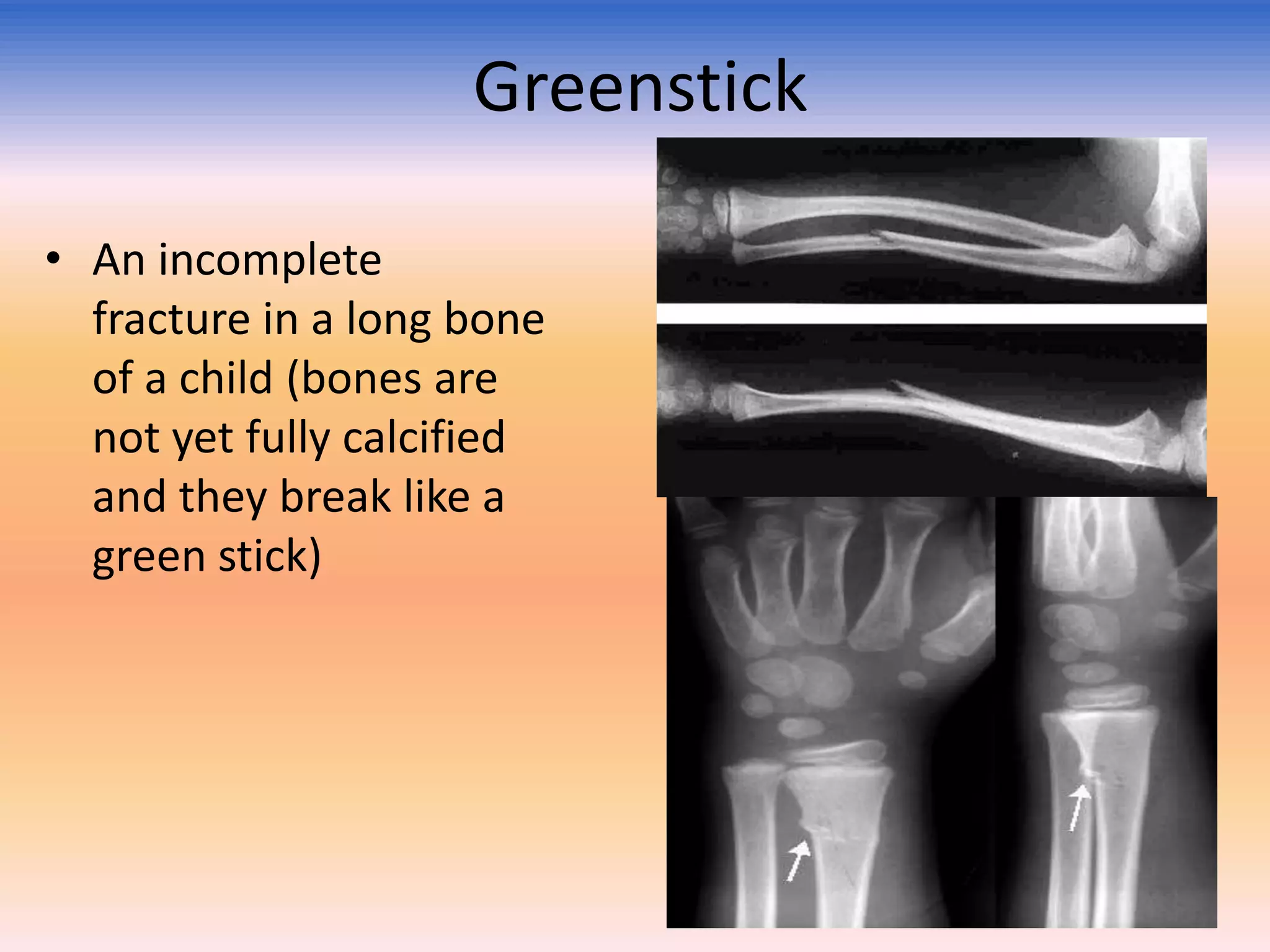
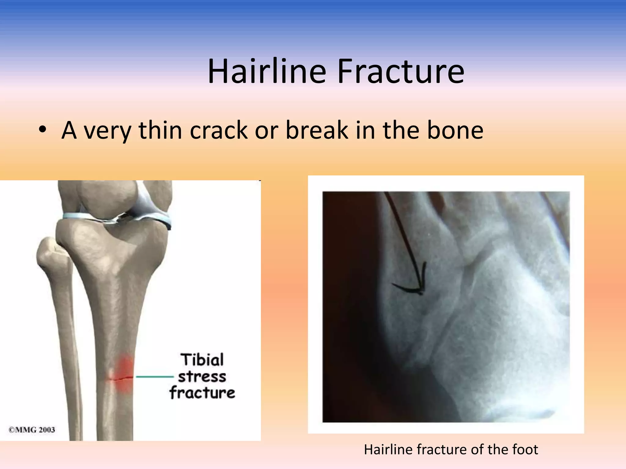
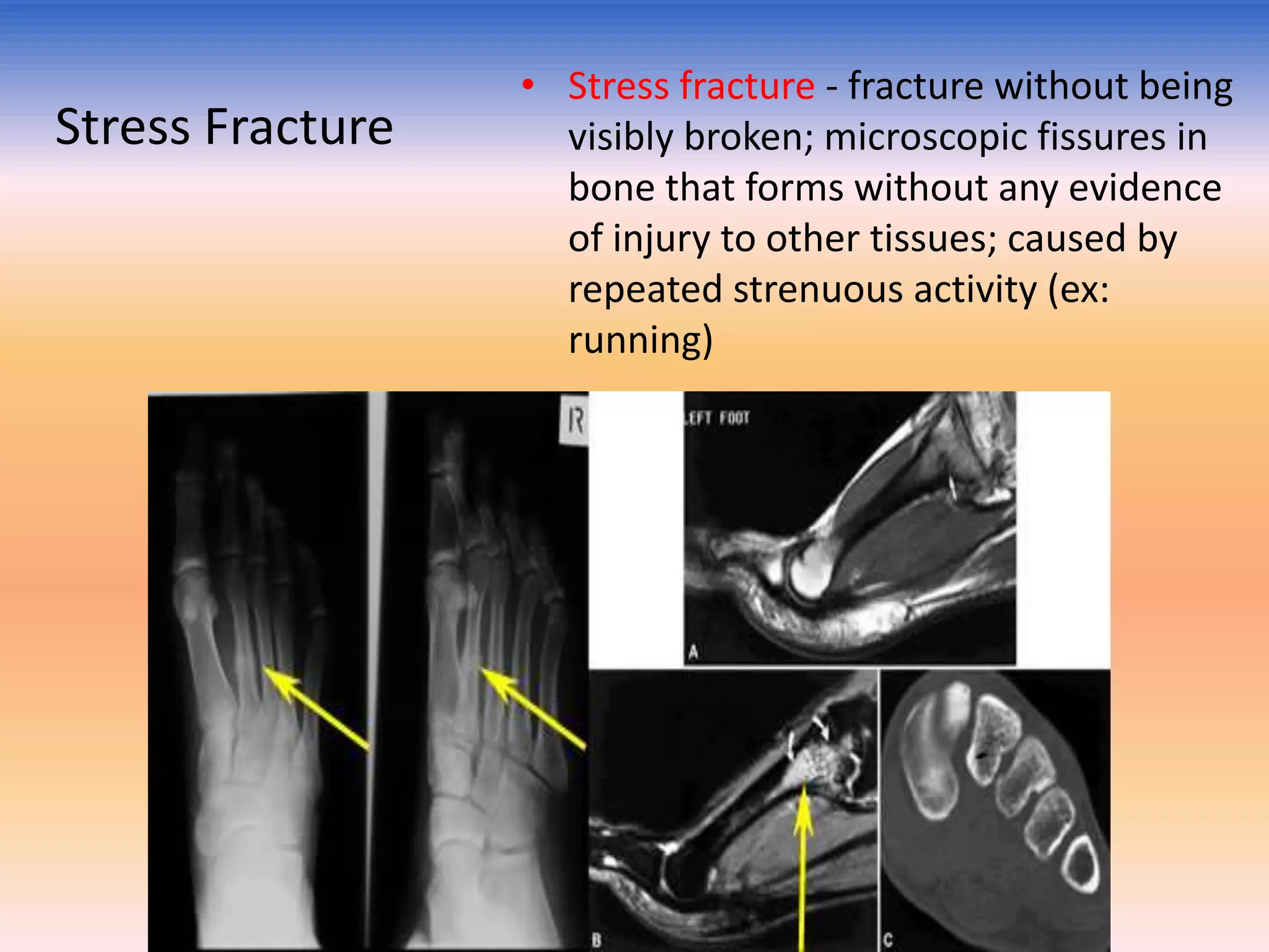
2) The document discusses many types of fractures including complete, incomplete, open, comminuted, spiral, oblique, transverse, impacted, greenstick, compression and stress fractures.
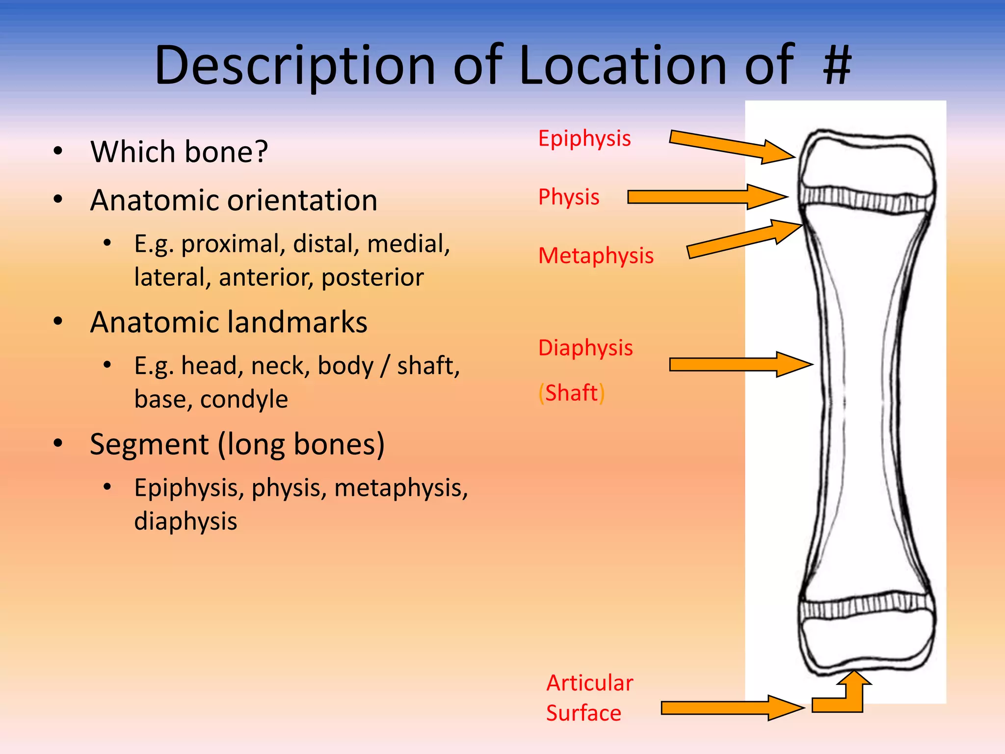
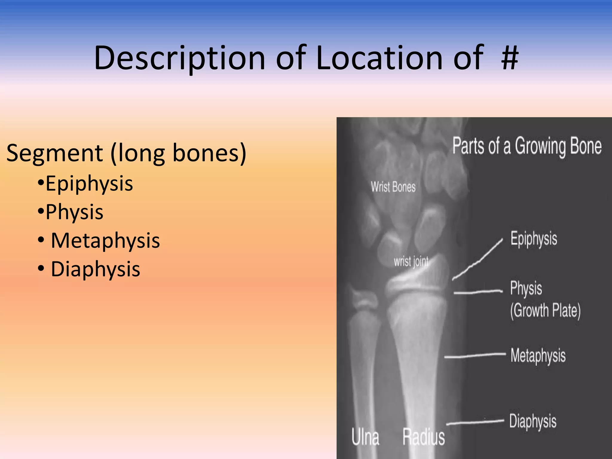
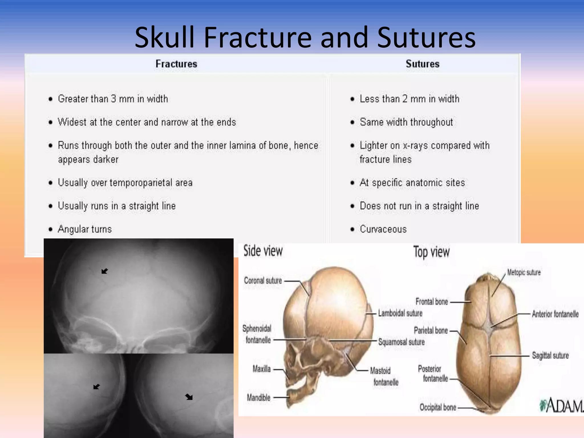
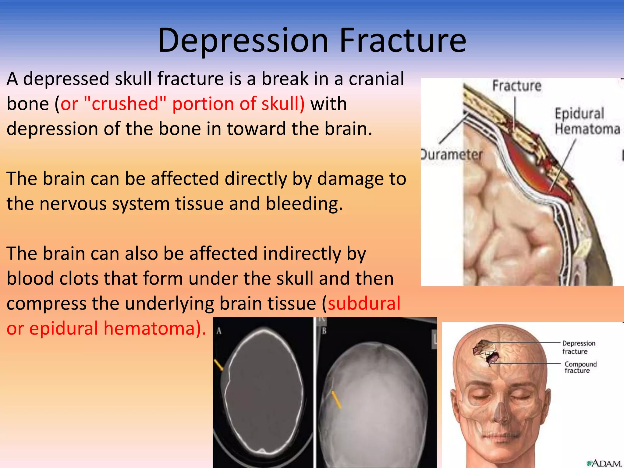
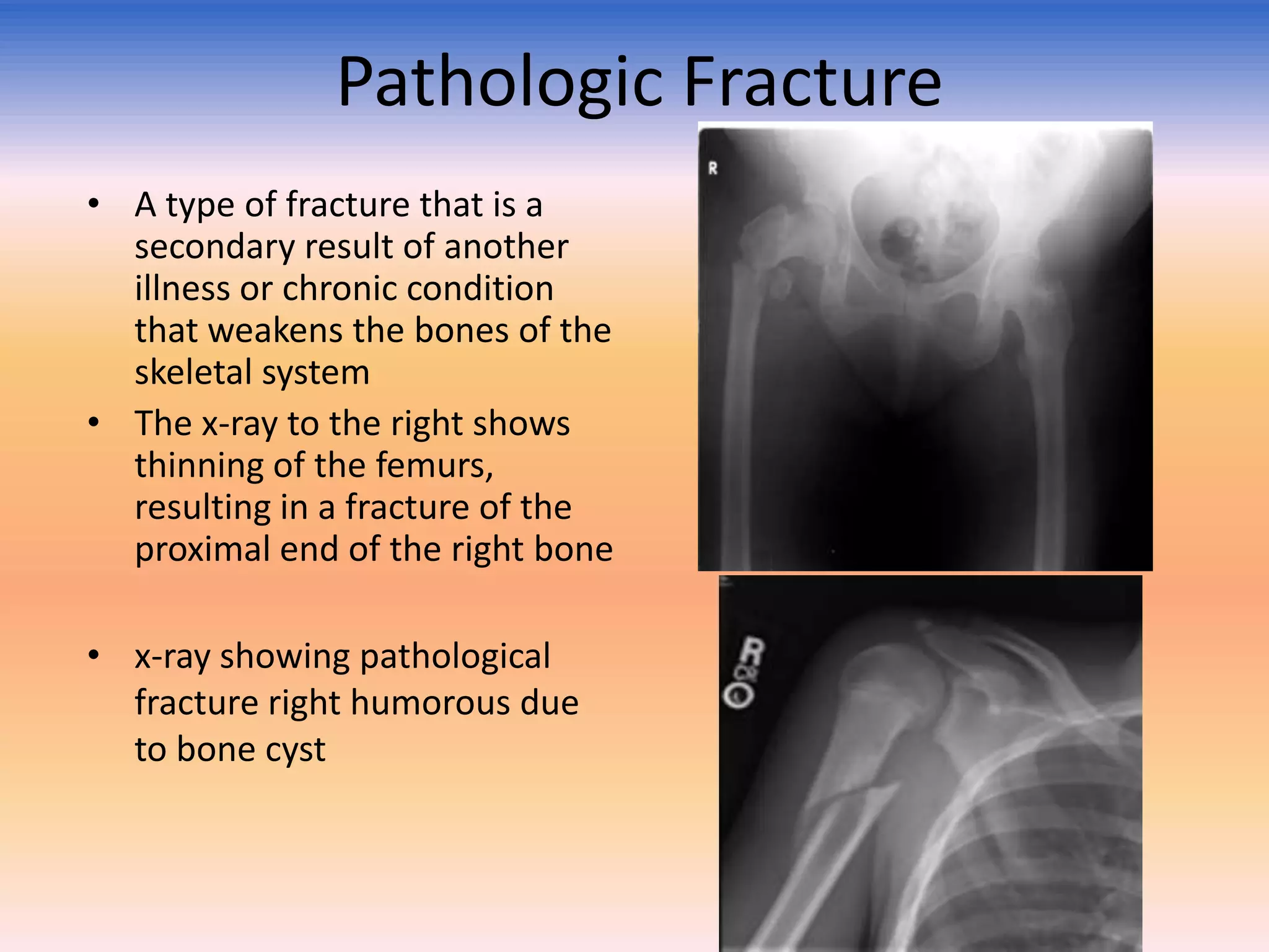
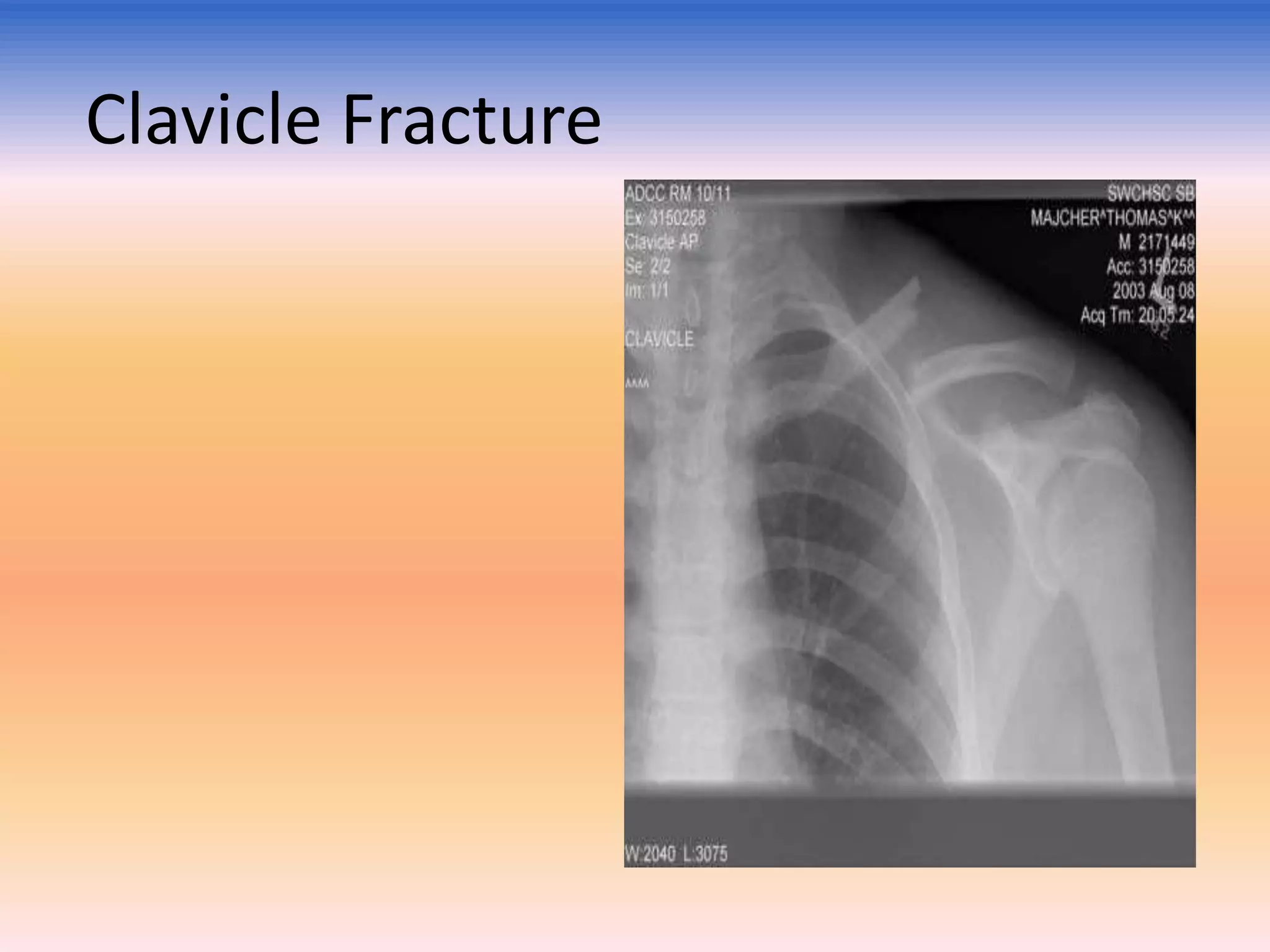
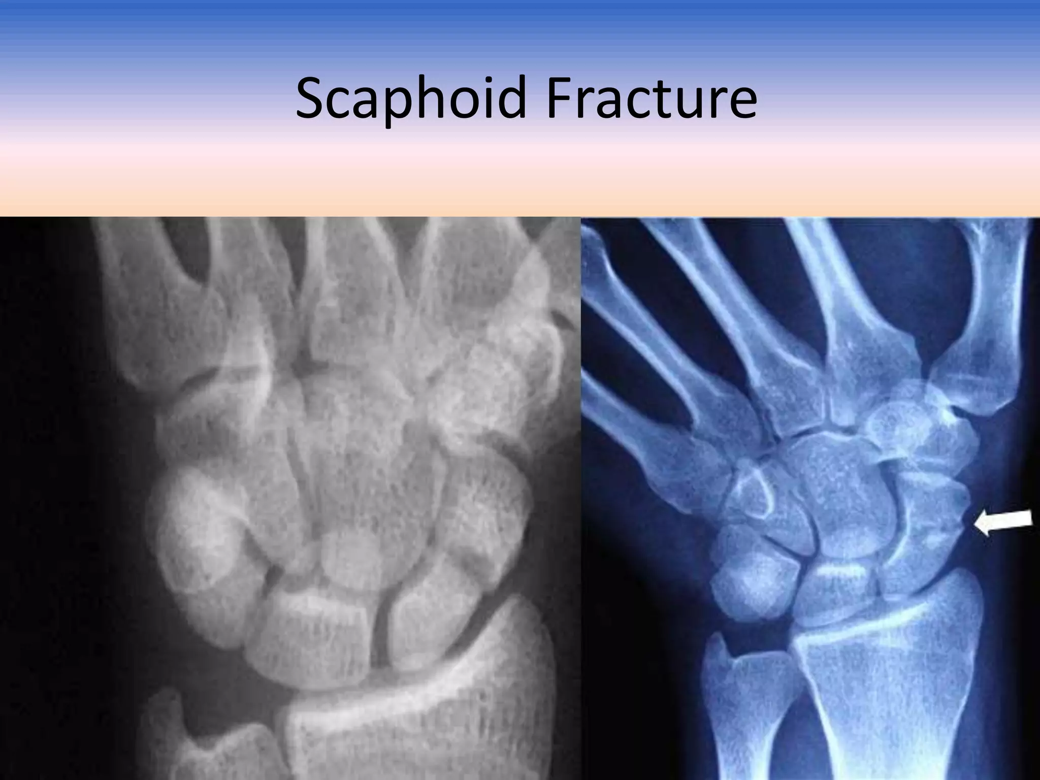
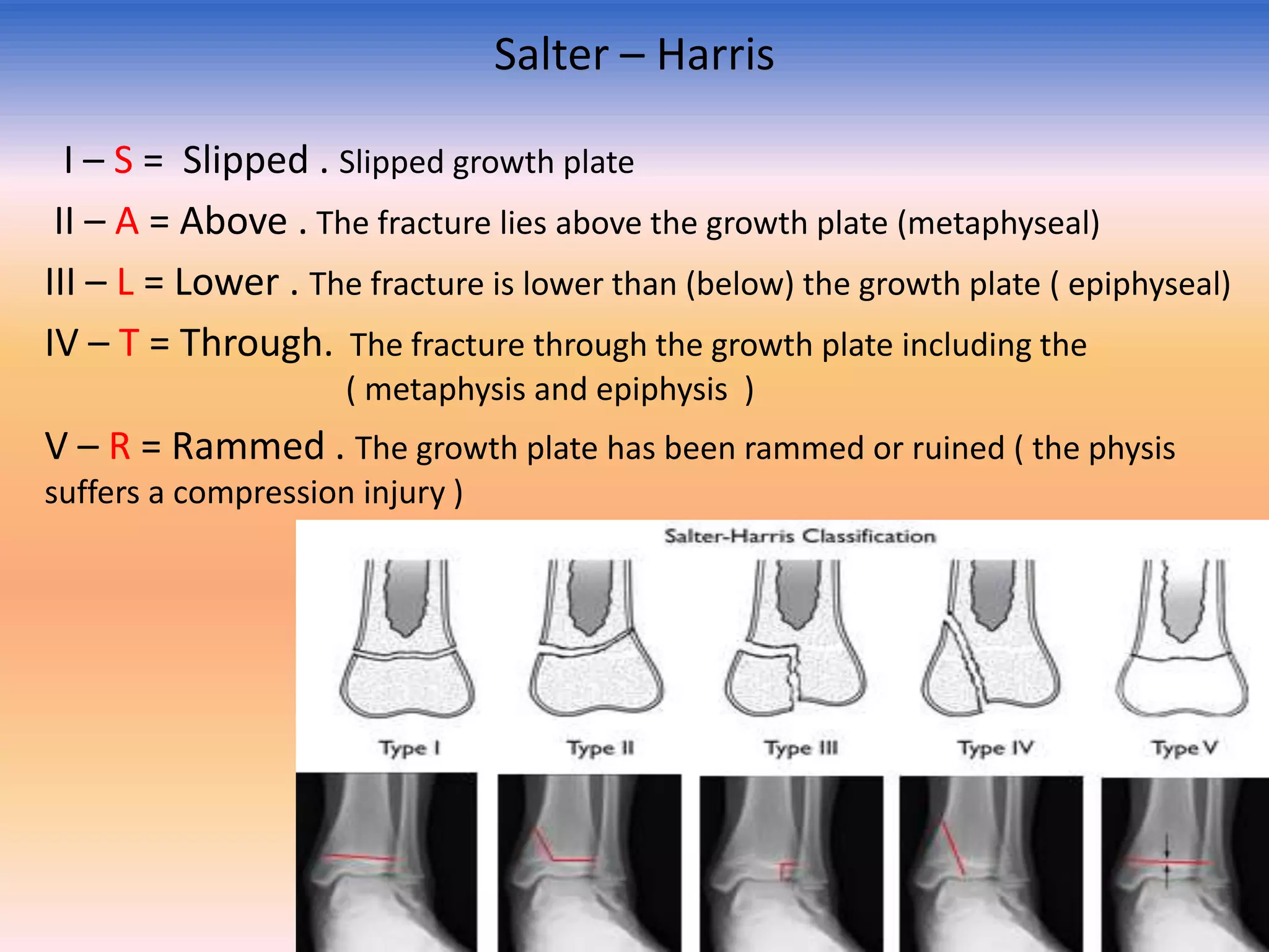
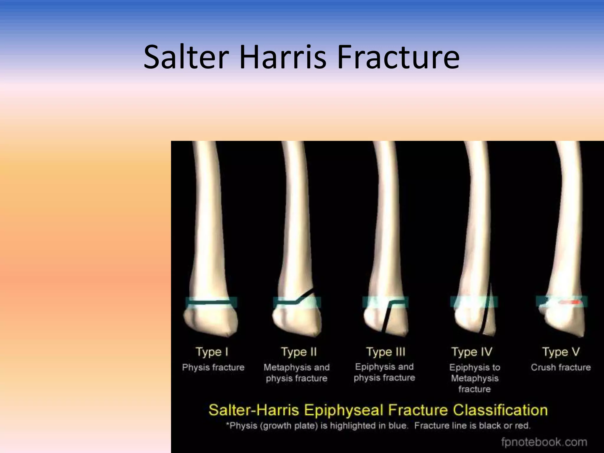
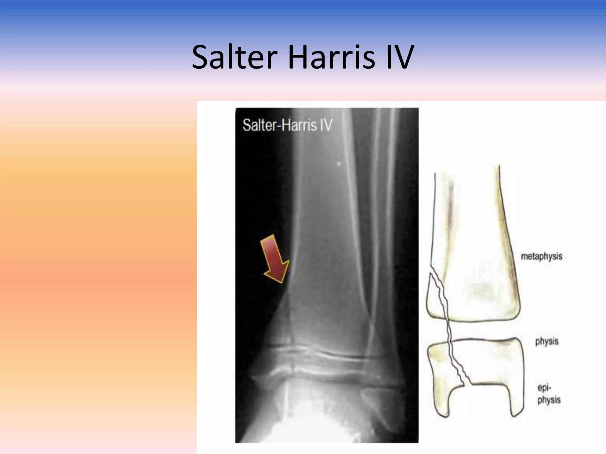
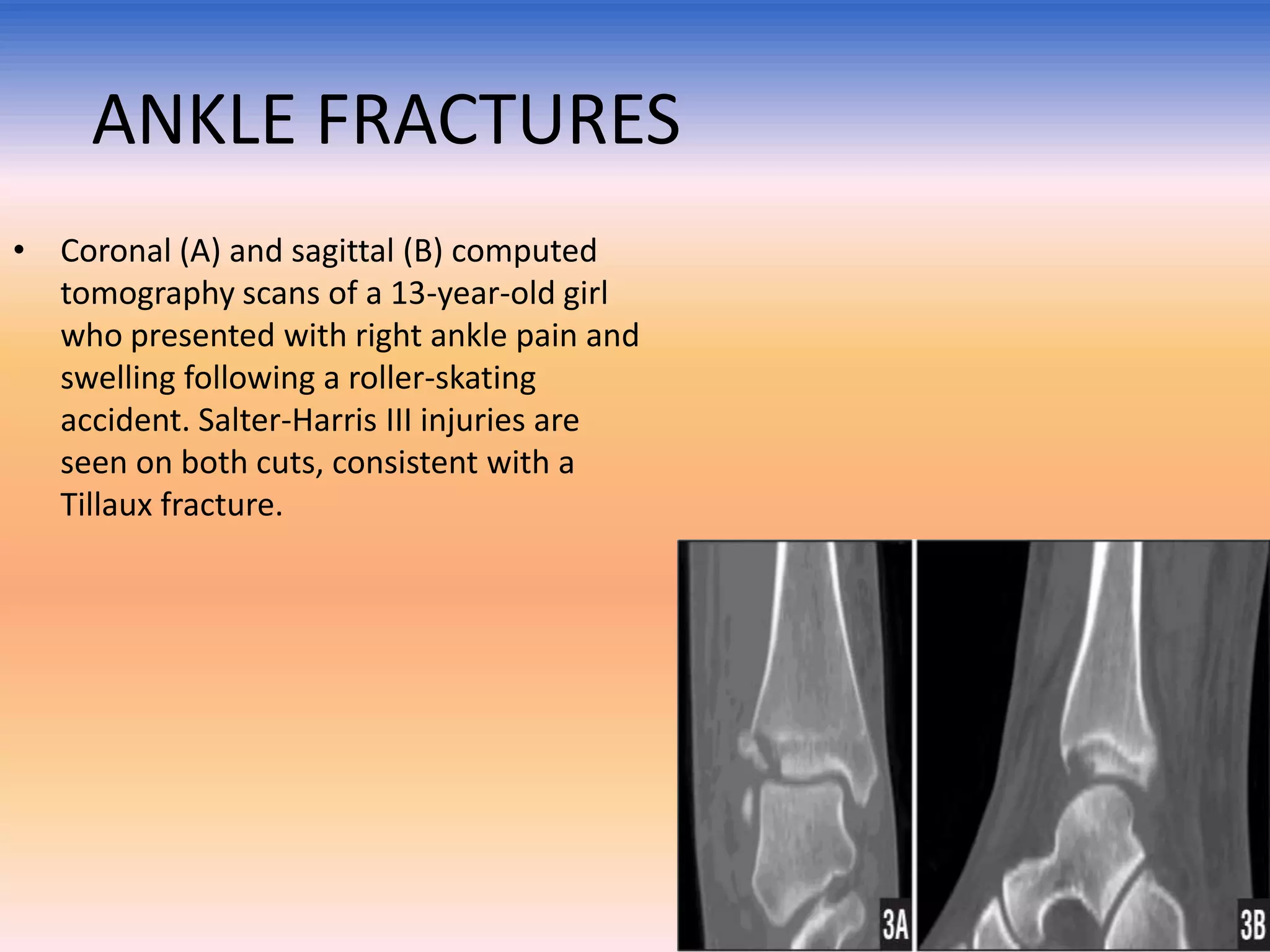
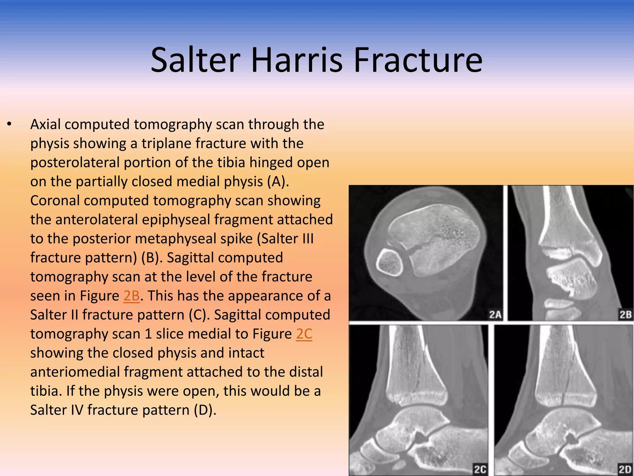
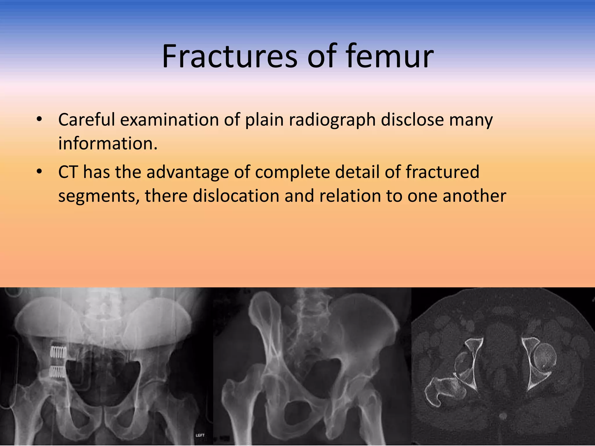
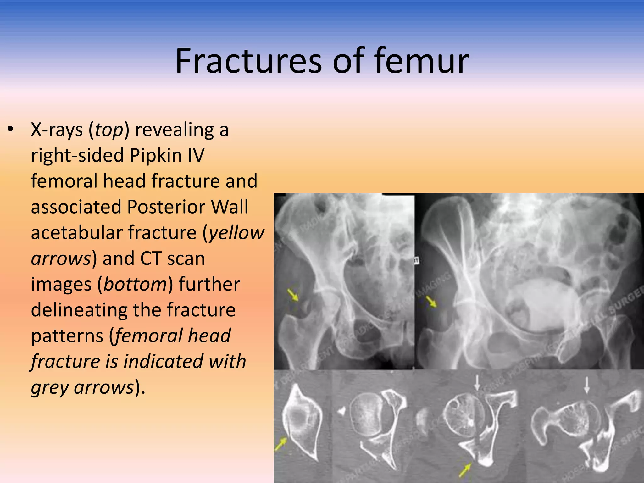
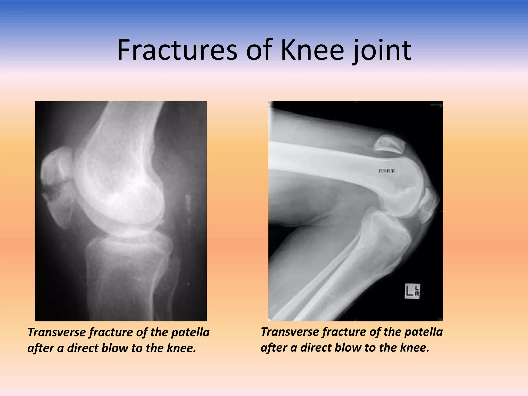
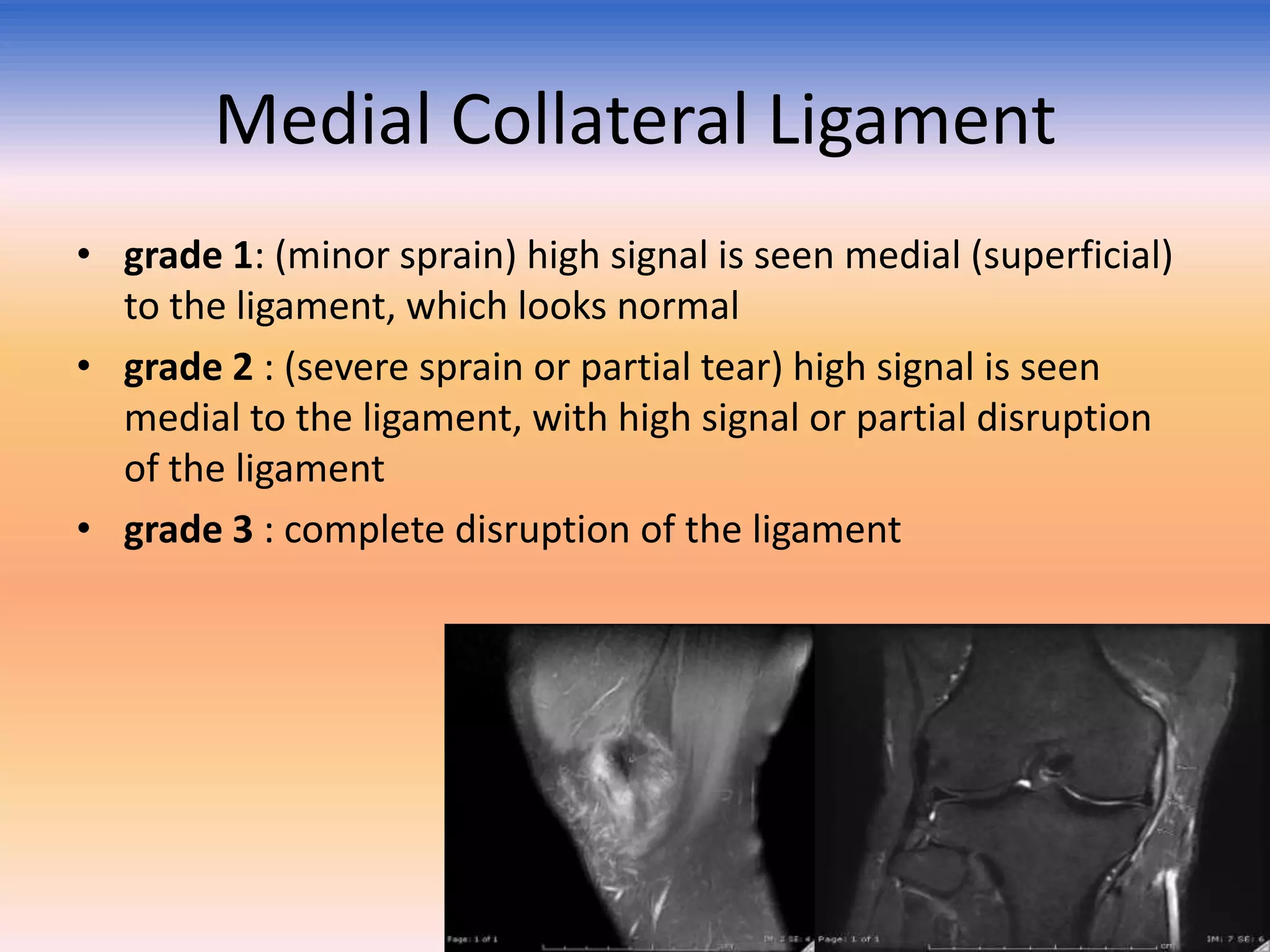
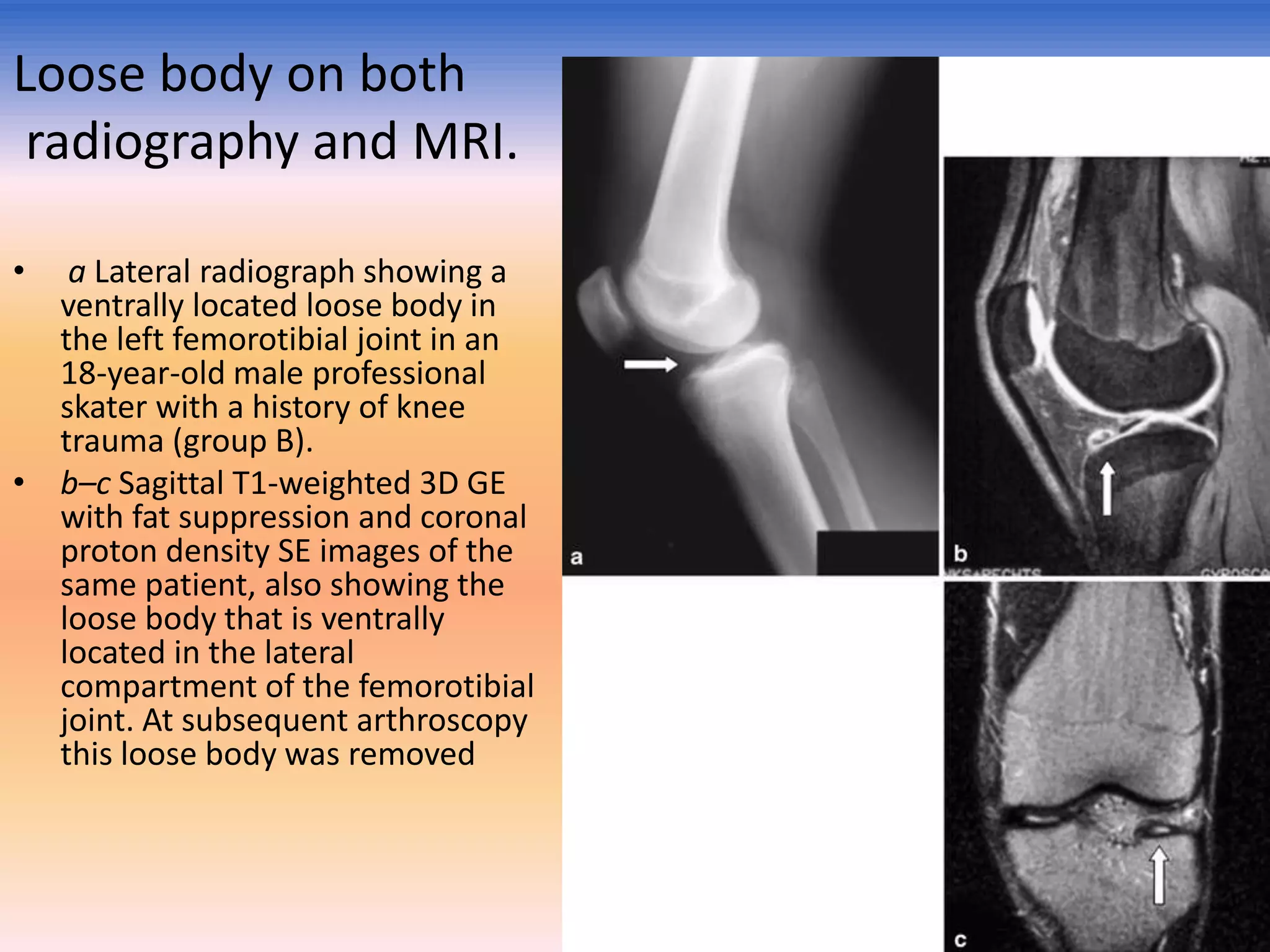
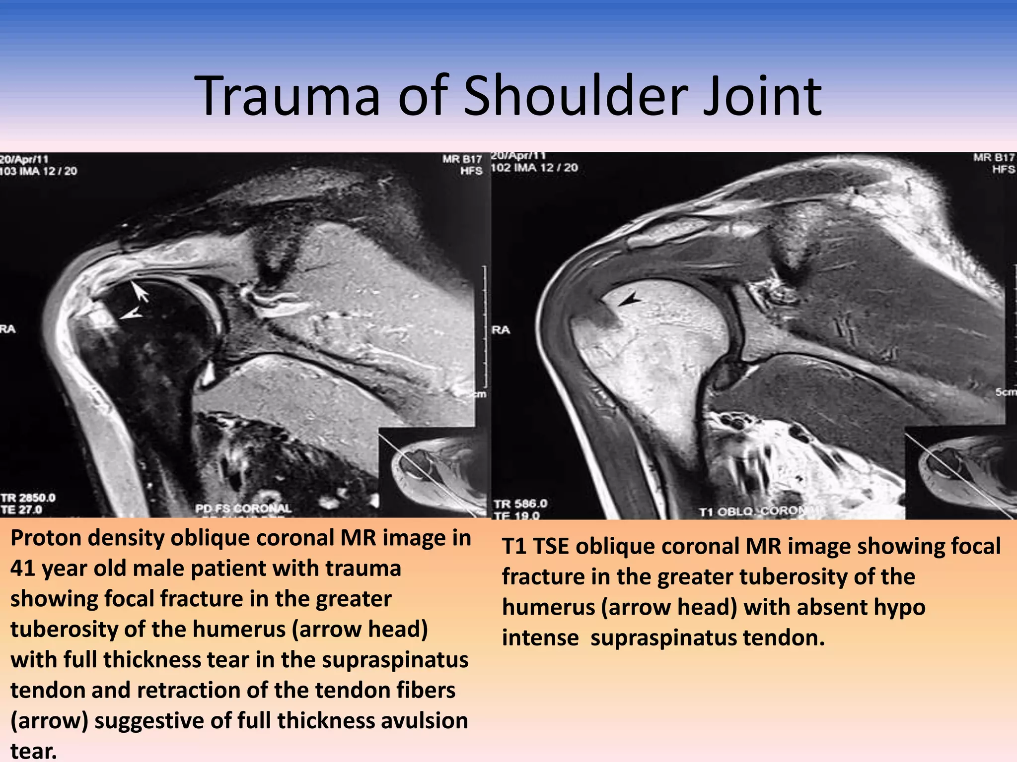
3) Imaging is also described for evaluating fractures in various bones including the skull, humerus, shoulder, wrist, hip and femur. Salter-Harris fractures in children and their classification are also covered.